medRxiv
  • Home
  • About
  • Submit
  • ALERTS / RSS
Advanced Search

Chloroquine Administration in Breastfeeding Mothers Associates with Increased HIV-1 Plasma Viral Loads

William A Paxton, Marloes A Naarding, Ferdinand WNM Wit, Nienke J Veldhuijzen, Matthew F Chersich, Brigitte Kankindi, Rene A Douma, Samuel Tuyizere, Suzanne Jurriaans, Rolf W Sparidans, Jos H Beijnen, Georgios Pollakis, Johan R Boelaert, Joep MA Lange, Joseph Vyankandondera, Stanley Luchters
doi: https://doi.org/10.1101/2020.04.29.20085308
William A Paxton
1Department of Clinical Infection, Microbiology and Immunology, Institute of Infection, Veterinary and Ecological Sciences, University of Liverpool, Liverpool, UK
2Laboratory of Experimental Virology, Department of Medical Microbiology, Amsterdam UMC, Locat AMC, University of Amsterdam, Amsterdam, Netherlands
  • Find this author on Google Scholar
  • Find this author on PubMed
  • Search for this author on this site
  • For correspondence: w.a.paxton{at}liverpool.ac.uk stanley.luchters{at}aku.edu
Marloes A Naarding
2Laboratory of Experimental Virology, Department of Medical Microbiology, Amsterdam UMC, Locat AMC, University of Amsterdam, Amsterdam, Netherlands
  • Find this author on Google Scholar
  • Find this author on PubMed
  • Search for this author on this site
Ferdinand WNM Wit
3Amsterdam Institute for Global Health and Development, Amsterdam, Netherlands
  • Find this author on Google Scholar
  • Find this author on PubMed
  • Search for this author on this site
Nienke J Veldhuijzen
3Amsterdam Institute for Global Health and Development, Amsterdam, Netherlands
  • Find this author on Google Scholar
  • Find this author on PubMed
  • Search for this author on this site
Matthew F Chersich
4Univ Witwatersrand, Fac Hlth Sci, Wits Reprod Hlth & HIV Inst, Johannesburg, South Africa
  • Find this author on Google Scholar
  • Find this author on PubMed
  • Search for this author on this site
Brigitte Kankindi
5CHUK, Centre Hospitalier Universitaire de Kigali and Belgian Technical Cooperation, Kigali, Rwanda
  • Find this author on Google Scholar
  • Find this author on PubMed
  • Search for this author on this site
Rene A Douma
5CHUK, Centre Hospitalier Universitaire de Kigali and Belgian Technical Cooperation, Kigali, Rwanda
  • Find this author on Google Scholar
  • Find this author on PubMed
  • Search for this author on this site
Samuel Tuyizere
5CHUK, Centre Hospitalier Universitaire de Kigali and Belgian Technical Cooperation, Kigali, Rwanda
  • Find this author on Google Scholar
  • Find this author on PubMed
  • Search for this author on this site
Suzanne Jurriaans
6Laboratory of Clinical Virology, Department of Medical Microbiology, Amsterdam UMC, Locat AMC, University of Amsterdam, Amsterdam, Netherlands
  • Find this author on Google Scholar
  • Find this author on PubMed
  • Search for this author on this site
Rolf W Sparidans
7Universiteit Utrecht, Department of Pharmaceutical Sciences, Section of Biomedical Analysis, Division of Drug Toxicology, Utrecht, The Netherlands
  • Find this author on Google Scholar
  • Find this author on PubMed
  • Search for this author on this site
Jos H Beijnen
7Universiteit Utrecht, Department of Pharmaceutical Sciences, Section of Biomedical Analysis, Division of Drug Toxicology, Utrecht, The Netherlands
  • Find this author on Google Scholar
  • Find this author on PubMed
  • Search for this author on this site
Georgios Pollakis
1Department of Clinical Infection, Microbiology and Immunology, Institute of Infection, Veterinary and Ecological Sciences, University of Liverpool, Liverpool, UK
2Laboratory of Experimental Virology, Department of Medical Microbiology, Amsterdam UMC, Locat AMC, University of Amsterdam, Amsterdam, Netherlands
  • Find this author on Google Scholar
  • Find this author on PubMed
  • Search for this author on this site
Johan R Boelaert
8Internal Medicine, Unit of Renal and Infectious Diseases, Algemeen Ziekenhuis St-Jan, Brugge, Belgium
  • Find this author on Google Scholar
  • Find this author on PubMed
  • Search for this author on this site
Joep MA Lange
3Amsterdam Institute for Global Health and Development, Amsterdam, Netherlands
  • Find this author on Google Scholar
  • Find this author on PubMed
  • Search for this author on this site
Joseph Vyankandondera
5CHUK, Centre Hospitalier Universitaire de Kigali and Belgian Technical Cooperation, Kigali, Rwanda
  • Find this author on Google Scholar
  • Find this author on PubMed
  • Search for this author on this site
Stanley Luchters
5CHUK, Centre Hospitalier Universitaire de Kigali and Belgian Technical Cooperation, Kigali, Rwanda
9Department of Population Health, Aga Khan University, Nairobi, Kenya
10Department of Public Health and Primary Care, International Centre for Reproductive Health (ICRH), Ghent University, Ghent, Belgium
11Department of Epidemiology and Preventive Medicine, Monash University, Australia
12Burnet Institute, Australia
  • Find this author on Google Scholar
  • Find this author on PubMed
  • Search for this author on this site
  • For correspondence: w.a.paxton{at}liverpool.ac.uk stanley.luchters{at}aku.edu
  • Abstract
  • Full Text
  • Info/History
  • Metrics
  • Data/Code
  • Preview PDF
Loading

Abstract

Chloroquine (CQ) and Hydroxychloroquine (HCQ) have been proposed to be effective at treating COVID-19 patients. We, and others, have previously reported on the capacity of CQ to reduce HIV-1 replication in vitro. We tested CQ administration in post-partum mothers on influencing HIV-1 viral loads in human milk as a means of lowering mother to child transmission. A Phase I/II, randomized, placebo-controlled study to evaluate chloroquine administration to reduce HIV-1 RNA levels in human milk: the CHARGE study. Thirty HIV-1 positive pregnant Rwandese women (CQ n = 20; placebo n = 10) were enrolled in a 16-week study, with the treatment group receiving a 200 mg oral dose of CQ daily. Base-line plasma viral load (pVL) measurements and CD4 counts were determined prior to delivery, and pVL, breast milk VL (bmVL) and CQ levels measured during treatment. For women receiving treatment, CQ concentration was higher in breast milk compared to plasma (over 2.5-fold), with a positive correlation between the levels in the two compartments (P < 0.003). A link between high CQ concentrations in plasma and high CD4 counts (P < 0.001) was observed. Surprisingly, we found a significant increase in pVL after CQ treatment in over half of the mothers (n=11; P < 0.001) and with no alteration to bmVL measurements. No specific amino acid alterations in the gp120 envelope sequences could be associated with CQ administration. CQ usage is associated with a significant increase to pVL in early breastfeeding mothers from Rwanda which cautions against the use of CQ in such individuals. Our results highlight a discrepancy between CQ effects on modulating HIV-1 replication in vitro versus in vivo and indicate caution when prescribing CQ to postpartum HIV-1 untreated mothers. This discrepancy should be taken into consideration when testing CQ or HCQ treatment in COVID-19 clinical trials, especially relating to the post-partum setting.

Introduction

The extent of COVID-19 disease (SARS-CoV-2) has accelerated efforts to identify readily-available, safe and effective or vaccines. Given its antiviral properties, studies are investigating the effectiveness of chloroquine (CQ) and hydroxychloroquine (HCQ) to treat people with confirmed coronavirus infection.

Chloroquine and HCQ are cheap, widely available and have been shown to reduce HIV-1 replication in vitro and in vivo [1–5]. We have previously shown that CQ can interfere with virus replication in CD4+ T lymphocytes and that the inhibition is likely conferred at the cellular level [6]. It has been shown that CQ treatment can interfere with the potential N-linked glycosylation (PNG) pattern of the gp120 envelope protein with the reported modulation of the 2G12 monoclonal antibody epitope [1] and the capacity by which HIV-1 can undergo viral transfer to CD4+ T lymphocytes via cells expressing DC-SIGN in vitro [6]. The endosomal pH increases with CQ treatment and is believed to reduce IL-6 production [7], thereby causing a down-regulation of HIV-1 production in chronically infected T-cells and monocyte cell lines [2,8]. Several clinical trials have previously shown that CQ and HCQ treatment can reduce pVL in vivo with no effect on CD4 counts [3,4].

Mother to child transmission of HIV-1 (MTCT) can occur either in-utero, intra-partum or via breastfeeding [9–11]. Transmission of HIV-1 to the infant via breastfeeding has been associated with high pVL and breast milk VL (bmVL), low CD4 count and the presence of sub-clinical mastitis [13,14]. CQ has been shown to be secreted in human milk [15–17] suggesting it may be effective at lowering bmVL. The presence of CQ in human milk has also been shown to be non-toxic to children [18], making it ideally suitable for such a purpose. This study was undertaken to test whether CQ treatment of HIV-1 infected breastfeeding mothers could be effective at lowering VL and potentially limiting MTCT, and may provide valuable lessons for its effect with other viral infections, such as SARS-CoV-2.

Methods

Study design, Participants and clinical evaluations

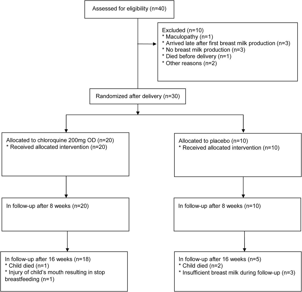
Between 2000–2001, we conducted a phase I/II randomized placebo-controlled pilot trial undertaken in Rwanda with 30 HIV-1 breastfeeding mothers randomized (2:1) to receive oral CQ 200 mg once daily or placebo (Pl) for 16 weeks (wks). All women received single-dose nevirapine (200 mg) at start of labor and children received single-dose nevirapine within 72 hrs. The study was approved by all the appropriate Ethic Committees (IEC) in both the Netherlands, the STEG-METC (ref no R01-089) and Rwanda and informed consent was obtained for all mothers. The CQ capsules, and matched placebo capsules, were prepared at the pharmacy department of the Slotervaart Hospital, Amsterdam, and concealed to the investigators I Rwanda (double-blinded). Patients were randomized on day 1 of breastfeeding (wk0) and the study un-blinded at wk18. All participants were confirmed HIV-1 infected (rapid tests), at least 18 years of age, therapy-naive, between 32–35 wks gestation and had not received CQ in the six wks prior. Pre-screen (psc) with follow-up and counseling visits were scheduled at wk0 (delivery), wk4, 8, 16, 17 and 18. All pVL were assessed at psc, wk0, 8 and 16 and bmVL at wk0, 8 and 16 and in both breasts. CQ concentrations were measured in blood plasma and from right (RB) and left (LB) breasts at wk0, 8 and 16. Both pVL and bmVL measurements were determined using the Roche diagnostics HIV-1 RNA PCR quantification assay.

Measuring CQ concentrations

To 1.0 ml plasma (supplemented with CQ free plasma if required) in a glass conical tube, 25 µl of internal standard solution (50 µg/ml diaminonaphthalene in methanol), 1 ml of 1M sodium hydroxide and 6 ml diethylether were added. After shaking for 10 min, the mixture was centrifuged for 5 min at ca. 2×10³ g. After storage of the tube for 1 h at −30°C, the organic layer was poured into a second tube and evaporated at 40°C under a gentle stream of nitrogen. Finally, the residue was reconstituted in 200 µl eluent using vortex-mixing and 50 µl was injected onto the HPLC. To 1.0 ml milk (supplemented with drug-free cow milk if required) in a glass conical tube, 6 ml n-hexane was added. The tube was shaken for 10 min and centrifuged for 5 min at ca. 2×10³ g. After storage of the tube for 1 h at −30°C, the organic layer was discarded. After thawing the aqueous layer, 1 ml of 1M sodium hydroxide and 6 ml diethylether were added and the procedure was continued according to the pre-treatment of plasma samples. Chromatographic conditions: Fifty µl injections were made on a Symmetry C18 column (100×4.6 mm, dp = 3.5 µm, average pore diameter = 10 nm, Waters), protected by a Symmetry C18 pre-column (20×3.8 mm, dp = 5 µm, Waters). The column temperature was maintained at ambient temperature (23–29°C). The eluent comprised 9% (v/v) acetonitrile and 91% (v/v) phosphate buffer (45 mM, pH 3.0) and the eluent flow rate was 1.0 mL/min. The UV detection wavelength was 340 nm.

PCR amplification and sequencing of gp120

Viral RNA was isolated from serum and the C1C4 gp120 region amplified by RT-PCR as previously described with the appropriate primers [19]. PCR products were cloned into the TOPO II vector (Invitrogen) and sequenced bi-directionally using the BigDye Terminator Cycle Sequencing kit (ABI). Primers A1360 GAG CCA ATT CCY ATA CAT TAT TG, SP6 ATT TAG GTG ACA CTA TAG and T7 TAA TAC GAC TCA CTA TAG GG were used for sequencing. Subtype determination and phylogenetic analysis was performed with the neighborhood-joining (N-J) analysis of MEGA [20]. PNG sites were determined using Glycosite (http://hiv-web.lanl.gov/content/hivdb/GLYCOSITE/glycosite.html).

Statistical analysis

Descriptive statistics for group comparison and regression analyses were performed utilizing the Prism package software (GraphPad software Inc., San Diego, California, USA, version 4.0). All available CD4 T cell counts, pVL and CQ level data were utilized, with the pVL values Log10-transformed. The Mann Whitney or the Wilcoxon signed rank tests were applied appropriately and the level of significance was set at P = 0.05.

Results

Clinical parameters of study individuals

The study participants are described in Table 1 (Placebo: n = 10, CQ: n = 20) and with a study schematic shown (Fig. 1). Of the 30 study individuals enrolled 7 did not finish due to early termination of breastfeeding or death of the child (Placebo: n = 5, CQ: n = 2). Infant pVL measurements taken at wk16 identify 6 infants to be infected, 1/5 in the Placebo group and 5/18 in the CQ group. From gp120 V3 region PCR analysis at wk0 we identified 3 infants to be HIV-1 positive at time of delivery with the remaining 3 infected either peri-partum or during the early breastfeeding period. Interestingly, the highest wk16 pVL were in those HIV-1 positive at time of delivery.

View this table:
  • View inline
  • View popup
  • Download powerpoint
Table 1 Study individuals
View this table:
  • View inline
  • View popup
  • Download powerpoint
Table 2.

Sequence analysis of gp120 of CQ and Placebo treated group

Fig. 1.
  • Download figure
  • Open in new tab
Fig. 1. Schematic of the proposal study design

30 mothers were enrolled with 20 belonging to the treatment arm and 10 to the placebo group.

CQ concentrations in breast milk are higher than in plasma

No differences in mean CD4 counts were identified between the Placebo and CQ groups at either wk0 or wk16, or between the different wks for both groups. The mean CQ concentration in plasma at wk8 is 759.7ng/ml (244–877ng/ml) and at wk16 478.2ng/ml (174– 414ng/ml). Of note, there was an association between high plasma CQ concentrations at wk8 with high CD4 counts, with those harboring concentrations of CQ over 1,200 ng/ml having the highest CD4 counts (P < 0.0001) but which did not correlate with pVL at wk16 (data not shown). CQ levels in breast milk are higher than in plasma at both wk8 and wk16 (Fig. 2a) with a positive correlation between CQ levels in plasma and breast milk at wk8 and wk16 (Fig. 2b). In summary, CQ levels are higher in breast milk than in plasma and at wk8 the CQ levels are on average 2.5 fold higher in breast milk.

Fig. 2.
  • Download figure
  • Open in new tab
Fig. 2. CQ levels after treatment

(a) CQ levels in the plasma, left breast and right breast of each study individual at wk8 and wk16 of treatment, with mean values depict. (b) correlation between plasma CQ levels and milk CQ levels at wk8

CQ treatment results in increased plasma but not breast milk HIV-1 viral loads

Mean pVL for CQ and Placebo groups are shown in Fig. 3a and 3c, respectively. No differences were observed between psc and wk0 or wk8 and wk16 for either group. The pVL values were, however, higher in the Placebo group compared to the CQ group at both psc and wk0 (2.5- and 2.1-fold, respectively). When comparing pVL measurements before and after treatment we observed a significant increase in the CQ group between psc and either wk8 or wk16 (P < 0.001) as well as wk0 and the later time-points (P < 0.001) (Fig. 3b). The mean pVL in the CQ treated group shows an increase of 1.0 log after wk8. Similar conclusions are drawn when we calculate the fold increase in pVL between wk8/wk0 or wk16/wk0, shown in Table 1 and Fig. 3b. The results show that over half of the CQ treated individuals harbor large increases in pVL by wk8, with the others showing little changes. We found bmVL to be consistently lower than pVL with many below the detection limit and with no differences between the CQ and Placebo groups (Fig 3a and 3c, respectively). When we compare the pVL with the bmVL in the CQ group we find a positive correlation (P<0.05). We also observe no difference in bmVL fold increase when comparing wk8 or wk16 with psc or wk0 for either the CQ or Pl group (Fig. 3b and Fig. 3d, respectively).

Fig. 3.
  • Download figure
  • Open in new tab
Fig. 3. HIV-1 viral load measurements before and after CQ treatment

(a) mean HIV-1 viral load measurements for plasma, LB and RB at psc, wk0, 8 and 16 for the CQ treated individuals (b) fold increase in viral load measurements in plasma, LB and RB between different weeks for the CQ treated individuals (* P < 0.05, ** P < 0.005) (c) mean HIV-1 viral load measurements for plasma, LB and RB at psc, wk0, 8 and 16 for the Pl treated individuals (d) fold increase in viral load measurements in plasma, LB and RB between the different weeks for the Pl treated individuals.

gp120 envelope genetic analysis before and after CQ treatment

Since we have previously reported genetic changes in the envelope of viruses passaged in the presence of CQ [6], we analyzed the gp120 C1C4 region DNA sequences in plasma from Pl (n = 4) and CQ individuals (n = 10) as designated in Table 1, before and after treatment (wk16). When comparing the overall amino acid charge of the V1V2 region we identified an increase over time for both the Pl and CQ treated groups and for the CQ treated individuals we found a link with increased pVL (P = 0.028), which was not observed for the Pl group. The V3 charge, which has been linked to coreceptor usage [21,22], was found to increase in 5/10 of the CQ treated individuals versus 1/4 of the Pl group, but was not statistically significant nor could be linked to increased pVL. A similar observation was found in the V4 regions. When comparing the alterations to the length of the variable regions no differences were found for either the Pl or CQ treated groups or with higher pVL in the CQ group. Similarly, alterations in the number of PNG sites did not alter in any of the variable regions in either the Placebo or CQ groups or between wk0 and wk16 of CQ treated individuals where pVL increased. Collectively, the results indicate no strong selection pressure exerted by CQ during the 16-week study period.

Discussion

High levels of CQ could accumulate in both plasma and breast milk, with substantially higher levels accumulating in breast milk. CQ treatment did not correlate with reduced HIV-1 bmVL, but surprisingly, did associate with increased pVL at both wk8 and wk16. This is in stark contrast to previous reports which have described decreases in pVL after CQ treatment [3,4]. Given the CQ concentration administered and a previous study showing that the drug dosage prescribed correlates to HCQ blood levels [23] it is unlikely that we have reached insufficient concentrations of the drug. Although the majority of in vitro studies to-date have described lower replication of HIV-1 in the presence of CQ, one study has indicated that high concentrations of CQ can be associated with increased viral entry [24]. It should also be noted that in animal models for various virus infections, including Semliki Forest Virus (SFV), CQ treatment can result in increased viral replication and modulate cytokine responses in vivo [25].

It has been reported that CQ treatment can decrease the glycosylation pattern of the HIV-1 gp120 protein [1]. When we compare the alterations to gp120 sequences from before and after CQ treatment we did not observe any major alteration to the V1-V2, V3 or V4 sequences, suggesting that CQ had no effect on selecting variants with altered replication phenotypes. Although glycosylation differences were not observed at the amino acid level it does not rule out the possibility that post-translational modification differences account for the results obtained i.e. altered recognition of HIV-1 by the neutralizing antibody repertoire leading to immune escape.

CD8+ T-lymphocyte responses have been shown to be modulated by CQ treatment, with higher levels of cellular activation mediated through an increase in antigen presentation by MHC class-I molecules [26]. The same scenario may hold true for CD4+ T-lymphocytes with an increase in antigenic presentation via MHC class-II molecules by antigen presenting cells, resulting in heightened cellular activation and increased viral replication. This, however, would not explain the lack of increase in pVL in previous CQ or HCQ studies [3,4]. The present results support a recent study where mothers receiving CQ did not show a decrease in pVL or bmVL [27]. The previous studies with CQ and HCQ have all been performed on male participants and we cannot rule out gender effects or differences due to child birth and/or lactation. It has been shown that bacterial translocation from the gut can be associated with heightened immune activation in HIV-1 infected individuals [28]. Whether CQ can influence bacterial growth in the gut or at the vaginal mucosa and ultimately modulate immune activation in treated mothers soon after delivery needs to be studied. It has been suggested CQ can restrict signaling via the toll like receptor 9 (TLR9), however, it has also been shown that with intestinal epithelial cells, CQ can augment cellular activation and thereby possibly upsetting colonic homeostasis [29].

It has been hypothesized that CQ could be used as a safe and cheap alternative to conventional HIV-1 antiretroviral pMTCT regimes and has been considered for use in resource-limited settings. More recently chloroquine as well as hydroxychloroquine treatment has been advocated for inhibiting SARS-CoV-2 replication and treating COVID-19 patients. Our results indicate caution when treating HIV-1 positive women in the post-natal period with CQ and especially those soon after childbirth where the likelihood of HIV-1 transmission through breastfeeding is present. Increased HIV-1 viral loads in mothers receiving CQ, opposed to the drugs observed inhibitory effects in vitro, provide caution in prescribing the drug as an effective antiviral to all patients. Increased viral loads in plasma of post-partum mothers may reflect hormonal effects which have been shown to associate with modulation of immune responses and where CQ treatment may have an effect on influencing HIV-1 activation and replication [30]. It has also been documented that CQ can have differential effects on activation of transcription factors, such as through NF-κB, with the potential to induce pro-inflammatory responses [31], with the potential consequences of increasing HIV-1 replication and plasma viral loads.

Several commentaries have proposed using CQ and HCQ to treat coronavirus COVID-19 as part of a ‘drug repositioning’ strategy [32–39]. CQ has displayed in vitro activity against the SARS-CoV-1 [40–43]. Middle East respiratory syndrome coronavirus (MERS-CoV) [44], human coronavirus 229E (HCoV-229E) [45] and HCoV-O43 [46,47]. While both HCQ and CQ display in vitro activity against SARS-CoV-2 [48,49], the evidence proposes HCQ as potentially exhibiting a stronger in vitro anti-SARS-CoV-2 activity, of the two drugs. The in vivo evidence is less distinct in that a study treating confirmed coronavirus patients with HCQ sulphate outperformed the control group [23] whereas a Shanghai [25] study reports the control having a slightly better outcome. It is apparent that more studies are required to inform the use of CQ or HCQ for SARS-CoV-2 treatment. Our findings of increased HIV-1 pVL with CQ in post-partum women should be taken into consideration when designing or administering CQ or HCQ in COVID-19 clinical trials, especially relating to the post-partum setting.

Data Availability

All data will be made available

Author Contributions

SL, JV, RWS, JHB, JRB, WAP and JML were involved with the clinical and scientific design of the study. SL was the Principal Investigator overseeing the study. SL, JV, FWW, NJV, BK and RD were involved with the clinical implementation of the study, data collection and clinical data analysis. MAN, RWS, SJ, JHB, GP and WAP were involved with experimental design and scientific data analysis. MAN, ST, performed experiments. WAP, MAN, MC, SL, wrote the manuscript.

Acknowledgements

This work was supported through Dutch AIDS Funds (6013) and Elizabeth Glaser Pediatric AIDS Foundation (27-PG-51269). We thank Dr Michel de Baar for critical review of the manuscript and we are indebted to the mothers and infants who participated in this study at the CHUK.

Footnotes

  • ↵+ Deceased

References

  1. 1.↵
    Savarino A, Gennero L, Chen HC, Serrano D, Malavasi F, Boelaert JR et al. Anti-HIV effects of chloroquine: mechanisms of inhibition and spectrum of activity. AIDS 2001; 15: 2221–2229.
    OpenUrlCrossRefPubMedWeb of Science
  2. 2.↵
    Sperber K, Kalb TH, Stecher VJ, Banerjee R, Mayer L. Inhibition of human immunodeficiency virus type 1 replication by hydroxychloroquine in T cells and monocytes. AIDS Res Hum Retroviruses 1993; 9: 91–98.
    OpenUrlCrossRefPubMedWeb of Science
  3. 3.↵
    Sperber K, Louie M, Kraus T, Proner J, Sapira E, Lin S et al. Hydroxychloroquine treatment of patients with human immunodeficiency virus type 1. Clin Ther 1995; 17: 622–636.
    OpenUrlCrossRefPubMedWeb of Science
  4. 4.↵
    Sperber K, Chiang G, Chen H, Ross W, Chusid E, Gonchar M et al. Comparison of hydroxychloroquine with zidovudine in asymptomatic patients infected with human immunodeficiency virus type 1. Clin Ther 1997; 19: 913–923.
    OpenUrlCrossRefPubMedWeb of Science
  5. 5.↵
    Tsai WP, Nara PL, Kung HF, Oroszlan S. Inhibition of human immunodeficiency virus infectivity by chloroquine. AIDS Res Hum Retroviruses 1990; 6: 481–489.
    OpenUrlCrossRefPubMedWeb of Science
  6. 6.↵
    Naarding MA, Baan E, Pollakis G, Paxton WA. Effect of chloroquine on reducing HIV-1 replication in vitro and the DC-SIGN mediated transfer of virus to CD4+ T-lymphocytes. Retrovirology 2007; 4: 6.
    OpenUrlCrossRefPubMed
  7. 7.↵
    Sperber K, Quraishi H, Kalb TH, Panja A, Stecher V, Mayer L. Selective regulation of cytokine secretion by hydroxychloroquine: inhibition of interleukin 1 alpha (IL-1-alpha) and IL-6 in human monocytes and T cells. J Rheumatol 1993; 20: 803–808.
    OpenUrlPubMedWeb of Science
  8. 8.↵
    Poli G, Bressler P, Kinter A, Duh E, Timmer WC, Rabson A et al. Interleukin 6 induces human immunodeficiency virus expression in infected monocytic cells alone and in synergy with tumor necrosis factor alpha by transcriptional and post-transcriptional mechanisms. J Exp Med 1990; 172: 151–158.
    OpenUrlAbstract/FREE Full Text
  9. 9.↵
    Nduati R, John G, Mbori-Ngacha D, Richardson B, Overbaugh J, Mwatha A et al. Effect of breastfeeding and formula feeding on transmission of HIV-1: a randomized clinical trial. Journal of American Medical Association 2000; 283: 1167–1174.
    OpenUrlCrossRefPubMedWeb of Science
  10. 10.
    Newell ML. Mechanisms and timing of mother-to-child transmission of HIV-1. AIDS 1998; 12: 831–837.
    OpenUrlCrossRefPubMedWeb of Science
  11. 11.↵
    Kourtis AP, Bulterys M, Nesheim SR, Lee FK. Understanding the timing of HIV transmission from mother to infant. Journal of American Medical Association 2001; 285: 709–712.
    OpenUrlCrossRefPubMedWeb of Science
  12. 12.
    Wiktor SZ, Ekpini E, Nduati RW. Prevention of mother-to-child transmission of HIV-1 in Africa. AIDS 1997; 11 (Suppl B):S79-S87.
    OpenUrlCrossRefWeb of Science
  13. 13.↵
    Bulterys M, Fowler MG, van Rompay KK, Kourtis AP. Prevention of mother-to-child transmission of HIV-1 through breast-feeding: past, present, and future. J Infect Dis. 2004; 189: 2149–2153.
    OpenUrlCrossRefPubMedWeb of Science
  14. 14.↵
    Shaffer N, Roongpisuthipong A, Siriwasin W, Chotpitayasunondh T, Chearskul S, Young NL et al. Maternal virus load and perinatal human immunodeficiency virus type 1 subtype E transmission, Thailand. Bangkok Collaborative Perinatal HIV Transmission Study Group. J InfectDis 1999; 179: 590–599.
    OpenUrlCrossRefPubMedWeb of Science
  15. 15.↵
    Ogunbona FA, Onyeji CO, Bolaji OO, Torimiro SE. Excretion of chloroquine and desethylchloroquine in human milk. BrJ Clin Pharmacol 1987; 23: 473–476.
    OpenUrlCrossRefPubMedWeb of Science
  16. 16.
    Nation RL, Hackett LP, Dusci LJ, Ilett KF. Excretion of hydroxychloroquine in human milk. Br.J Clin Pharmacol 1984; 17: 368–369.
    OpenUrlCrossRefPubMedWeb of Science
  17. 17.↵
    Boelaert JR, Yaro S, Augustijns P, Meda N, Schneider YJ, Schols D et al. Chloroquine accumulates in breast-milk cells: potential impact in the prophylaxis of postnatal mother-to-child transmission of HIV-1. AIDS 2001; 15: 2205–2207.
    OpenUrlCrossRefPubMedWeb of Science
  18. 18.↵
    Motta M, Tincani A, Faden D, Zinzini E, Lojacono A, Marchesi A et al. Follow-up of infants exposed to hydroxychloroquine given to mothers during pregnancy and lactation. J Perinatol 2005; 25: 86–89.
    OpenUrlCrossRefPubMed
  19. 19.↵
    Geels MJ, Dubey SA, Anderson K, Baan E, Bakker M, Pollakis G et al. Broad cross-clade T-cell responses to gag in individuals infected with human immunodeficiency virus type 1 non-B clades (A to G): importance of HLA anchor residue conservation. J Virol 2005; 79: 11247–11258.
    OpenUrlAbstract/FREE Full Text
  20. 20.↵
    Kumar S, Tamura K, Nei M. Molecular evolutionary genetics analysis (MEGA). Comput Appl Biosc 1994; 10: 189–191.
    OpenUrl
  21. 21.↵
    Pastore C, Ramos A, Mosier DE. Intrinsic obstacles to human immunodeficiency virus type 1 coreceptor switching. J Virol 2004; 78: 7565–7574.
    OpenUrlAbstract/FREE Full Text
  22. 22.↵
    Pastore C, Nedellec R, Ramos A, Pontow S, Ratner L, Mosier DE. Human immunodeficiency virus type 1 coreceptor switching: V1/V2 gain-of-fitness mutations compensate for V3 loss-offitness mutations. J Virol 2006; 80: 750–758.
    OpenUrlAbstract/FREE Full Text
  23. 23.↵
    Tett SE, Cutler DJ, Beck C, Day RO. Concentration-effect relationship of hydroxychloroquine in patients with rheumatoid arthritis--a prospective, dose ranging study. J Rheumatol 2000; 27: 1656–1660.
    OpenUrlPubMedWeb of Science
  24. 24.↵
    Fredericksen BL, Wei BL, Yao J, Luo T, Garcia JV. Inhibition of endosomal/lysosomal degradation increases the infectivity of human immunodeficiency virus. J Virol 2002; 76: 1144011446.
    OpenUrl
  25. 25.↵
    Seth P, Mani H, Singh AK, Banaudha KK, Madhavan S, Sidhu GS et al. Acceleration of viral replication and up-regulation of cytokine levels by antimalarials: implications in malaria-endemic areas. Am J TropMed Hyg 1999; 61: 180–186.
    OpenUrl
  26. 26.↵
    Schultz KR, Bader S, Paquet J, Li W. Chloroquine treatment affects T-cell priming to minor histocompatibility antigens and graft-versus-host disease. Blood 1995; 86: 4344–4352.
    OpenUrlAbstract/FREE Full Text
  27. 27.↵
    Semrau K, Kuhn L, Kasonde P, Sinkala M, Kankasa C, Shutes E et al. Impact of chloroquine on viral load in breast milk. Tro Med Int.Health 2006; 11: 800–803.
    OpenUrl
  28. 28.↵
    Brenchley JM, Price DA, Schacker TW, Asher TE, Silvestri G, Rao S et al. Microbial translocation is a cause of systemic immune activation in chronic HIV infection. Nat Med 2006; 12: 1365–1371.
    OpenUrlCrossRefPubMedWeb of Science
  29. 29.↵
    Lee J, Mo JH, Katakura K, Alkalay I, Rucker AN, Liu YT et al. Maintenance of colonic homeostasis by distinctive apical TLR9 signalling in intestinal epithelial cells. Nat Cell Biol 2006; 8: 1327–1336.
    OpenUrlCrossRefPubMedWeb of Science
  30. 30.↵
    Redelman D, Welniak LA, Taub D, Murphy WJ. Neuroendocrine Hormones such as Growth Hormone and Prolactin are Integral Members of the Immunological Cytokine Network. Cell Immunol 2008; 252: 111–121.
    OpenUrlCrossRefPubMedWeb of Science
  31. 31.↵
    Park J, Kwon D, Choi C, Oh J-H. Benveniste EN. Chloroquine induces activation of nuclear factor-κB and subsequent expression of pro-inflammatory cytokines by human astroglial cells. Journal of Neurochemistry 2003; 84: 1266–1274.
    OpenUrlCrossRefPubMed
  32. 32.↵
    Touret F, de Lamballerie X. Of chloroquine and COVID-19. Antiviral research. 2020; 177: 104762.
    OpenUrlCrossRefPubMed
  33. 33.
    Cunningham AC, Goh HP, Koh D. Treatment of COVID-19: old tricks for new challenges. Critical care (London, England). 2020; 24: 91.
    OpenUrl
  34. 34.
    Colson P, Rolain JM, Raoult D. Chloroquine for the 2019 novel coronavirus SARS-CoV-2. International journal of antimicrobial agents 2020; 55: 105923.
    OpenUrlCrossRefPubMed
  35. 35.
    Chang R, Wei-Zen S. Repositioning chloroquine as an ideal antiviral prophylaxis against COVID-19—Time is now. Preprints. 2020;https://www.preprints.org/manuscript/202003.0279/v1.
  36. 36.
    Zhou D, Dai SM, Tong Q. COVID-19: a recommendation to examine the effect of hydroxychloroquine in preventing infection and progression. J Antimicrob Chemother 2020.
  37. 37.
    Devaux CA, Rolain JM, Colson P, Raoult D. New insights on the antiviral effects of chloroquine against coronavirus: what to expect for COVID-19? International journal of antimicrobial agents. 2020: 105938.
  38. 38.
    Colson P, Rolain JM, Lagier JC, Brouqui P, Raoult D. Chloroquine and hydroxychloroquine as available weapons to fight COVID-19. International journal of antimicrobial agents 2020: 105932.
  39. 39.↵
    Kearney JC. Chloroquine as a Potential Treatment and Prevention Measure for the 2019 Novel Coronavirus: A Review. 2020; 10.20944/preprints202003.0275.v1.
  40. 40.↵
    Chen Q, Quan B, Li X, Gao G, Zheng W, Zhang J, et al. A report of clinical diagnosis and treatment of nine cases of coronavirus disease 2019. J Med Virol. 2020.
  41. 41.
    Keyaerts E, Vijgen L, Maes P, Neyts J, Van Ranst M. In vitro inhibition of severe acute respiratory syndrome coronavirus by chloroquine. Biochem Biophys Res Commun.2004;323:264268.
    OpenUrl
  42. 42.
    Vincent MJ, Bergeron E, Benjannet S, Erickson BR, Rollin PE, Ksiazek TG, et al. Chloroquine is a potent inhibitor of SARS coronavirus infection and spread. Virol J 2005; 2: 69.
    OpenUrlCrossRefPubMed
  43. 43.↵
    Biot C, Daher W, Chavain N, Fandeur T, Khalife J, Dive D, et al. Design and synthesis of hydroxyferroquine derivatives with antimalarial and antiviral activities. J Med Chem. 2006; 49: 2845–2849.
    OpenUrlCrossRefPubMed
  44. 44.↵
    de Wilde AH, Jochmans D, Posthuma CC, Zevenhoven-Dobbe JC, van Nieuwkoop S, Bestebroer TM, et al. Screening of an FDA-approved compound library identifies four small-molecule inhibitors of Middle East respiratory syndrome coronavirus replication in cell culture. Antimicrob Agents Chemother 2014;58:4875–4884.
    OpenUrlAbstract/FREE Full Text
  45. 45.↵
    Kono M, Tatsumi K, Imai AM, Saito K, Kuriyama T, Shirasawa H. Inhibition of human coronavirus 229E infection in human epithelial lung cells (L132) by chloroquine: involvement of p38 MAPK and ERK. Antiviral Res 2008; 77: 150–152.
    OpenUrlCrossRefPubMed
  46. 46.↵
    Keyaerts E, Li S, Vijgen L, Rysman E, Verbeeck J, Van Ranst M, et al. Antiviral activity of chloroquine against human coronavirus OC43 infection in newborn mice. Antimicrob Agents Chemother. 2009; 53: 3416–3421.
    OpenUrlAbstract/FREE Full Text
  47. 47.↵
    Shen L, Yang Y, Ye F, Liu G, Desforges M, Talbot PJ, et al. Safe and Sensitive Antiviral Screening Platform Based on Recombinant Human Coronavirus OC43 Expressing the Luciferase Reporter Gene. Antimicrob Agents Chemother 2016; 60: 5492–503.
    OpenUrlAbstract/FREE Full Text
  48. 48.↵
    Yao X, Ye F, Zhang M, Cui C, Huang B, Niu P, et al. In Vitro Antiviral Activity and Projection of Optimized Dosing Design of Hydroxychloroquine for the Treatment of Severe Acute Respiratory Syndrome Coronavirus 2 (SARS-CoV-2). Clinical infectious diseases: an official publication of the Infectious Diseases Society of America. 2020.
  49. 49.↵
    Liu J, Cao R, Xu M, Wang X, Zhang H, Hu H, et al. Hydroxychloroquine, a less toxic derivative of chloroquine, is effective in inhibiting SARS-CoV-2 infection in vitro. Cell Discov 2020; 6: 16.
    OpenUrl
Back to top
PreviousNext
Posted May 07, 2020.
Download PDF
Data/Code
Email

Thank you for your interest in spreading the word about medRxiv.

NOTE: Your email address is requested solely to identify you as the sender of this article.

Enter multiple addresses on separate lines or separate them with commas.
Chloroquine Administration in Breastfeeding Mothers Associates with Increased HIV-1 Plasma Viral Loads
(Your Name) has forwarded a page to you from medRxiv
(Your Name) thought you would like to see this page from the medRxiv website.
CAPTCHA
This question is for testing whether or not you are a human visitor and to prevent automated spam submissions.
Share
Chloroquine Administration in Breastfeeding Mothers Associates with Increased HIV-1 Plasma Viral Loads
William A Paxton, Marloes A Naarding, Ferdinand WNM Wit, Nienke J Veldhuijzen, Matthew F Chersich, Brigitte Kankindi, Rene A Douma, Samuel Tuyizere, Suzanne Jurriaans, Rolf W Sparidans, Jos H Beijnen, Georgios Pollakis, Johan R Boelaert, Joep MA Lange, Joseph Vyankandondera, Stanley Luchters
medRxiv 2020.04.29.20085308; doi: https://doi.org/10.1101/2020.04.29.20085308
Twitter logo Facebook logo LinkedIn logo Mendeley logo
Citation Tools
Chloroquine Administration in Breastfeeding Mothers Associates with Increased HIV-1 Plasma Viral Loads
William A Paxton, Marloes A Naarding, Ferdinand WNM Wit, Nienke J Veldhuijzen, Matthew F Chersich, Brigitte Kankindi, Rene A Douma, Samuel Tuyizere, Suzanne Jurriaans, Rolf W Sparidans, Jos H Beijnen, Georgios Pollakis, Johan R Boelaert, Joep MA Lange, Joseph Vyankandondera, Stanley Luchters
medRxiv 2020.04.29.20085308; doi: https://doi.org/10.1101/2020.04.29.20085308

Citation Manager Formats

  • BibTeX
  • Bookends
  • EasyBib
  • EndNote (tagged)
  • EndNote 8 (xml)
  • Medlars
  • Mendeley
  • Papers
  • RefWorks Tagged
  • Ref Manager
  • RIS
  • Zotero
  • Tweet Widget
  • Facebook Like
  • Google Plus One

Subject Area

  • HIV/AIDS
Subject Areas
All Articles
  • Addiction Medicine (349)
  • Allergy and Immunology (668)
  • Allergy and Immunology (668)
  • Anesthesia (181)
  • Cardiovascular Medicine (2648)
  • Dentistry and Oral Medicine (316)
  • Dermatology (223)
  • Emergency Medicine (399)
  • Endocrinology (including Diabetes Mellitus and Metabolic Disease) (942)
  • Epidemiology (12228)
  • Forensic Medicine (10)
  • Gastroenterology (759)
  • Genetic and Genomic Medicine (4103)
  • Geriatric Medicine (387)
  • Health Economics (680)
  • Health Informatics (2657)
  • Health Policy (1005)
  • Health Systems and Quality Improvement (985)
  • Hematology (363)
  • HIV/AIDS (851)
  • Infectious Diseases (except HIV/AIDS) (13695)
  • Intensive Care and Critical Care Medicine (797)
  • Medical Education (399)
  • Medical Ethics (109)
  • Nephrology (436)
  • Neurology (3882)
  • Nursing (209)
  • Nutrition (577)
  • Obstetrics and Gynecology (739)
  • Occupational and Environmental Health (695)
  • Oncology (2030)
  • Ophthalmology (585)
  • Orthopedics (240)
  • Otolaryngology (306)
  • Pain Medicine (250)
  • Palliative Medicine (75)
  • Pathology (473)
  • Pediatrics (1115)
  • Pharmacology and Therapeutics (466)
  • Primary Care Research (452)
  • Psychiatry and Clinical Psychology (3432)
  • Public and Global Health (6527)
  • Radiology and Imaging (1403)
  • Rehabilitation Medicine and Physical Therapy (814)
  • Respiratory Medicine (871)
  • Rheumatology (409)
  • Sexual and Reproductive Health (410)
  • Sports Medicine (342)
  • Surgery (448)
  • Toxicology (53)
  • Transplantation (185)
  • Urology (165)