medRxiv
  • Home
  • About
  • Submit
  • ALERTS / RSS
Advanced Search

The Role of Host Genetic Factors in Coronavirus Susceptibility: Review of Animal and Systematic Review of Human Literature

Marissa LoPresti, David B. Beck, Priya Duggal, Derek A. T. Cummings, Benjamin D. Solomon
doi: https://doi.org/10.1101/2020.05.30.20117788
Marissa LoPresti
1University of Florida College of Veterinary Medicine, Gainesville, Florida, United States of America
  • Find this author on Google Scholar
  • Find this author on PubMed
  • Search for this author on this site
David B. Beck
2Inflammatory Disease Section, National Human Genome Research Institute, National Institutes of Health, Bethesda, Maryland, United States of America
  • Find this author on Google Scholar
  • Find this author on PubMed
  • Search for this author on this site
Priya Duggal
3Department of Epidemiology, Johns Hopkins Bloomberg School of Public Health, Baltimore, Maryland, United States of America
  • Find this author on Google Scholar
  • Find this author on PubMed
  • Search for this author on this site
Derek A. T. Cummings
4Department of Biology, University of Florida, Gainesville, Florida, United States of America
5Emerging Pathogens Institute, University of Florida, Gainesville, Florida, United States of America
  • Find this author on Google Scholar
  • Find this author on PubMed
  • Search for this author on this site
Benjamin D. Solomon
6Office of the Clinical Director, National Human Genome Research Institute, National Institutes of Health, Bethesda, Maryland, United States America
  • Find this author on Google Scholar
  • Find this author on PubMed
  • Search for this author on this site
  • For correspondence: solomonb{at}mail.nih.gov
  • Abstract
  • Full Text
  • Info/History
  • Metrics
  • Supplementary material
  • Data/Code
  • Preview PDF
Loading

Abstract

Background The recent SARS-CoV-2 pandemic raises many scientific and clinical questions. One set of questions involves host genetic factors that may affect disease susceptibility and pathogenesis. New work is emerging related to SARS-CoV-2; previous work on other coronaviruses in humans or other host species may be relevant.

Objectives To review existing literature on host genetic factors and their association with infection and disease with coronaviruses in humans and in other host species.

Methods We conducted a systematic review of literature on host genetic factors in humans associated with coronavirus outcomes. We also reviewed studies of host genetic factors associated with coronavirus outcomes in non-human species. We categorized articles, summarized themes related to animal studies, and extracted data from human studies for analyses.

Results We identified 1,187 articles of potential relevance. Forty-five studies examined human host genetic factors related to coronavirus, of which 35 involved analysis of specific genes or loci; aside from one meta-analysis on respiratory infections, all were candidate-driven studies, typically investigating small numbers of research subjects and loci. Multiple significant loci were identified, including 16 related to susceptibility to coronavirus (of which 7 identified protective alleles), and 16 related to outcomes or clinical variables (of which 3 identified protective alleles). The types of cases and controls used varied considerably; four studies used traditional replication/validation cohorts. Of the other studies, 28 involved both human and non-human host genetic factors related to coronavirus, and 174 involved study of non-human (animal) host genetic factors related to coronavirus.

Key findings We have outlined key genes and loci from animal and human host genetic studies that may bear investigation in the nascent host genetic factor studies of COVID-19. Previous human studies have been limited by relatively low numbers of eligible participants and limited availability of advanced genomic methods. These limitations may be less important to studies of SARS-CoV-2.

Introduction

The ongoing severe acute respiratory syndrome coronavirus 2 (SARS-CoV-2) pandemic raises many scientific and clinical questions. One unknown is the extent to which individuals vary in susceptibility to infection and disease (COVID-19). Various hypotheses have been suggested to explain observed differences, including sex, age, comorbidities, and genetic factors.1 As with many complex diseases, the explanations likely involve a combination of genetic and non-genetic factors. In this context, genetic factors involve an interplay between virus and host genetics.2

Large, international studies and collaborations have formed to investigate host genetic factors related to COVID-19, including disease severity and susceptibility. These investigations include analyses of existing public and private datasets, as well as the establishment of new cohorts (e.g., https://blog.23andme.com/23andme-research/genetics-and-covid-19-severity/).3

While SARS-CoV-2 has seized recent attention, there are many other coronaviruses and a significant related body of literature exists about host genetic factors and their association with infection and outcomes in both humans and non-human host species. The Coronavirinae subfamily of the Coronaviridae family consists of four genera. The alphacoronaviruses include two major human coronaviruses, HCoV-229E (of which multiple HCoV-229E-like strains have been identified) and HCoV- NL63. Alphacoronaviruses that affect other species include mouse hepatitis virus (MHV), feline coronavirus (FCoV), which includes feline infectious peritonitis virus (FIPV) and feline enteric coronavirus (FECV), canine coronavirus (CCoV), and transmissible gastroenteritis coronavirus (TGEV) and porcine transmissible gastroenteritis coronavirus (TGEV) in pigs. The betacoronaviruses consist of four lineages: lineage A (HCoV-OC43 and HCoV-HKU1), lineage B (SARS-CoV-1 and SARS-CoV-2), lineage C (Middle East Respiratory Syndrome (MERS) and many bat coronaviruses), and lineage D (coronaviruses only identified in bats to date). HCoV-OC43, HCoV-229E, HCoV-HKU1, and HCoV-NL63 can result in a variety of presentations, including “common cold” and severe but rarely fatal disease; they are also frequently detected as co-infections with other viruses.4 There are other rare coronaviruses observed in humans as well as in other species.5,6 Relative to other coronaviruses, SARS-CoV-2 has unique biological properties and related clinical impact, but data regarding other coronaviruses may be relevant.

In various species, much work has focused on the genes encoding the relevant coronavirus receptor, including effects of viral and host genetic changes. Among other cell surface determinants,7 these receptor genes include ACE2 for HCoV-NL63,8 SARS-CoV-1,9,10 and SARS-CoV-2,11 ANPEP for HCoV- 229,12,13 FIPV,14 CCoV,15 and TGEV,16 DPP4 for MERS,17–19 and Ceacam1 for MHV.20 Host genetic studies have - to varying degrees and in different ways - analyzed these genes, as well as other genes identified through targeted and agnostic methods. Studies to date have been disparate in terms of the virus and species studied, as well as the aims of the particular study. This has resulted in a rich body of literature that is difficult to efficiently leverage for SARS-CoV-2-related work.

We aimed to perform a review of the literature to outline previous studies of host genetic factors related to coronaviruses, with the objective of performing a systematic review to encapsulate genes and loci interrogated through these efforts. We do not attempt to fully describe the findings nor recapitulate what is known about the underlying host biology related to coronavirus infection and disease. As the majority of studies are candidate-driven, we did not attempt to conduct a metaanalysis. However, one goal is that the data presented here can help populate lists of genes that - along with data from related work21–23 - may bear scrutiny in the developing and important large-scale host genetic studies related to SARS-CoV-2.24,25 We present an overview of themes and interrogated genes/loci from animal studies, and perform a systematic review on human studies.

Methods

We conducted an initial search of the PubMed database (last queried May 4, 2020) using each of the following phrases: “host genetics”; “genetic resistance”; “genetic susceptibility”; “genetic factors”; “genetics”; “GWAS” along with each of the following terms: “coronavirus”; “SARS”; “MERS”; “COVID- 19”; “COVID19”. We also identified additional articles by searching for specific coronaviruses or coronavirus-associated conditions (e.g., “canine coronavirus”; “middle east respiratory syndrome”) along with the term “genetics”. Articles were included in the search regardless of publication date. Articles included electronic, ahead-of-print publications available in the PubMed database. We also identified and categorized relevant articles from the references of initially selected articles. We did not include articles only available on non-peer reviewed preprint servers, though recognize that a substantial number of these manuscripts will be on PubMed soon.

Each abstract was reviewed by a single reviewer. Full articles were reviewed when insufficient data were available in the abstract, or when no abstract was available. Publications were classified into the following categories: 1) Study of human host genetic factors related to coronavirus; 2) Study of nonhuman (animal) host genetic factors related to coronavirus; 3) Study of non-genetic (including non-DNA- based analyses - see further explanation below) host factors related to coronavirus, including involving immunopathogenesis; 4) Study of other pathogens (not coronavirus); 5) Other studies of coronavirus. Articles containing information in both categories 1 and 2 were identified as such; articles were otherwise categorized according to the lowest numerical category (e.g., an article involving both human host genetic factors to coronavirus as well as immunopathogenesis would be categorized into group 1. Articles that did not involve investigations of specific DNA-based genetic changes (e.g., transcriptomic or proteomic studies) were categorized into group 3, as were studies that only included analyses of sex without other genetic analyses. Other publications, including: 6) Untranslated studies in another language (not English); 7) Not relevant (unrelated to coronavirus or other pathogens); 8) No data available; were removed from further analysis after categorization into these latter four categories. Data from category 1 publications were manually extracted for relevant information pertaining to: coronavirus studied; general methods and questions analyzed; gene(s), variant(s), or loci analyzed; size of cohorts studied; geographic or ancestral composition of cohorts; statistical results, including (where available) odds ratios, confidence intervals, and p-values.

Results

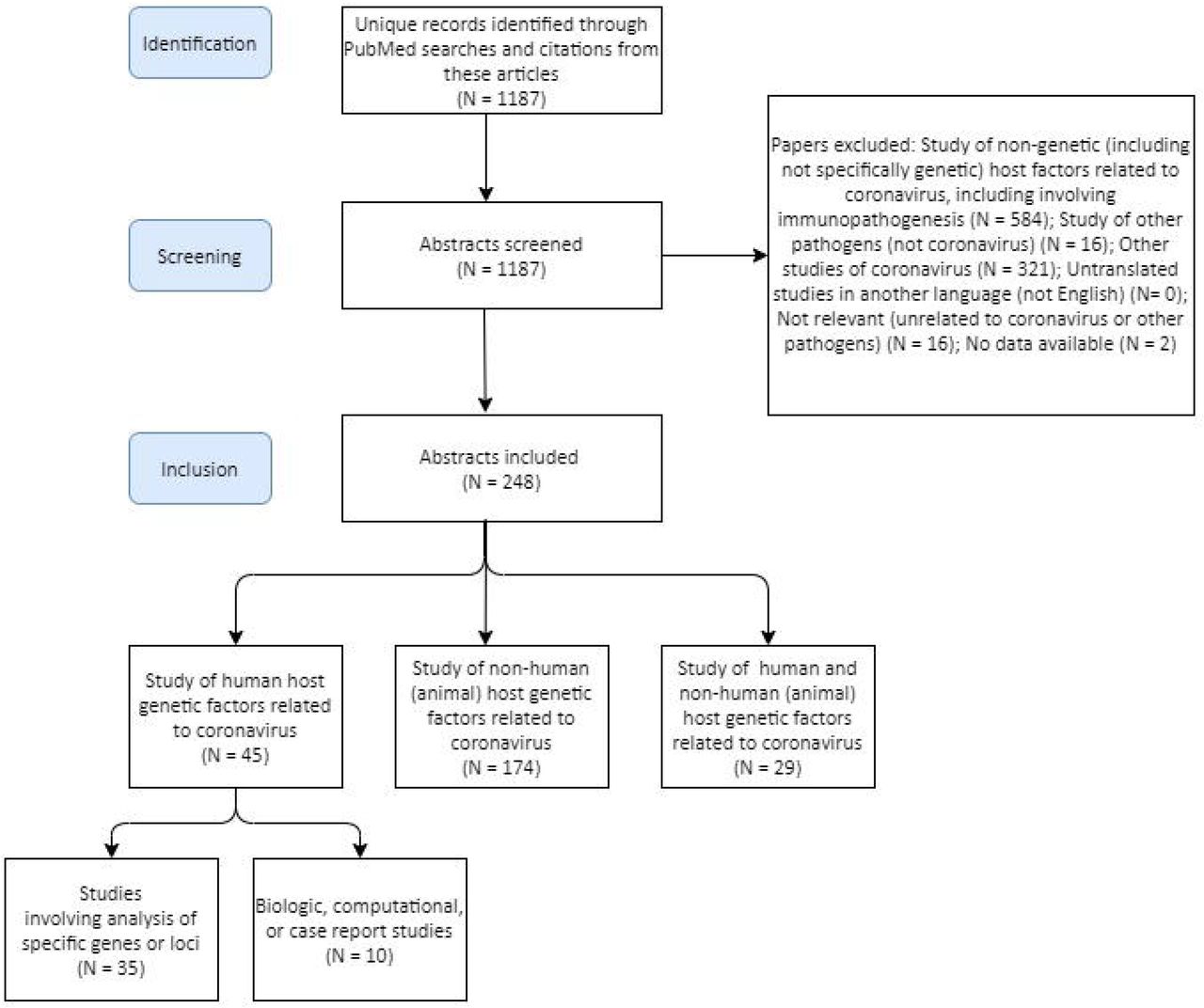
Our search identified 1,187 articles of potential relevance (Figure 1, Supplementary Table 1). Of these, 45 involved study of human host genetic factors related to coronavirus (Table 1); 35 of the 45 human studies involved analysis of specific genes or loci (only one was a non-candidate study), while 10 involved biological, computational, or case report studies of human host genetic factors. Twenty-eight involved both human and non-human host genetic factors related to coronavirus (these largely investigated inter-species differences in disease susceptibility and pathogenesis, such as related to differences in ACE2); 174 involved study of non-human (animal) host genetic factors related to coronavirus; 584 involved study of non-genetic host factors related to coronavirus, including involving immunopathogenesis; 16 involved study of other pathogens (not coronavirus); 321 involved other studies of coronavirus. 18 studies were assigned to the other categories and removed.

Figure 1.
  • Download figure
  • Open in new tab
Figure 1.

Description of articles identified through PubMed searches described in Methods.

View this table:
  • View inline
  • View popup
Table 1.

Summary of human studies (related to specific genes or loci) on host genetic factors related to coronaviruses. More details are available in Supplementary Table 2.

We organized our analysis and findings into the schema presented below.

Animal studies

Coronaviruses affect many species, from Beluga whales to spotted hyenas to turkeys, and sequelae of disease can range from apparently asymptomatic infections to severe or lethal effects on different organ systems, potentially manifesting as diarrheal, encephalitic, nephritic, respiratory, and other types of disease.26,27 There are numerous non-observational animal studies of coronaviruses, such as involving hamsters,28–30 guinea pigs,31 rats,10,32–35 and non-human primates.36–38 However, formal host genetic studies have been described for some but not all species. Many studies have involved examination of differences in species susceptibility and pathogenesis to human and non-human coronaviruses.14,39–41

Among the host genetic studies in animals, the objectives and methods used differ significantly depending on the species studied. For example, in chickens and other livestock, the types of published studies predictably differ from those conducted on experimental mice. That is, while MHV represents a problem for mouse colonies, the rationale of the livestock studies may focus more purely on economic repercussions versus attempts to use a model organism to understand immunopathogenesis of infectious disease.42 The degree to which results may be reported through the scientific literature (versus other routes) is also anticipated to differ depending on the species studied and the reason for the study. See Figure 2 for a summary of interrogated loci in animal studies.

Figure 2.
  • Download figure
  • Open in new tab
Figure 2.

Genes investigated in animal studies related to coronavirus disease. Human genes are shown only for those studies in multiple species analyses; other human gene details are presented elsewhere.

We describe representative studies and key findings below, but the descriptions should not be considered as truly comprehensive; additionally, as noted above, many studies compared susceptibility across species, both through cell-based assays and experimental animals. Many investigations using other methods (e.g., transcriptomics or proteomics) have identified key molecules involved in coronavirus susceptibility and pathogenesis. Though beyond the scope of this article, these molecules should also be considered in future SARS-CoV-2 host genetic studies.

Model animal strains, experimental animals, and domesticated animals

Chicken

In chickens, the infectious bronchitis virus (IBV) coronavirus can cause disease affecting different organ systems and tissues, such as IBV-associated nephritis. As with other species, inbred status and specific chicken lines impact host susceptibility, immune response, and outcomes, and virus/host genetic interactions have been described.43–47 Breeding experiments have suggested different inheritance patterns related to susceptibility and outcomes, and have implicated both MHC and non-MHC loci.48,49 Multiple GWAS investigating the immune response to IBV have identified significantly-associated polymorphisms in the breeds studied;50,51 the implicated or nearest genes include: AKT1, AvBD12, CEP170B, CRYL1, CWF19L2, DHRSX, FAM19A2, GABRB3, INTS9, NMNAT3, PINX1, RAB39A, VRK1, YEATS2; and SETBP1:50,51

Domestic cat

Felines can be infected by feline coronavirus (FCoV), which include feline infectious peritonitis (FIPV) and feline enteric coronavirus (FECV).52 As with other species, cats demonstrate a range of potential effects. In addition to association with traits such as age, sex, and reproductive status, purebred status and loss of heterozygosity has been shown to be associated with the effects of disease. Susceptibility and outcomes also appear to vary between different breeds.52–60 A small study of feline leukocyte antigen (FLA)-DRB alleles did not show a statistically significant association between the number of FLA-DRB alleles and FCoV infection outcome.61 Polymorphisms in IFNG (investigated as FIP can result in decreased interferon-gamma levels) were shown to correlate with plasma interferon-gamma levels and outcomes.62 Polymorphisms in TNFA and CD209 were also shown to be associated with outcomes in one inbred line.63

In addition to candidate studies, several GWAS have been performed in cats. One small study on outcomes in experimentally-induced infections in random-bred cats identified one associated genomic region (which did not harbor any obvious candidate genes).52 Another small study on an inbred breed identified multiple candidate genes (ELMO1, ERAP1, ERAP2, RRAGA, TNSF10) but none was fully concordant with the FIP disease phenotype.64 Recent studies on SARS-CoV-1 and SARS-CoV-2 have investigated the susceptibility of cats as well as other animals;65 see further details below (under Ferrets).

Dromedary camel

Camels are an important reservoir of coronaviruses that can infect humans; this became especially relevant in the context of MERS.66–68 Many studies have analyzed factors that contribute to spread,69 though the searches employed in this analysis identified relatively few host genetic studies separate from analyses of DPP4 receptor characteristics and tropism, including comparisons between camels, humans, and other species.70–74

Ferret

Several studies have investigated the susceptibility of various species to coronaviruses. One objective relates to identifying useful animal models of disease, in which non-human species show similar infection and disease outcomes to humans upon exposure to coronaviruses.65,75,76 For example bat, camel, and humans can be infected by MERS, unlike mouse, ferret, hamster, and guinea pig. SARS-CoV- 2 replicates better in ferrets and cats than in dogs, pigs, chickens, and ducks. One explanation involves genetic characteristics of the host receptor for the relevant virus.76,77 Additionally, within an infected animal, the site of viral replication appears to vary according to the species and coronavirus, and is additionally potentially related to tissue-specific receptor expression.78 This line of reasoning may also be relevant to age-specific differences observed with SARS-CoV-2 and human infections.79

Hamster

As noted above, hamsters have been used as model organisms to study coronaviruses, including studies of host receptors. This includes studies using standard hamster cell lines as well as other approaches involving hamster models.80–85 For example, hamster models have been used to study species susceptibility to MHV (related to Ceacam1),86 how alterations of specific Dpp4 amino acids in hamster affect susceptibility to MERS,71,87 and the roles of ACE2 and CD209L in SARS-CoV-1 susceptibility.82

Mouse

MHV has represented a challenge for the health of mouse colonies, though relatively recent improvements in animal care practices have been beneficial.88 Differences in the susceptibility of different mouse strains to MHV has been noted for seven decades.89–91 Studies have examined a number of different MHV strains. These strains demonstrate different tissue tropism and have different effects on various mouse lines.92 One distinct example is the JHM strain of MHV, which causes encephalitis in susceptible animals.93,94 In the discussion below, though susceptibility and outcome findings will be summarized, it is important to note that studies generally focus on the interactions between certain MHV strains and mouse lines, and it is not always clear how well these findings extrapolate to other strains and lines.

Many studies have investigated biological explanations for differences in MHV susceptibility and pathogenesis.95–97 Studies examining different laboratory mouse strains have suggested that multiple loci are involved.98–111 Early studies suggested various models, including potential monogenic/Mendelian explanations as well as more complex explanations involving interacting loci.92,112–115

Among many studies aiming to understand the underlying pathophysiology, mouse studies originally focused on strains believed to be involved in host susceptibility and reaction to infection. Importantly, these studies have identified interactions of host genetic factors with other factors, such as the cellular environment,116,117 cell and tissue-specific effects related to viral as well as host genetics,118–123 and host age.124–126 Unsurprisingly, some aspects of the disease process appear to be independent of observed strain differences.127 These studies also showed that host genetic factors influence different parts of the disease process, from initial virus-receptor binding,117 to cellular viral spreading128,129 and multiple aspects of the immune response.101,130–134 These studies enabled the cloning of Ceacam1, the MHV receptor gene,81 as well as related work regarding how genetic changes affecting this receptor confers MHV resistance in SJL mouse lines via inhibition of viral integration into host cells.103,106,135,136

In addition to the above studies, MHV-based mouse studies have used transgenic models to directly test the role of implicated pathways (summarized in Table 2). Not surprisingly, the majority of work in mouse models have focused on pathways already implicated in viral infection susceptibility including adaptive immune responses including both humoral and cellular, specific cytokine and immune receptor pathways, viral receptors, complement pathway, apoptosis, autophagy, and tissue repair. These studies have prominently implicated Type I (β) and II (γ) interferon responses in host response and predominantly protection against MHV infection. However, not all pro-inflammatory pathways are protective. For example, complement activation promotes tissue damage caused by MHV infection, highlighting the complex interplay between the host and virus. In addition to targeted gene disruptions described above, a GWAS using a recombinant inbred mouse panel implicated Trim55, which is involved in vascular cuffing and inflammation in response to SARS-CoV-1.137

View this table:
  • View inline
  • View popup
Table 2.

Summary of relevant mouse studies related to coronavirus. Note that the different studies have disparate objectives, many of which more directly involve aspects of immunopathogenesis versus standard host genetic questions regarding why specific genetic variants may affect disease susceptibility and outcomes.

Additional transgenic studies have investigated multiple biologic effects as well as returning to questions regarding susceptibility of different strains.138 Other mouse models (including knockouts, specific knockin mutations, humanized mice, and other models involving genetic manipulation) have been used to study human pathogens such as SARS-CoV and MERS; revealing similar properties for viral receptors, Dpp4 for MERS, Ace2 for SARS-CoV, cytokine and immune receptor pathways, and complement pathway as with mouse models of MHV. Intriguingly, there are differences between the importance of interferon pathways in host response to SARS-CoV1, where these pathways are dispensable as compared to MHV, where they are protective. Together, these different pathogen models have shown overlapping and unique pathways of host response between coronaviruses and highlight the potential relevance for SARS-CoV-2. See also the Additional papers on humans and other species section regarding further examples of studies involving mice and humans, as well as other species.

Pigs

Pigs can be infected by transmissible gastroenteritis virus (TGEV) and porcine epidemic diarrhea virus (PEDV), as well as the more recently-identified porcine deltacoronavirus (PDCoV). Like coronavirus disease in chickens, these diseases can have economic effects on the food industry,139 and analyses aim to address ways to ameliorate disease, such as the development of vaccines. Importantly, variants (both natural and experimentally-induced) may have different effects on different coronaviruses. For example, aminopeptidase N, encoded by ANPEP (also called APN) was reported as a functional receptor for TGEV and PEDV (as well as HCoV-229E), but multiple models, including CRISPR/Cas9-generated knock-outs, show differences in cellular susceptibility to TGEV and PEDV.139,140 In another study, infection by PEDV and TGEV correlated positively with ANPEP expression, but PEDV and TGEV could infect ANPEP-positive and negative enterocytes, with differences observed between viral strains.

Overall, the results suggest the presence of an additional receptor.141 Building on this type of work, site-specific editing of ANPEP has been suggested as a potential means to breed resistant animals.142 Studies focusing on PEDV have shown that knock-out of CMAH (hypothesized to affect cellular binding) does not result in immunity, but may improve outcomes.143

Rats

Rats can be affected by rat coronaviruses, and can be hosts to a number of different coronaviruses that affect other species.144,145 Rats have been used as model systems to investigate MHV, including through cellular-based assays.146,147 Several studies have examined rat susceptibility to various coronaviruses. As with many other studies, these have implicated key interactions between viral and host genetics that affect species and tissue tropism [17151094].33 In addition to computational approaches examining receptor characteristics, such as involving ACE2 in the context of SARS-CoV-110 experimental studies suggest that rats are not susceptible to MERS based on Dpp4 characteristics.148

Non-domesticated animals

As described, many species can be infected by coronaviruses. These species include wild as well as domesticated animals. The below section provides select examples of genetic studies on wild animals. Others studies been conducted on coronaviruses (as well as other pathogens),149 especially related to host ranges or reservoirs and involving host/pathogen co-evolution.150,151 Related to host genetic studies that are particularly relevant to the current SARS-CoV-2 pandemic (e.g., pangolin), our searches did not identify relevant articles.

Cheetah

Among wild animals, severe population bottlenecks (resulting in reduced genetic diversity) in cheetahs has been used to explain their increased susceptibility to infection by FIPV as well as other infectious diseases. Several such bottlenecks appear to have occurred in cheetah, due to a combination of factors.152–154 Among possible explanations for this susceptibility, genetic uniformity of the major histocompatibility complex (MHC) has been suspected to be involved.155

Civet

Studies have focused on palm civets (as well as other species) related to zoonotic implications as this species has been implicated as the reservoir associated with introduction of SARS-CoV-1 into humans. Specifically, questions about host receptor characteristics (ACE2) have been described in the context of SARS-CoV-1.156–158 As with other coronaviruses and species, the interactions of viral and host genetics have been shown to be important.159,160

Bat

As a natural reservoir for many coronaviruses, bats have been studied more extensively than other species outside of laboratory-based animals and livestock. Studies have included co-evolutionary studies between coronaviruses and the genomes of bat hosts (e.g., by correlating phylogenetic analyses of bat coronaviruses with CYTB in multiple bat species)161 as well as genetic/biologic studies related to host genetic factors. These have involved well-studied genes such as ACE2 with SARS-CoV-1162,163 and DPP4 with MERS.164,165 In addition to allowing analyses of host susceptibility, these and similar studies help provide estimates for the time-frame of coronavirus circulation in species and populations, and explore cross-species transmission.150

Human

Details of the human studies are presented in Table 1, Figure 3, and Supplementary Table 2. Forty-five studies were initially identified by the methods described. Of these, 35 involved association or other studies related to human host genetic factors (see summary in the next paragraph). Ten others involved biological, computational, or other non-genetic association studies. Many other studies were identified that used a combination of human and animal models, but were categorized separately; additionally, many studies that might be considered genetic studies - if the definition were applied less stringently - were grouped in category 3. For example, studies have examined how specific genes are involved in aspects of viral disease but did not strictly study how DNA-based host genetic variants affect this process. In summary, these ten included mapping of a susceptibility locus to HCoV-229E to chromosome 15.166 Multiple studies examined the biological effects of mutant genes. Studying the effects of mutant ACE2 on SARS-CoV-1 entry provided evidence that the cytoplasmic tail of ACE2 is not required for SARS-CoV-1 penetration.167 Studies of mutant TRIM56 on antiviral activity against HCoV-OC43 and other viruses showed that anti-HCoV-OC43 activity relies solely upon TRIM56 E3 ligase activity; this appears different from the mechanisms related to other viral pathogens.168 Knockout culture cells and nonsynonymous variant PPIA models result in limitation of HCoV-229E replication.169 (Please note that we did not separately or exhaustively investigate human genetic experiments involving cell culture systems.) Specific variants in IFITM genes (IFITM1 and IFITM3 were studied) modulate the entry of multiple human coronaviruses (HCoV-229E; HCoV-NL63; HCoV OC43; MERS-CoV; and SARS-CoV- 1 were studied).170 Computational models suggest that, while most ACE2 variants have similar binding affinity for SARS-CoV-2 spike protein, certain variants (rs73635825 and rs143936283) demonstrate different intermolecular interactions with the spike protein.171 An in silico analysis of viral peptide-MHC class I binding affinity related to HLA genotypes for SARS-CoV-2 peptides, as well as potential crossprotective immunity related to four common human coronaviruses, provides evidence that HLA-B*46:01 may be associated COVID-19 vulnerability, while HLA-B*15:03 may enable cross-protective T-cell based immunity.172 A recent study on viral cell entry showed that SARS-CoV-2 uses ACE2 for cell entry and TMPRSS2 for S protein priming; potential interventions based on these results include TMPRSS2 inhibition and convalescent sera.173 In addition to these examples, there are undoubtedly other biological, computational, and other studies examining how changes in and affecting key proteins may modulate disease.

Figure 3.
  • Download figure
  • Open in new tab
Figure 3.
  • Download figure
  • Open in new tab
Figure 3.

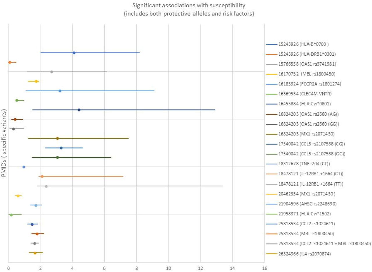
3A: Significant genetic associations with human susceptibility to coronavirus disease. Both protective and permissive genes are shown. Only studies reporting odds ratios (OR) and confidence intervals are shown. 3B: Significant genetic associations with human clinical variables and outcomes related to coronavirus disease. Both protective and permissive genes are shown. Only studies reporting odds ratios (OR) and confidence intervals (Cl) are shown (PMID 32348495 did not include Cl).

Of the 35 human studies meeting the host genetic study criteria described above, 32 (91%) involved SARS-CoV-1, while 3 (9%) involved SARS-CoV-2. Two of the three SARS-CoV-2 studies were case reports (one on a single family, the other on two patients with a rare immunodeficiency) without specific studies related to host factors; it is anticipated that many more studies on SARS-CoV-2 will be published soon.

All of the association studies except one were candidate-gene analyses based on genes hypothesized to be important in disease susceptibility or clinical variables/outcome. The exception was a meta-analysis of 386 studies on susceptibility to tuberculosis, influenza, respiratory syncytial virus, SARS-CoV-1, and pneumonia.174

Candidate studies ranged from studies of single variants to studies of over 50 genes selected due to biological plausibility; seven of these studies focused on HLA alleles. Sixteen significant loci related to susceptibility to coronavirus were reported (of which 7 identified protective alleles). Sixteen significant loci related to outcomes or clinical variables were reported (of which 3 identified protective alleles). The types of cases and controls used varied. Only four studies used separate cohorts for replication/validation. However, the studies used many different types of cases and controls, including within the same study. For example, some studies compared healthcare workers with SARS-CoV-1 infection with healthcare workers who tested negative. Others compared data from individuals with documented infection with data from control samples taken from blood donors. Four studies conducted laboratory-based biological studies in addition to association analyses. These studies are summarized in Table 1 and Figure 3; more details are available in Supplementary Table 2.

Additional papers on humans and other species

As described, human and animal studies have examined various host factors related to coronavirus infection. For example, human175 and animal33,52,125 studies have implicated age as having significant associations with outcomes; age appears to be strongly correlated with COVID-19 outcomes.176 The overall explanations remain unclear, but could at least partially involve age-related gene expression. Sex also appears to have a role. Human studies of SARS-CoV-1 and SARS-CoV2 suggest a correlation between sex and certain clinical parameters, perhaps rooted in sex-based or related immunologic differences.175,177,178 However, separating biological differences from sex-related cultural practices (e.g., different rates of social distancing) may be difficult.

Animal studies also suggest sex effects in multiple species, such as related to disease severity.57,179 Multiple studies examined different genes/proteins to determine disease susceptibility, transmissibility, and pathogenesis in various species. In addition to humanized genes, such as used in mouse models, studies have involved a combination of computational and biological approaches, and have investigated the viral entry receptors ACE2 in SARS-CoV-110,35,157,158,180–183 and SARS-CoV-2184 (for which there already exists a large body of unpublished and preprint work) and DPP4 in MERS.71,148,185–188 Among other findings, these studies examined specific protein residues that are critical in viral-host interactions [18448527],157 Other studies examined manipulations of various genes/proteins to study the functional biological effects, including of ANPEP,189 GLTSCR2,190 IFITM1, IFITM2, and IFITM3,191 and MAVS.192

Discussion: human studies

Traditional genome-wide methods have been applied to human viral infections generally,174,193 but results have not been specific to coronaviruses, and it is unclear to what extent the observations are relevant to the current pandemic. Several dozen studies have investigated human genetic factors related to coronavirus infection. However, these studies have been limited by several potential factors. For endemic human coronaviruses, the mildness of disease may have deprioritized these studies; similar observations may explain the relative dearth of serologic knowledge related to these pathogens.6 For coronaviruses associated with more severe human disease, such as MERS and SARS-CoV-1, the fact that these epidemics were limited more than the current pandemic crisis may have fortunately led to a lack of cases with which one might conduct traditional association studies (unlike some other respiratory infections leading to more widespread disease).194,195 Additionally, these two severe conditions primarily affected human populations prior to the technological developments that led to wide availability of much cheaper and faster genomic sequencing.196

As shown (Supplementary Table 2), the small sample sizes of previous studies may have led to the preponderance of candidate gene studies. The sample sizes may also have precluded significant findings due to limitations of statistical power and the ability to replicate or validate findings. As previous human studies occurred in areas of the world affected by the coronavirus studied, it is possible that results from these studies would not extrapolate to other populations. Finally, different genes and loci are involved than those previously hypothesized. That is, hypothesis-free approaches may identify significant loci that were not identified by candidate approaches.

Based on announcements about multiple large-scale projects on host genetic factors and SARS-CoV-2, as well as the existence of larger genomic datasets that can be mined quickly and new methods that can be used to address biological questions,197 it is anticipated that considerable efforts - and an unfortunately large pool of research subjects - and may yield significant new results quickly.

Limitations

There are multiple limitations to our summaries and analyses. First, it is likely that relevant articles were missed by our search process, and that key findings - including the study of certain genes - were therefore omitted. Along these lines, important findings within identified articles may also have been missed. Second, this analysis focused on DNA-based variants. These DNA-based genetic changes include those studied and identified through association studies as well as genes that were manipulated in experimental approaches, such as via knockout models to understand disease pathogenesis. Related ‘omic approaches, such as targeted or broad transcriptomic or proteomic studies, are frequently used to understand important aspects of disease. These approaches can lead to knowledge regarding specific genetic changes. For example, observed transcriptomic changes may enable the identification of important DNA-based variants that explain disease by correlating transcriptomic data with results of DNA sequencing.198 However, we categorized non-DNA based ‘omic approaches separately from DNA- based studies, and did not attempt to comprehensively recapitulate what is known about host reaction to disease. Finally, as the studies varied in many aspects, such as how cases and controls were defined, and which loci were interrogated, we were careful about comparing or combining data between different studies.

Data Availability

All data referred to in the manuscript are available either in the main manuscript or in supporting files.

Acknowledgments

This research was supported [in part] by the Intramural Research Program of the National Human Genome Research Institute, National Institutes of Health. PD was supported by the US National Institutes of Health award number 2R01AI148049–21A1. DATC was supported by the US National Institutes of Health award number R01-AI114703–01.

REFERENCE

  1. 1.↵
    Zhang X, Tan Y, Ling Y, et al. Viral and host factors related to the clinical outcome of COVID-19. Nature 2020.
  2. 2.↵
    Letko M, Miazgowicz K, McMinn R, et al. Adaptive Evolution of MERS-CoV to Species Variation in DPP4. Cell Rep 2018;24:1730–7.
    OpenUrlCrossRefPubMed
  3. 3.↵
    Initiative C-HG. The COVID-19 Host Genetics Initiative, a global initiative to elucidate the role of host genetic factors in susceptibility and severity of the SARS-CoV-2 virus pandemic. Eur J Hum Genet 2020.
  4. 4.↵
    Gaunt ER, Hardie A, Claas EC, Simmonds P, Templeton KE. Epidemiology and clinical presentations of the four human coronaviruses 229E, HKU1, NL63, and OC43 detected over 3 years using a novel multiplex real-time PCR method. J Clin Microbiol 2010;48:2940–7.
    OpenUrlAbstract/FREE Full Text
  5. 5.↵
    Perlman S, Dandekar AA. Immunopathogenesis of coronavirus infections: implications for SARS. Nat Rev Immunol 2005;5:917–27.
    OpenUrlCrossRefPubMed
  6. 6.↵
    Huang AT, Garcia-Carrera B, Hitchings MDT, et al. A systematic review of antibody immunity to coronaviruses: antibody kinetics, correlates of protection, and association of antibody responses with severity of disease. medRxiv 2020.
  7. 7.↵
    Bakkers MJ, Zeng Q, Feitsma LJ, et al. Coronavirus receptor switch explained from the stereochemistry of protein-carbohydrate interactions and a single mutation. Proc Natl Acad Sci U S A 2016;113:E3111–9.
    OpenUrlAbstract/FREE Full Text
  8. 8.↵
    Hofmann H, Pyre K, van der Hoek L, Geier M, Berkhout B, Pohlmann S. Human coronavirus NL63 employs the severe acute respiratory syndrome coronavirus receptor for cellular entry. Proc Natl Acad Sci US A 2005;102:7988–93.
    OpenUrlAbstract/FREE Full Text
  9. 9.↵
    Li W, Moore MJ, Vasilieva N, et al. Angiotensin-converting enzyme 2 is a functional receptor for the SARS coronavirus. Nature 2003;426:450–4.
    OpenUrlCrossRefPubMed
  10. 10.↵
    Li KK, Yip CW, Hon CC, Lam CY, Zeng F, Leung FC. Characterisation of animal angiotensinconverting enzyme 2 receptors and use of pseudotyped virus to correlate receptor binding with susceptibility of SARS-CoV infection. Hong Kong Med J 2012;18 Suppl 3:35–8.
    OpenUrl
  11. 11.↵
    Yan R, Zhang Y, Li Y, Xia L, Guo Y, Zhou Q. Structural basis for the recognition of SARS-CoV-2 by full-length human ACE2. Science 2020;367:1444–8.
    OpenUrlAbstract/FREE Full Text
  12. 12.↵
    Yeager CL, Ashmun RA, Williams RK, et al. Human aminopeptidase N is a receptor for human coronavirus 229E. Nature 1992;357:420–2.
    OpenUrlCrossRefPubMed
  13. 13.↵
    Li Z, Tomlinson AC, Wong AH, et al. The human coronavirus HCoV-229E S-protein structure and receptor binding. Elife 2019;8.
  14. 14.↵
    Tresnan DB, Levis R, Holmes KV. Feline aminopeptidase N serves as a receptor for feline, canine, porcine, and human coronaviruses in serogroup I. J Virol 1996;70:8669–74.
    OpenUrlAbstract/FREE Full Text
  15. 15.↵
    Benbacer L, Kut E, Besnardeau L, Laude H, Delmas B. Interspecies aminopeptidase-N chimeras reveal species-specific receptor recognition by canine coronavirus, feline infectious peritonitis virus, and transmissible gastroenteritis virus. J Virol 1997;71:734–7.
    OpenUrlAbstract/FREE Full Text
  16. 16.↵
    Delmas B, Gelfi J, L’Haridon R, et al. Aminopeptidase N is a major receptor for the entero-pathogenic coronavirus TGEV. Nature 1992;357:417–20.
    OpenUrlCrossRefPubMed
  17. 17.↵
    Wang N, Shi X, Jiang L, et al. Structure of MERS-CoV spike receptor-binding domain complexed with human receptor DPP4. Cell Res 2013;23:986–93.
    OpenUrlCrossRefPubMedWeb of Science
  18. 18.
    Raj VS, Mou H, Smits SL, et al. Dipeptidyl peptidase 4 is a functional receptor for the emerging human coronavirus-EMC. Nature 2013;495:251–4.
    OpenUrlCrossRefPubMedWeb of Science
  19. 19.↵
    Peck KM, Scobey T, Swanstrom J, et al. Permissivity of Dipeptidyl Peptidase 4 Orthologs to Middle East Respiratory Syndrome Coronavirus Is Governed by Glycosylation and Other Complex Determinants. J Virol 2017;91.
  20. 20.↵
    Williams RK, Jiang GS, Holmes KV. Receptor for mouse hepatitis virus is a member of the carcinoembryonic antigen family of glycoproteins. Proc Natl Acad Sci U S A 1991;88:5533–6.
    OpenUrlAbstract/FREE Full Text
  21. 21.↵
    Rockx B, Baas T, Zornetzer GA, et al. Early upregulation of acute respiratory distress syndrome-associated cytokines promotes lethal disease in an aged-mouse model of severe acute respiratory syndrome coronavirus infection. J Virol 2009;83:7062–74.
    OpenUrlAbstract/FREE Full Text
  22. 22.
    Ostaszewski M, Mazein A, Gillespie ME, et al. COVID-19 Disease Map, building a computational repository of SARS-CoV-2 virus-host interaction mechanisms. Sci Data 2020;7:136.
    OpenUrl
  23. 23.↵
    de Lang A, Baas T, Teal T, et al. Functional genomics highlights differential induction of antiviral pathways in the lungs of SARS-CoV-infected macaques. PLoS Pathog 2007;3:e112.
    OpenUrlCrossRefPubMed
  24. 24.↵
    Murray MF, Kenny EE, Ritchie MD, et al. COVID-19 outcomes and the human genome. Genet Med 2020.
  25. 25.↵
    Zhou W, Zhao Z, Nielsen JB, et al. Scalable generalized linear mixed model for region-based association tests in large biobanks and cohorts. Nat Genet 2020.
  26. 26.↵
    East ML, Moestl K, Benetka V, et al. Coronavirus infection of spotted hyenas in the Serengeti ecosystem. Vet Microbiol 2004;102:1–9.
    OpenUrlCrossRefPubMedWeb of Science
  27. 27.↵
    Le Poder S. Feline and canine coronaviruses: common genetic and pathobiological features. Adv Virol 2011;2011:609465.
  28. 28.↵
    Roberts A, Vogel L, Guarner J, et al. Severe acute respiratory syndrome coronavirus infection of golden Syrian hamsters. J Virol 2005;79:503–11.
    OpenUrlAbstract/FREE Full Text
  29. 29.
    Chan JF, Zhang AJ, Yuan S, et al. Simulation of the clinical and pathological manifestations of Coronavirus Disease 2019 (COVID-19) in golden Syrian hamster model: implications for disease pathogenesis and transmissibility. Clin Infect Dis 2020.
  30. 30.↵
    Lau SY, Wang P, Mok BW, et al. Attenuated SARS-CoV-2 variants with deletions at the S1/S2 junction. Emerg Microbes Infect 2020;9:837–42.
    OpenUrl
  31. 31.↵
    Liang L, He C, Lei M, et al. Pathology of guinea pigs experimentally infected with a novel reovirus and coronavirus isolated from SARS patients. DNA Cell Biol 2005;24:485–90.
    OpenUrlCrossRefPubMed
  32. 32.↵
    Liu RY, Wu LZ, Huang BJ, et al. Adenoviral expression of a truncated S1 subunit of SARS-CoV spike protein results in specific humoral immune responses against SARS-CoV in rats. Virus Res 2005;112:24–31.
    OpenUrlCrossRefPubMedWeb of Science
  33. 33.↵
    Nagata N, Iwata N, Hasegawa H, et al. Participation of both host and virus factors in induction of severe acute respiratory syndrome (SARS) in F344 rats infected with SARS coronavirus. J Virol 2007;81:1848–57.
    OpenUrlAbstract/FREE Full Text
  34. 34.
    Fukushi S, Mizutani T, Sakai K, et al. Amino acid substitutions in the s2 region enhance severe acute respiratory syndrome coronavirus infectivity in rat angiotensin-converting enzyme 2-expressing cells. J Virol 2007;81:10831–4.
    OpenUrlAbstract/FREE Full Text
  35. 35.↵
    Ren W, Qu X, Li W, et al. Difference in receptor usage between severe acute respiratory syndrome (SARS) coronavirus and SARS-like coronavirus of bat origin. J Virol 2008;82:1899–907.
    OpenUrlAbstract/FREE Full Text
  36. 36.↵
    Fouchier RA, Kuiken T, Schutten M, et al. Aetiology: Koch’s postulates fulfilled for SARS virus. Nature 2003;423:240.
    OpenUrlCrossRefPubMedWeb of Science
  37. 37.
    Rowe T, Gao G, Hogan RJ, et al. Macaque model for severe acute respiratory syndrome. J Virol 2004;78:11401–4.
    OpenUrlAbstract/FREE Full Text
  38. 38.↵
    McAuliffe J, Vogel L, Roberts A, et al. Replication of SARS coronavirus administered into the respiratory tract of African Green, rhesus and cynomolgus monkeys. Virology 2004;330:8–15.
    OpenUrlCrossRefPubMedWeb of Science
  39. 39.↵
    Compton SR, Stephensen CB, Snyder SW, Weismiller DG, Holmes KV. Coronavirus species specificity: murine coronavirus binds to a mouse-specific epitope on its carcinoembryonic antigen-related receptor glycoprotein. J Virol 1992;66:7420–8.
    OpenUrlAbstract/FREE Full Text
  40. 40.
    Gagneten S, Scanga CA, Dveksler GS, Beauchemin N, Percy D, Holmes KV. Attachment glycoproteins and receptor specificity of rat coronaviruses. Lab Anim Sci 1996;46:159–66.
    OpenUrlPubMed
  41. 41.↵
    Baric RS, Yount B, Hensley L, Peel SA, Chen W. Episodic evolution mediates interspecies transfer of a murine coronavirus. J Virol 1997;71:1946–55.
    OpenUrlAbstract/FREE Full Text
  42. 42.↵
    Bumstead N. Genetic resistance to avian viruses. Rev Sci Tech 1998;17:249–55.
    OpenUrlPubMedWeb of Science
  43. 43.↵
    Cook J, Otsuki K, Huggins M, Bumstead N. Investigations into resistance of chicken lines to infection with infectious bronchitis virus. Adv Exp Med Biol 1990;276:491–6.
    OpenUrlPubMed
  44. 44.
    Ignjatovic J, Reece R, Ashton F. Susceptibility of three genetic lines of chicks to infection with a nephropathogenic T strain of avian infectious bronchitis virus. J Comp Pathol 2003;128:92–8.
    OpenUrlCrossRefPubMed
  45. 45.
    Asif M, Lowenthal JW, Ford ME, Schat KA, Kimpton WG, Bean AG. Interleukin-6 expression after infectious bronchitis virus infection in chickens. Viral Immunol 2007;20:479–86.
    OpenUrlCrossRefPubMedWeb of Science
  46. 46.
    Dawes ME, Griggs LM, Collisson EW, Briles WE, Drechsler Y. Dramatic differences in the response of macrophages from B2 and B19 MHC-defined haplotypes to interferon gamma and polyinosinic:polycytidylic acid stimulation. Poult Sci 2014;93:830–8.
    OpenUrlCrossRefPubMed
  47. 47.↵
    da Silva AP, Hauck R, Zhou H, Gallardo RA. Understanding Immune Resistance to Infectious Bronchitis Using Major Histocompatibility Complex Chicken Lines. Avian Dis 2017;61:358–65.
    OpenUrl
  48. 48.↵
    Bumstead N, Huggins MB, Cook JK. Genetic differences in susceptibility to a mixture of avian infectious bronchitis virus and Escherichia coli. Br Poult Sci 1989;30:39–48.
    OpenUrlCrossRefPubMed
  49. 49.↵
    Bacon LD, Hunter DB, Zhang HM, Brand K, Etches R. Retrospective evidence that the MHC (B haplotype) of chickens influences genetic resistance to attenuated infectious bronchitis vaccine strains in chickens. Avian Pathol 2004;33:605–9.
    OpenUrlCrossRefPubMedWeb of Science
  50. 50.↵
    Luo C, Qu H, Ma J, et al. A genome-wide association study identifies major loci affecting the immune response against infectious bronchitis virus in chicken. Infect Genet Evol 2014;21:351–8.
    OpenUrl
  51. 51.↵
    Wang W, Zhang T, Zhang G, et al. Genome-wide association study of antibody level response to NDV and IBV in Jinghai yellow chicken based on SLAF-seq technology. J Appl Genet 2015;56:365–73.
    OpenUrl
  52. 52.↵
    Pedersen NC, Liu H, Gandolfi B, Lyons LA. The influence of age and genetics on natural resistance to experimentally induced feline infectious peritonitis. Vet Immunol Immunopathol 2014;162:33–40.
    OpenUrlCrossRefPubMed
  53. 53.
    Robison RL, Holzworth J, Gilmore CE. Naturally occurring feline infectious peritonitis: signs and clinical diagnosis. J Am Vet Med Assoc 1971;158:Suppl 2:981–6.
    OpenUrl
  54. 54.
    Foley JE, Poland A, Carlson J, Pedersen NC. Risk factors for feline infectious peritonitis among cats in multiple-cat environments with endemic feline enteric coronavirus. J Am Vet Med Assoc 1997;210:1313–8.
    OpenUrlPubMed
  55. 55.
    Rohrbach BW, Legendre AM, Baldwin CA, Lein DH, Reed WM, Wilson RB. Epidemiology of feline infectious peritonitis among cats examined at veterinary medical teaching hospitals. J Am Vet Med Assoc 2001;218:1111–5.
    OpenUrlCrossRefPubMedWeb of Science
  56. 56.
    Pesteanu-Somogyi LD, Radzai C, Pressler BM. Prevalence of feline infectious peritonitis in specific cat breeds. J Feline Med Surg 2006;8:1–5.
    OpenUrlCrossRefPubMed
  57. 57.↵
    Norris JM, Bosward KL, White JD, Baral RM, Catt MJ, Malik R. Clinicopathological findings associated with feline infectious peritonitis in Sydney, Australia: 42 cases (1990–2002). Aust Vet J 2005;83:666–73.
    OpenUrlCrossRefPubMedWeb of Science
  58. 58.
    Worthing KA, Wigney Dl, Dhand NK, et al. Risk factors for feline infectious peritonitis in Australian cats. J Feline Med Surg 2012;14:405–12.
    OpenUrlCrossRefPubMed
  59. 59.
    Judd F, Newman LK, Komiti AA. Time for a new zeitgeist in perinatal mental health. Aust N Z J Psychiatry 2018;52:112–6.
    OpenUrl
  60. 60.↵
    Bell ET, Malik R, Norris JM. The relationship between the feline coronavirus antibody titre and the age, breed, gender and health status of Australian cats. Aust Vet J 2006;84:2–7.
    OpenUrlCrossRefPubMedWeb of Science
  61. 61.↵
    Addie DD, Kennedy LJ, Ryvar R, et al. Feline leucocyte antigen class II polymorphism and susceptibility to feline infectious peritonitis. J Feline Med Surg 2004;6:59–62.
    OpenUrlCrossRefPubMed
  62. 62.↵
    Hsieh LE, Chueh LL. Identification and genotyping of feline infectious peritonitis-associated single nucleotide polymorphisms in the feline interferon-gamma gene. Vet Res 2014;45:57.
    OpenUrlCrossRefPubMed
  63. 63.↵
    Wang YT, Hsieh LE, Dai YR, Chueh LL. Polymorphisms in the feline TNFA and CD209 genes are associated with the outcome of feline coronavirus infection. Vet Res 2014;45:123.
    OpenUrl
  64. 64.↵
    Golovko U Lyons LA, Liu H, Sorensen A, Wehnert S, Pedersen NC. Genetic susceptibility to feline infectious peritonitis in Birman cats. Virus Res 2013;175:58–63.
    OpenUrlCrossRefPubMed
  65. 65.↵
    Martina BE, Haagmans BL, Kuiken T, et al. Virology: SARS virus infection of cats and ferrets. Nature 2003;425:915.
    OpenUrlCrossRefPubMed
  66. 66.↵
    Sabir JS, Lam TT, Ahmed MM, et al. Co-circulation of three camel coronavirus species and recombination of MERS-CoVs in Saudi Arabia. Science 2016;351:81–4.
    OpenUrlAbstract/FREE Full Text
  67. 67.
    Dudas G, Carvalho LM, Rambaut A, Bedford T. MERS-CoV spillover at the camel-human interface. Elife 2018;7.
  68. 68.↵
    Chu DKW, Hui KPY, Perera R, et al. MERS coronaviruses from camels in Africa exhibit region-dependent genetic diversity. Proc Natl Acad Sci U S A 2018;115:3144–9.
    OpenUrlAbstract/FREE Full Text
  69. 69.↵
    Miguel E, Chevalier V, Ayelet G, et al. Risk factors for MERS coronavirus infection in dromedary camels in Burkina Faso, Ethiopia, and Morocco, 2015. Euro Surveill 2017;22.
  70. 70.↵
    Eckerle I, Corman VM, Muller MA, Lenk M, Ulrich RG, Drosten C. Replicative Capacity of MERS Coronavirus in Livestock Cell Lines. Emerg Infect Dis 2014;20:276–9.
    OpenUrlCrossRefPubMed
  71. 71.↵
    van Doremalen N, Miazgowicz KL, Milne-Price S, et al. Host species restriction of Middle East respiratory syndrome coronavirus through its receptor, dipeptidyl peptidase 4. J Virol 2014;88:9220–32.
    OpenUrlAbstract/FREE Full Text
  72. 72.
    Widagdo W, Raj VS, Schipper D, et al. Differential Expression of the Middle East Respiratory Syndrome Coronavirus Receptor in the Upper Respiratory Tracts of Humans and Dromedary Camels. J Virol 2016;90:4838–42.
    OpenUrlAbstract/FREE Full Text
  73. 73.
    Zhang Z, Shen L, Gu X. Evolutionary Dynamics of MERS-CoV: Potential Recombination, Positive Selection and Transmission. Sci Rep 2016;6:25049.
    OpenUrlCrossRef
  74. 74.↵
    Corman VM, Eckerle I, Memish ZA, et al. Link of a ubiquitous human coronavirus to dromedary camels. Proc Natl Acad Sci U S A 2016;113:9864–9.
    OpenUrlAbstract/FREE Full Text
  75. 75.↵
    Chu YK, Ali GD, Jia F, et al. The SARS-CoV ferret model in an infection-challenge study. Virology 2008;374:151–63.
    OpenUrlCrossRefPubMed
  76. 76.↵
    Kim YI, Kim SG, Kim SM, et al. Infection and Rapid Transmission of SARS-CoV-2 in Ferrets. Cell Host Microbe 2020;27:704–9 e2.
    OpenUrl
  77. 77.↵
    Shi J, Wen Z, Zhong G, et al. Susceptibility of ferrets, cats, dogs, and other domesticated animals to SARS-coronavirus 2. Science 2020.
  78. 78.↵
    van den Brand JM, Haagmans BL, Leijten L, et al. Pathology of experimental SARS coronavirus infection in cats and ferrets. Vet Pathol 2008;45:551–62.
    OpenUrlCrossRefPubMed
  79. 79.↵
    Bunyavanich S, Do A, Vicencio A. Nasal Gene Expression of Angiotensin-Converting Enzyme 2 in Children and Adults. JAMA 2020.
  80. 80.↵
    Evans MR, Simpson RW. The coronavirus avian infectious bronchitis virus requires the cell nucleus and host transcriptional factors. Virology 1980;105:582–91.
    OpenUrlCrossRefPubMedWeb of Science
  81. 81.↵
    Dveksler GS, Pensiero MN, Cardellichio CB, et al. Cloning of the mouse hepatitis virus (MHV) receptor: expression in human and hamster cell lines confers susceptibility to MHV. J Virol 1991;65:6881–91.
    OpenUrlAbstract/FREE Full Text
  82. 82.↵
    Jeffers SA, Tusell SM, Gillim-Ross L, et al. CD209L (L-SIGN) is a receptor for severe acute respiratory syndrome coronavirus. Proc Natl Acad Sci U S A 2004;101:15748–53.
    OpenUrlAbstract/FREE Full Text
  83. 83.
    Lai ZW, Lew RA, Yarski MA, Mu FT, Andrews RK, Smith AI. The identification of a calmodulinbinding domain within the cytoplasmic tail of angiotensin-converting enzyme-2. Endocrinology 2009;150:2376–81.
    OpenUrlCrossRefPubMedWeb of Science
  84. 84.
    Lin SC, Leng CH, Wu SC. Generating stable Chinese hamster ovary cell clones to produce a truncated SARS-CoV spike protein for vaccine development. Biotechnol Prog 2010;26:1733–40.
    OpenUrlPubMed
  85. 85.↵
    Chan CM, Lau SK, Woo PC, et al. Identification of major histocompatibility complex class I C molecule as an attachment factor that facilitates coronavirus HKU1 spike-mediated infection. J Virol 2009;83:1026–35.
    OpenUrlAbstract/FREE Full Text
  86. 86.↵
    Schickli JH, Thackray LB, Sawicki SG, Holmes KV. The N-terminal region of the murine coronavirus spike glycoprotein is associated with the extended host range of viruses from persistently infected murine cells. J Virol 2004;78:9073–83.
    OpenUrlAbstract/FREE Full Text
  87. 87.↵
    van Doremalen N, Miazgowicz KL, Munster VJ. Mapping the Specific Amino Acid Residues That Make Hamster DPP4 Functional as a Receptor for Middle East Respiratory Syndrome Coronavirus. J Virol 2016;90:5499–502.
    OpenUrlAbstract/FREE Full Text
  88. 88.↵
    Guenet JL. Assessing the genetic component of the susceptibility of mice to viral infections. Brief Funct Genomic Proteomic 2005;4:225–40.
    OpenUrlCrossRefPubMed
  89. 89.↵
    Gledhill AW, Andrewes CH. A hepatitis virus of mice. Br J Exp Pathol 1951;32:559–68.
    OpenUrlPubMedWeb of Science
  90. 90.
    Gledhill AW, Andrewes CH, Dick GW. Production of hepatitis in mice by the combined action of two filterable agents. Lancet 1952;2:509–11.
    OpenUrlPubMed
  91. 91.↵
    Gallily R, Warwick A, Bang FB. Effect of Cortisone of Genetic Resistance to Mouse Hepatitis Virus in Vivo and in Vitro. Proc Natl Acad Sci U S A 1964;51:1158–64.
    OpenUrlFREE Full Text
  92. 92.↵
    Kantoch M, Warwick A, Bang FB. The cellular nature of genetic susceptibility to a virus. J Exp Med 1963;117:781–98.
    OpenUrlAbstract/FREE Full Text
  93. 93.↵
    Wilson GA, Dales S. In vivo and in vitro models of demyelinating disease: efficiency of virus spread and formation of infectious centers among glial cells is genetically determined by the murine host. J Virol 1988;62:3371–7.
    OpenUrlAbstract/FREE Full Text
  94. 94.↵
    Tardieu M, Boespflug O, Barbe T. Selective tropism of a neurotropic coronavirus for ependymal cells, neurons, and meningeal cells. J Virol 1986;60:574–82.
    OpenUrlAbstract/FREE Full Text
  95. 95.↵
    Weiser W, Bang FB. Macrophages genetically resistant to mouse hepatitis virus converted in vitro to susceptible macrophages. J Exp Med 1976;143:690–5.
    OpenUrlAbstract/FREE Full Text
  96. 96.
    Bang FB. The use of a genetically incompatible combination of host and virus (MHV) for the study of mechanisms of host resistance. Adv Exp Med Biol 1981;142:359–73.
    OpenUrlPubMed
  97. 97.↵
    Yokomori K, Lai MM. The receptor for mouse hepatitis virus in the resistant mouse strain SJL is functional: implications for the requirement of a second factor for viral infection. J Virol 1992;66:6931–8.
    OpenUrlAbstract/FREE Full Text
  98. 98.↵
    Dindzans VJ, Skamene E, Levy GA. Susceptibility/resistance to mouse hepatitis virus strain 3 and macrophage procoagulant activity are genetically linked and controlled by two non-H-2-linked genes. J Immunol 1986;137:2355–60.
    OpenUrlAbstract
  99. 99.
    Shif I, Bang FB. In vitro interaction of mouse hepatitis virus and macrophages from genetically resistant mice. I. Adsorption of virus and growth curves. J Exp Med 1970;131:843–50.
    OpenUrl
  100. 100.
    Levy-Leblond E, Oth D, Dupuy JM. Genetic study of mouse sensitivity to MHV3 infection: influence of the H-2 complex. J Immunol 1979;122:1359–62.
    OpenUrlAbstract/FREE Full Text
  101. 101.↵
    Knobler RL, Tunison LA, Oldstone MB. Host genetic control of mouse hepatitis virus type 4 (JHM strain) replication. I. Restriction of virus amplification and spread in macrophages from resistant mice. J Gen Virol 1984;65 (Pt 9):1543–8.
    OpenUrl
  102. 102.
    Knobler RL, Taylor BA, Wooddell MK, Beamer WG, Oldstone MB. Host genetic control of mouse hepatitis virus type-4 (JHM strain) replication. II. The gene locus for susceptibility is linked to the Svp-2 locus on mouse chromosome 7. Exp Clin Immunogenet 1984;1:217–22.
    OpenUrl
  103. 103.↵
    Stohlman SA, Frelinger JA, Weiner LP. Resistance to fatal central nervous system disease by mouse hepatitis virus, strain JHM. II. Adherent cell-mediated protection. J Immunol 1980;124:1733–9.
    OpenUrl
  104. 104.
    Smith MS, Click RE, Plagemann PG. Control of mouse hepatitis virus replication in macrophages by a recessive gene on chromosome 7. J Immunol 1984;133:428–32.
    OpenUrlAbstract
  105. 105.
    Pereira CA, Lucchiari MA, Modolell M, Kuhn L, Lefkovits I. An attempt to identify gene products related to the induction of an antiviral state in macrophages resistant and sensitive to IFN-gamma. Res Virol 1993;144:479–86.
    OpenUrlPubMed
  106. 106.↵
    Dveksler G, Nedellec P, Lu JH, et al. Characterization of a new gene that encodes a functional MHV receptor and progress in the identification of the virus-binding site(s). Adv Exp Med Biol 1995;380:345–50.
    OpenUrlPubMed
  107. 107.
    Weiser WY, Bang FB. Blocking of in vitro and in vivo susceptibility to mouse hepatitis virus. J Exp Med 1977;146:1467–72.
    OpenUrlAbstract/FREE Full Text
  108. 108.
    Kyuwa S, Yamaguchi K, Toyoda Y, Fujiwara K, Hilgers J. Acute and late disease induced by murine coronavirus, strain JHM, in a series of recombinant inbred strains between BALB/cHeA and STS/A mice. Microb Pathog 1992;12:95–104.
    OpenUrlPubMed
  109. 109.
    Robbins J, Robbins PF, Kozak CA, Callahan R. The mouse biliary glycoprotein gene (Bgp): partial nucleotide sequence, expression, and chromosomal assignment. Genomics 1991;10:583–7.
    OpenUrlCrossRefPubMed
  110. 110.
    Sussman MA, Shubin RA, Kyuwa S, Stohlman SA. T-cell-mediated clearance of mouse hepatitis virus strain JHM from the central nervous system. J Virol 1989;63:3051–6.
    OpenUrlAbstract/FREE Full Text
  111. 111.↵
    Castro RF, Evans GD, Jaszewski A, Perlman S. Coronavirus-induced demyelination occurs in the presence of virus-specific cytotoxic T cells. Virology 1994;200:733–43.
    OpenUrlCrossRefPubMed
  112. 112.↵
    Daya M, Wong F, Cervin M, et al. Mouse fibroblast mutants selected for survival against mouse hepatitis virus infection show increased resistance to infection and virus-induced cell fusion. Adv Exp Med Biol 1990;276:59–66.
    OpenUrlPubMed
  113. 113.
    Damy SB, Vassao RC, Lucchiari MA, Pereira CA, Sant’Anna OA. A comparative study of resistance to MHV3 infection in genetically homogeneous and heterogeneous mouse populations. Braz J Med Biol Res 1992;25:1025–7.
    OpenUrlPubMed
  114. 114.
    Daya M, Wong F, Cervin M, et al. Mutation of host cell determinants which discriminate between lytic and persistent mouse hepatitis virus infection results in a fusion-resistant phenotype. J Gen Virol 1989;70 (Pt 12):3335–46.
    OpenUrlPubMed
  115. 115.↵
    Knobler RL, Linthicum DS, Cohn M. Host genetic regulation of acute MHV-4 viral encephalomyelitis and acute experimental autoimmune encephalomyelitis in (BALB/cKe x SJL/J) recombinant-inbred mice. J Neuroimmunol 1985;8:15–28.
    OpenUrlCrossRefPubMed
  116. 116.↵
    Lavelle GC, Bang FB. Influence of type and concentration of sera in vitro on susceptibility of genetically resistant cells to mouse hepatitis virus. J Gen Virol 1971;12:233–8.
    OpenUrlPubMed
  117. 117.↵
    Boyle JF, Weismiller DG, Holmes KV. Genetic resistance to mouse hepatitis virus correlates with absence of virus-binding activity on target tissues. J Virol 1987;61:185–9.
    OpenUrlAbstract/FREE Full Text
  118. 118.↵
    Arnheiter H, Haller O. Inborn resistance of mice to mouse hepatitis virus type 3 (MHV3): liver parenchymal cells express phenotype in culture. Adv Exp Med Biol 1981;142:409–17.
    OpenUrlPubMed
  119. 119.
    Arnheiter H, Baechi T, Haller O. Adult mouse hepatocytes in primary monolayer culture express genetic resistance to mouse hepatitis virus type 3. J Immunol 1982;129:1275–81.
    OpenUrlPubMedWeb of Science
  120. 120.
    Decimo D, Boespflug O, Meunier-Rotival M, Hadchouel M, Tardieu M. Genetic restriction of murine hepatitis virus type 3 expression in liver and brain: comparative study in BALB/c and C3H mice by immunochemistry and hybridization in situ. Arch Virol 1993;130:269–77.
    OpenUrlCrossRefPubMed
  121. 121.
    Wang Y, Burnier M, Detrick B, Hooks JJ. Genetic predisposition to coronavirus-induced retinal disease. Invest Ophthalmol Vis Sci 1996;37:250–4.
    OpenUrlAbstract/FREE Full Text
  122. 122.
    Wang Y, Detrick B, Yu ZX, Zhang J, Chesky L, Hooks JJ. The role of apoptosis within the retina of coronavirus-infected mice. Invest Ophthalmol Vis Sci 2000;41:3011–8.
    OpenUrlAbstract/FREE Full Text
  123. 123.↵
    Lassnig C, Kolb A, Strobl B, Enjuanes L, Muller M. Studying human pathogens in animal models: fine tuning the humanized mouse. Transgenic Res 2005;14:803–6.
    OpenUrlCrossRefPubMed
  124. 124.↵
    Sorensen O, Dugre R, Percy D, Dales S. In vivo and in vitro models of demyelinating disease: endogenous factors influencing demyelinating disease caused by mouse hepatitis virus in rats and mice. Infect Immun 1982;37:1248–60.
    OpenUrlAbstract/FREE Full Text
  125. 125.↵
    Barthold SW, Smith AL. Role of host age and genotype in murine enterotropic coronavirus infection. Adv Exp Med Biol 1993;342:371–6.
    OpenUrlPubMed
  126. 126.↵
    MacNamara KC, Chua MM, Phillips JJ, Weiss SR. Contributions of the viral genetic background and a single amino acid substitution in an immunodominant CD8+ T-cell epitope to murine coronavirus neurovirulence. J Virol 2005;79:9108–18.
    OpenUrlAbstract/FREE Full Text
  127. 127.↵
    Mansour S, Mercier G, Oth D. Lymphokine release as measurement of anti-mouse hepatitis virus type 3 (MHV3) cellular reactions in various mouse lines exhibiting differential susceptibilities to MHV3-induced paralysis. Acta Virol 1990;34:423–32.
    OpenUrlPubMed
  128. 128.↵
    Barthold SW, Smith AL. Viremic dissemination of mouse hepatitis virus-JHM following intranasal inoculation of mice. Arch Virol 1992;122:35–44.
    OpenUrlCrossRefPubMedWeb of Science
  129. 129.↵
    Lamontagne L, Decarie D, Dupuy JM. Host cell resistance to mouse hepatitis virus type 3 is expressed in vitro in macrophages and lymphocytes. Viral Immunol 1989;2:37–45.
    OpenUrlPubMed
  130. 130.↵
    Knobler RL, Haspel MV, Oldstone MB. Mouse hepatitis virus type 4 (JHM strains), induced fatal central nervous system disease. I. genetic control and murine neuron as the susceptible site of disease. J Exp Med 1981;153:832–43.
    OpenUrl
  131. 131.
    Vassao RC, Cabrera WH, Ibanez OC, Pereira CA. Specific T-cell response correlates with resistance of genetic heterogeneous mouse populations to mouse hepatitis virus 3 infection. Arch Virol 1995;140:1235–45.
    OpenUrlPubMed
  132. 132.
    Vassao RC, Sant’ Anna OA, Pereira CA. A genetic analysis of macrophage activation and specific antibodies in relation to the resistance of heterogeneous mouse populations to MHV3 infection. Arch Virol 1994;139:417–25.
    OpenUrlPubMed
  133. 133.
    Vassao RC, Mello IG, Pereira CA. Role of macrophages, interferon gamma and procoagulant activity in the resistance of genetic heterogeneous mouse populations to mouse hepatitis virus infection. Arch Virol 1994;137:277–88.
    OpenUrlCrossRefPubMed
  134. 134.↵
    Chung S, Sinclair S, Leibowitz J, Skamene E, Fung LS, Levy G. Cellular and metabolic requirements for induction of macrophage procoagulant activity by murine hepatitis virus strain 3 in vitro. J Immunol 1991;146:271–8.
    OpenUrlPubMed
  135. 135.↵
    Chen W, Madden VJ, Bagnell CR, Jr.., Baric RS. Host-derived intracellular immunization against mouse hepatitis virus infection. Virology 1997;228:318–32.
    OpenUrlCrossRefPubMed
  136. 136.↵
    Williams RK, Snyder SW, Holmes KV. MHV-resistant SJL/J mice express a non-functional homolog to the MHV receptor glycoprotein. Adv Exp Med Biol 1990;276:45–50.
    OpenUrlPubMedWeb of Science
  137. 137.↵
    Gralinski LE, Ferris MT, Aylor DL, et al. Genome Wide Identification of SARS-CoV Susceptibility Loci Using the Collaborative Cross. PLoS Genet 2015;ll:el005504.
    OpenUrl
  138. 138.↵
    Kyuwa S, Shibata S, Tagawa Y, Iwakura Y, Machii K, Urano T. Acute hepatic failure in IFN-gamma-deficient BALB/c mice after murine coronavirus infection. Virus Res 2002;83:169–77.
    OpenUrlCrossRefPubMedWeb of Science
  139. 139.↵
    Li W, Luo R, He Q, van Kuppeveld FJM, Rottier PJM, Bosch BJ. Aminopeptidase N is not required for porcine epidemic diarrhea virus cell entry. Virus Res 2017;235:6–13.
    OpenUrlCrossRef
  140. 140.↵
    Whitworth KM, Rowland RRR, Petrovan V, et al. Resistance to coronavirus infection in amino peptidase N-deficient pigs. Transgenic Res 2019;28:21–32.
    OpenUrlCrossRef
  141. 141.↵
    Cui T, Theuns S, Xie J, Van den Broeck W, Nauwynck HJ. Role of Porcine Aminopeptidase N and Sialic Acids in Porcine Coronavirus Infections in Primary Porcine Enterocytes. Viruses 2020;12.
  142. 142.↵
    Chen J, Pan K, Chen Z, et al. Production of porcine aminopeptidase N (pAPN) site-specific edited pigs. Anim Sci J 2019;90:366–71.
    OpenUrl
  143. 143.↵
    Tu CF, Chuang CK, Hsiao KH, et al. Lessening of porcine epidemic diarrhoea virus susceptibility in piglets after editing of the CMP-N-glycolylneuraminic acid hydroxylase gene with CRISPR/Cas9 to nullify N-glycolylneuraminic acid expression. PLoS One 2019;14:e0217236.
    OpenUrl
  144. 144.↵
    Compton SR, Barthold SW, Smith AL. The cellular and molecular pathogenesis of coronaviruses. Lab Anim Sci 1993;43:15–28.
    OpenUrlPubMedWeb of Science
  145. 145.↵
    Lau SK, Woo PC, Li KS, et al. Discovery of a novel coronavirus, China Rattus coronavirus HKU24, from Norway rats supports the murine origin of Betacoronavirus 1 and has implications for the ancestor of Betacoronavirus lineage A. J Virol 2015;89:3076–92.
    OpenUrlAbstract/FREE Full Text
  146. 146.↵
    Liu Y, Cai Y, Zhang X. Induction of caspase-dependent apoptosis in cultured rat oligodendrocytes by murine coronavirus is mediated during cell entry and does not require virus replication. J Virol 2003;77:11952–63.
    OpenUrlAbstract/FREE Full Text
  147. 147.↵
    Liu Y, Zhang X. Expression of cellular oncogene Bcl-xL prevents coronavirus-induced cell death and converts acute infection to persistent infection in progenitor rat oligodendrocytes. J Virol 2005;79:47–56.
    OpenUrlAbstract/FREE Full Text
  148. 148.↵
    Fukuma A, Tani H, Taniguchi S, Shimojima M, Saijo M, Fukushi S. Inability of rat DPP4 to allow MERS-CoV infection revealed by using a VSV pseudotype bearing truncated MERS-CoV spike protein. Arch Virol 2015;160:2293–300.
    OpenUrlCrossRef
  149. 149.↵
    Fish I, Boissinot S. Contrasted patterns of variation and evolutionary convergence at the antiviral OAS1 gene in old world primates. Immunogenetics 2015;67:487–99.
    OpenUrl
  150. 150.↵
    Leopardi S, Holmes EC, Gastaldelli M, et al. Interplay between co-divergence and cross-species transmission in the evolutionary history of bat coronaviruses. Infect Genet Evol 2018;58:279–89.
    OpenUrl
  151. 151.↵
    Ar Gouilh M, Puechmaille SJ, Diancourt L, et al. SARS-CoV related Betacoronavirus and diverse Alphacoronavirus members found in western old-world. Virology 2018;517:88–97.
    OpenUrlCrossRef
  152. 152.↵
    Evermann JF, Heeney JL, Roelke ME, McKeirnan AJ, O’Brien SJ. Biological and pathological consequences of feline infectious peritonitis virus infection in the cheetah. Arch Virol 1988;102:155–71.
    OpenUrlCrossRefPubMedWeb of Science
  153. 153.
    O’Brien SJ, Troyer JL, Roelke M, Marker L, Pecon-Slattery J. Plagues and adaptation: Lessons from the Felidae models for SARS and AIDS. Biol Conserv 2006;131:255–67.
    OpenUrl
  154. 154.↵
    Dobrynin P, Liu S, Tamazian G, et al. Genomic legacy of the African cheetah, Acinonyx jubatus. Genome Biol 2015;16:277.
    OpenUrlCrossRef
  155. 155.↵
    O’Brien SJ, Roelke ME, Marker L, et al. Genetic basis for species vulnerability in the cheetah. Science 1985;227:1428–34.
    OpenUrlAbstract/FREE Full Text
  156. 156.↵
    Li W, Zhang C, Sui J, et al. Receptor and viral determinants of SARS-coronavirus adaptation to human ACE2. EMBO J 2005;24:1634–43.
    OpenUrlAbstract/FREE Full Text
  157. 157.↵
    Li F. Structural analysis of major species barriers between humans and palm civets for severe acute respiratory syndrome coronavirus infections. J Virol 2008;82:6984–91.
    OpenUrlAbstract/FREE Full Text
  158. 158.↵
    Sheahan T, Rockx B, Donaldson E, Corti D, Baric R. Pathways of cross-species transmission of synthetically reconstructed zoonotic severe acute respiratory syndrome coronavirus. J Virol 2008;82:8721–32.
    OpenUrlAbstract/FREE Full Text
  159. 159.↵
    Song HD, Tu CC, Zhang GW, et al. Cross-host evolution of severe acute respiratory syndrome coronavirus in palm civet and human. Proc Natl Acad Sci U S A 2005;102:2430–5.
    OpenUrlAbstract/FREE Full Text
  160. 160.↵
    Haagmans BL, Andeweg AC, Osterhaus AD. The application of genomics to emerging zoonotic viral diseases. PLoS Pathog 2009;5:el000557.
    OpenUrl
  161. 161.↵
    Cui J, Han N, Streicker D, et al. Evolutionary relationships between bat coronaviruses and their hosts. Emerg Infect Dis 2007;13:1526–32.
    OpenUrlCrossRefPubMed
  162. 162.↵
    Yu M, Tachedjian M, Crameri G, Shi Z, Wang LF. Identification of key amino acid residues required for horseshoe bat angiotensin-l converting enzyme 2 to function as a receptor for severe acute respiratory syndrome coronavirus. J Gen Virol 2010;91:1708–12.
    OpenUrlCrossRefPubMed
  163. 163.↵
    Hou Y, Peng C, Yu M, et al. Angiotensin-converting enzyme 2 (ACE2) proteins of different bat species confer variable susceptibility to SARS-CoV entry. Arch Virol 2010;155:1563–9.
    OpenUrlCrossRefPubMed
  164. 164.↵
    Cui J, Eden JS, Holmes EC, Wang LF. Adaptive evolution of bat dipeptidyl peptidase 4 (dpp4): implications for the origin and emergence of Middle East respiratory syndrome coronavirus. Virol J 2013;10:304.
    OpenUrlCrossRefPubMed
  165. 165.↵
    Lau SKP, Fan RYY, Luk HKH, et al. Replication of MERS and SARS coronaviruses in bat cells offers insights to their ancestral origins. Emerg Microbes Infect 2018;7:209.
    OpenUrl
  166. 166.↵
    Sakaguchi AY, Shows TB. Coronavirus 229E susceptibility in man-mouse hybrids is located on human chromosome 15. Somatic Cell Genet 1982;8:83–94.
    OpenUrlCrossRefPubMedWeb of Science
  167. 167.↵
    Inoue Y, Tanaka N, Tanaka Y, et al. Clathrin-dependent entry of severe acute respiratory syndrome coronavirus into target cells expressing ACE2 with the cytoplasmic tail deleted. J Virol 2007;81:8722–9.
    OpenUrlAbstract/FREE Full Text
  168. 168.↵
    Liu B, Li NL, Wang J, et al. Overlapping and distinct molecular determinants dictating the antiviral activities of TRIM56 against flaviviruses and coronavirus. J Virol 2014;88:13821–35.
    OpenUrlAbstract/FREE Full Text
  169. 169.↵
    von Brunn A, Ciesek S, von Brunn B, Carbajo-Lozoya J. Genetic deficiency and polymorphisms of cyclophilin A reveal its essential role for Human Coronavirus 229E replication. Curr Opin Virol 2015;14:56–61.
    OpenUrlCrossRef
  170. 170.↵
    Zhao X, Sehgal M, Hou Z, et al. Identification of Residues Controlling Restriction versus Enhancing Activities of IFITM Proteins on Entry of Human Coronaviruses. J Virol 2018;92.
  171. 171.↵
    Hussain M, Jabeen N, Raza F, et al. Structural variations in human ACE2 may influence its binding with SARS-CoV-2 spike protein. J Med Virol 2020.
  172. 172.↵
    Nguyen A, David JK, Maden SK, et al. Human leukocyte antigen susceptibility map for SARS-CoV-2. J Virol 2020.
  173. 173.↵
    Hoffmann M, Kleine-Weber H, Schroeder S, et al. SARS-CoV-2 Cell Entry Depends on ACE2 and TMPRSS2 and Is Blocked by a Clinically Proven Protease Inhibitor. Cell 2020;181:271–80 e8.
    OpenUrlCrossRefPubMed
  174. 174.↵
    Patarcic I, Gelemanovic A, Kirin M, et al. The role of host genetic factors in respiratory tract infectious diseases: systematic review, meta-analyses and field synopsis. Sci Rep 2015;5:16119.
    OpenUrlCrossRef
  175. 175.↵
    Chan KC, Tang NL, Hui DS, et al. Absence of association between angiotensin converting enzyme polymorphism and development of adult respiratory distress syndrome in patients with severe acute respiratory syndrome: a case control study. BMC Infect Dis 2005;5:26.
    OpenUrlCrossRefPubMed
  176. 176.↵
    Verity R, Okell LC, Dorigatti I, et al. Estimates of the severity of coronavirus disease 2019: a model-based analysis. Lancet Infect Dis 2020.
  177. 177.↵
    La Vignera S, Cannarella R, Condorelli RA, Torre F, Aversa A, Calogero AE. Sex-Specific SARS-CoV-2 Mortality: Among Hormone-Modulated ACE2 Expression, Risk of Venous Thromboembolism and Hypovitaminosis D. Int J Mol Sci 2020;21.
  178. 178.↵
    Li MY, Li L, Zhang Y, Wang XS. Expression of the SARS-CoV-2 cell receptor gene ACE2 in a wide variety of human tissues. Infect Dis Poverty 2020;9:45.
    OpenUrlCrossRefPubMed
  179. 179.↵
    Karnam G, Rygiel TP, Raaben M, et al. CD200 receptor controls sex-specific TLR7 responses to viral infection. PLoS Pathog 2012;8:el002710.
    OpenUrl
  180. 180.↵
    Zhang Y, Zheng N, Nan P, Cao Y, Hasegawa M, Zhong Y. Computational simulation of interactions between SARS coronavirus spike mutants and host species-specific receptors. Comput Biol Chem 2007;31:134–7.
    OpenUrlPubMed
  181. 181.
    Yang XH, Deng W, Tong Z, et al. Mice transgenic for human angiotensin-converting enzyme 2 provide a model for SARS coronavirus infection. Comp Med 2007;57:450–9.
    OpenUrlPubMed
  182. 182.
    Yoshikawa N, Yoshikawa T, Hill T, et al. Differential virological and immunological outcome of severe acute respiratory syndrome coronavirus infection in susceptible and resistant transgenic mice expressing human angiotensin-converting enzyme 2. J Virol 2009;83:5451–65.
    OpenUrlAbstract/FREE Full Text
  183. 183.↵
    Miyoshi-Akiyama T, Ishida I, Fukushi M, et al. Fully human monoclonal antibody directed to proteolytic cleavage site in severe acute respiratory syndrome (SARS) coronavirus S protein neutralizes the virus in a rhesus macaque SARS model. J Infect Dis 2011;203:1574–81.
    OpenUrlCrossRefPubMed
  184. 184.↵
    Bao L, Deng W, Fluang B, et al. The pathogenicity of SARS-CoV-2 in hACE2 transgenic mice. Nature 2020.
  185. 185.↵
    Raj VS, Smits SL, Provacia LB, et al. Adenosine deaminase acts as a natural antagonist for dipeptidyl peptidase 4-mediated entry of the Middle East respiratory syndrome coronavirus. J Virol 2014;88:1834–8.
    OpenUrlAbstract/FREE Full Text
  186. 186.
    Kandeel M, Elaiziz MA, Kandeel A, Altaher AA, Kitade Y. Association of host tropism of Middle East syndrome coronavirus with the amino acid structure of host cell receptor dipeptidyl peptidase 4. Acta Virol 2014;58:359–63.
    OpenUrl
  187. 187.
    Agrawal AS, Garron T, Tao X, et al. Generation of a transgenic mouse model of Middle East respiratory syndrome coronavirus infection and disease. J Virol 2015;89:3659–70.
    OpenUrlAbstract/FREE Full Text
  188. 188.↵
    Algaissi A, Agrawal AS, Han S, et al. Elevated Human Dipeptidyl Peptidase 4 Expression Reduces the Susceptibility of hDPP4 Transgenic Mice to Middle East Respiratory Syndrome Coronavirus Infection and Disease. J Infect Dis 2019;219:829–35.
    OpenUrl
  189. 189.↵
    Tusell SM, Schittone SA, Holmes KV. Mutational analysis of aminopeptidase N, a receptor for several group 1 coronaviruses, identifies key determinants of viral host range. J Virol 2007;81:1261–73.
    OpenUrlAbstract/FREE Full Text
  190. 190.↵
    Wang P, Meng W, Han SC, Li CC, Wang XJ, Wang XJ. The nucleolar protein GLTSCR2 is required for efficient viral replication. Sci Rep 2016;6:36226.
    OpenUrlCrossRefPubMed
  191. 191.↵
    Zhao X, Guo F, Liu F, et al. Interferon induction of IFITM proteins promotes infection by human coronavirus OC43. Proc Natl Acad Sci U S A 2014;111:6756–61.
    OpenUrlAbstract/FREE Full Text
  192. 192.↵
    Lei Y, Moore CB, Liesman RM, et al. MAVS-mediated apoptosis and its inhibition by viral proteins. PLoS One 2009;4:e5466.
    OpenUrlCrossRefPubMed
  193. 193.↵
    Fumagalli M, Pozzoli U, Cagliani R, et al. Genome-wide identification of susceptibility alleles for viral infections through a population genetics approach. PLoS Genet 2010;6:e1000849.
    OpenUrlCrossRefPubMed
  194. 194.↵
    Curtis J, Luo Y, Zenner HL, et al. Susceptibility to tuberculosis is associated with variants in the ASAP1 gene encoding a regulator of dendritic cell migration. Nat Genet 2015;47:523–7.
    OpenUrlCrossRefPubMed
  195. 195.↵
    Sveinbjornsson G, Gudbjartsson DF, Halldorsson BV, et al. HLA class II sequence variants influence tuberculosis risk in populations of European ancestry. Nat Genet 2016;48:318–22.
    OpenUrlCrossRef
  196. 196.↵
    Wheeler DA, Srinivasan M, Egholm M, et al. The complete genome of an individual by massively parallel DNA sequencing. Nature 2008;452:872–6.
    OpenUrlCrossRefPubMedWeb of Science
  197. 197.↵
    Li B, Clohisey SM, Chia BS, et al. Genome-wide CRISPR screen identifies host dependency factors for influenza A virus infection. Nat Commun 2020;11:164.
    OpenUrlCrossRefPubMed
  198. 198.↵
    Fresard L, Smail C, Ferraro NM, et al. Identification of rare-disease genes using blood transcriptome sequencing and large control cohorts. Nat Med 2019;25:911–9.
    OpenUrlCrossRefPubMed
  199. 199.
    Lin M, Tseng HK, Trejaut JA, et al. Association of HLA class I with severe acute respiratory syndrome coronavirus infection. BMC Med Genet 2003;4:9.
    OpenUrlCrossRefPubMed
  200. 200.
    Ng MH, Lau KM, Li L, et al. Association of human-leukocyte-antigen class I (B*0703) and class II (DRB1*0301) genotypes with susceptibility and resistance to the development of severe acute respiratory syndrome. J Infect Dis 2004;190:515–8.
    OpenUrlCrossRefPubMedWeb of Science
  201. 201.
    Chiu RW, Tang NL, Hui DS, et al. ACE2 gene polymorphisms do not affect outcome of severe acute respiratory syndrome. Clin Chem 2004;50:1683–6.
    OpenUrlFREE Full Text
  202. 202.
    Itoyama S, Keicho N, Quy T, et al. ACE1 polymorphism and progression of SARS. Biochem Biophys Res Commun 2004;323:1124–9.
    OpenUrlCrossRefPubMedWeb of Science
  203. 203.
    Hamano E, Hijikata M, Itoyama S, et al. Polymorphisms of interferon-inducible genes OAS-1 and MxA associated with SARS in the Vietnamese population. Biochem Biophys Res Commun 2005;329:1234–9.
    OpenUrlCrossRefPubMedWeb of Science
  204. 204.
    Ip WK, Chan KH, Law HK, et al. Mannose-binding lectin in severe acute respiratory syndrome coronavirus infection. J Infect Dis 2005;191:1697–704.
    OpenUrlCrossRefPubMedWeb of Science
  205. 205.
    Itoyama S, Keicho N, Hijikata M, et al. Identification of an alternative 5’-untranslated exon and new polymorphisms of angiotensin-converting enzyme 2 gene: lack of association with SARS in the Vietnamese population. Am J Med Genet A 2005;136:52–7.
    OpenUrlPubMed
  206. 206.
    Zhang H, Zhou G, Zhi L, et al. Association between mannose-binding lectin gene polymorphisms and susceptibility to severe acute respiratory syndrome coronavirus infection. J Infect Dis 2005;192:1355–61.
    OpenUrlCrossRefPubMedWeb of Science
  207. 207.
    Yuan FF, Tanner J, Chan PK, et al. Influence of FcgammaRIIA and MBL polymorphisms on severe acute respiratory syndrome. Tissue Antigens 2005;66:291–6.
    OpenUrlCrossRefPubMed
  208. 208.
    Chan VS, Chan KY, Chen Y, et al. Homozygous L-SIGN (CLEC4M) plays a protective role in SARS coronavirus infection. Nat Genet 2006;38:38–46.
    OpenUrlCrossRefPubMedWeb of Science
  209. 209.
    Chen YM, Liang SY, Shih YP, et al. Epidemiological and genetic correlates of severe acute respiratory syndrome coronavirus infection in the hospital with the highest nosocomial infection rate in Taiwan in 2003. J Clin Microbiol 2006;44:359–65.
    OpenUrlAbstract/FREE Full Text
  210. 210.
    Chen WJ, Yang JY, Lin JH, et al. Nasopharyngeal shedding of severe acute respiratory syndrome-associated coronavirus is associated with genetic polymorphisms. Clin Infect Dis 2006;42:1561–9.
    OpenUrlCrossRefPubMed
  211. 211.
    He J, Feng D, de Vlas SJ, et al. Association of SARS susceptibility with single nucleic acid polymorphisms of OAS1 and MxA genes: a case-control study. BMC Infect Dis 2006;6:106.
    OpenUrlCrossRefPubMed
  212. 212.
    Tang NL, Chan PK, Hui DS, et al. Lack of support for an association between CLEC4M homozygosity and protection against SARS coronavirus infection. Nat Genet 2007;39:691–2; author reply 4-6.
    OpenUrlCrossRefPubMed
  213. 213.
    Zhi L, Zhou G, Zhang H, et al. Lack of support for an association between CLEC4M homozygosity and protection against SARS coronavirus infection. Nat Genet 2007;39:692–4; author reply 4-6.
    OpenUrlCrossRefPubMed
  214. 214.
    Ng MW, Zhou G, Chong WP, et al. The association of RANTES polymorphism with severe acute respiratory syndrome in Hong Kong and Beijing Chinese. BMC Infect Dis 2007;7:50.
    OpenUrlCrossRefPubMed
  215. 215.
    Chan KY, Ching JC, Xu MS, et al. Association of ICAM3 genetic variant with severe acute respiratory syndrome. J Infect Dis 2007;196:271–80.
    OpenUrlCrossRefPubMedWeb of Science
  216. 216.
    Yuan FF, Boehm I, Chan PK, et al. High prevalence of the CD14-159CC genotype in patients infected with severe acute respiratory syndrome-associated coronavirus. Clin Vaccine Immunol 2007;14:1644–5.
    OpenUrlAbstract/FREE Full Text
  217. 217.
    Wang S, Wei M, Han Y, et al. Roles of TNF-alpha gene polymorphisms in the occurrence and progress of SARS-Cov infection: a case-control study. BMC Infect Dis 2008;8:27.
    OpenUrlCrossRefPubMed
  218. 218.
    Tang F, Liu W, Zhang F, et al. IL-12 RB1 genetic variants contribute to human susceptibility to severe acute respiratory syndrome infection among Chinese. PLoS One 2008;3:e2183.
    OpenUrlCrossRefPubMed
  219. 219.
    Li H, Tang NL, Chan PK, et al. Polymorphisms in the C-type lectin genes cluster in chromosome 19 and predisposition to severe acute respiratory syndrome coronavirus (SARS-CoV) infection. J Med Genet 2008;45:752–8.
    OpenUrlAbstract/FREE Full Text
  220. 220.
    Khoo US, Chan KY, Chan VS, et al. Role of polymorphisms of the inflammatory response genes and DC-SIGNR in genetic susceptibility to SARS and other infections. Hong Kong Med J 2008;14 Suppl 4:31–5.
    OpenUrl
  221. 221.
    Wang Y, Yan J, Shi Y, et al. Lack of association between polymorphisms of MASP2 and susceptibility to SARS coronavirus infection. BMC Infect Dis 2009;9:51.
    OpenUrlCrossRefPubMed
  222. 222.
    Keicho N, Itoyama S, Kashiwase K, et al. Association of human leukocyte antigen class II alleles with severe acute respiratory syndrome in the Vietnamese population. Hum Immunol 2009;70:527–31.
    OpenUrlPubMed
  223. 223.
    Hsieh YH, Chen CW, Schmitz SF, et al. Candidate genes associated with susceptibility for SARS-coronavirus. Bull Math Biol 2010;72:122–32.
    OpenUrlPubMed
  224. 224.
    Chan KY, Xu MS, Ching JC, et al. CD209 (DC-SIGN) −336A>G promoter polymorphism and severe acute respiratory syndrome in Hong Kong Chinese. Hum Immunol 2010;71:702–7.
    OpenUrlPubMed
  225. 225.
    Ching JC, Chan KY, Lee EH, et al. Significance of the myxovirus resistance A (MxA) gene −123C>a single-nucleotide polymorphism in suppressed interferon beta induction of severe acute respiratory syndrome coronavirus infection. J Infect Dis 2010;201:1899–908.
    OpenUrlCrossRefPubMed
  226. 226.
    Ng MH, Cheng SH, Lau KM, et al. Immunogenetics in SARS: a case-control study. Hong Kong Med J 2010;16:29–33.
    OpenUrlPubMed
  227. 227.
    Chan KY, Xu MS, Ching JC, et al. Association of a single nucleotide polymorphism in the CD209 (DC-SIGN) promoter with SARS severity. Hong Kong Med J 2010;16:37–42.
    OpenUrlPubMed
  228. 228.
    Zhu X, Wang Y, Zhang H, et al. Genetic variation of the human alpha-2-Heremans-Schmid glycoprotein (AHSG) gene associated with the risk of SARS-CoV infection. PLoS One 2011;6:e23730.
    OpenUrlCrossRefPubMed
  229. 229.
    Wang SF, Chen KH, Chen M, et al. Human-leukocyte antigen class I Cw 1502 and class II DR 0301 genotypes are associated with resistance to severe acute respiratory syndrome (SARS) infection. Viral Immunol 2011;24:421–6.
    OpenUrlCrossRefPubMed
  230. 230.
    Yuan FF, Velickovic Z, Ashton LJ, et al. Influence of HLA gene polymorphisms on susceptibility and outcome post infection with the SARS-CoV virus. Virol Sin 2014;29:128–30.
    OpenUrl
  231. 231.
    Tu X, Chong WP, Zhai Y, et al. Functional polymorphisms of the CCL2 and MBL genes cumulatively increase susceptibility to severe acute respiratory syndrome coronavirus infection. J Infect 2015;71:101–9.
    OpenUrlCrossRefPubMed
  232. 232.
    Yousefzadegan S, Rezaei N. Case Report: Death Due to Novel Coronavirus Disease (COVID-19) in Three Brothers. Am J Trop Med Hyg 2020.
  233. 233.
    Soresina A, Moratto D, Chiarini M, et al. Two X-linked agammaglobulinemia patients develop pneumonia as COVID-19 manifestation but recover. Pediatr Allergy Immunol 2020.
  234. 234.
    Zhang Y, Qin L, Zhao Y, et al. Interferon-induced transmembrane protein-3 genetic variant rs12252-C is associated with disease severity in COVID-19. J Infect Dis 2020.
  235. 235.
    Netland J, Meyerholz DK, Moore S, Cassell M, Perlman S. Severe acute respiratory syndrome coronavirus infection causes neuronal death in the absence of encephalitis in mice transgenic for human ACE2. J Virol 2008;82:7264–75.
    OpenUrlAbstract/FREE Full Text
  236. 236.
    Prentice E, Jerome WG, Yoshimori T, Mizushima N, Denison MR. Coronavirus replication complex formation utilizes components of cellular autophagy. J Biol Chem 2004;279:10136–41.
    OpenUrlAbstract/FREE Full Text
  237. 237.
    Burkard C, Verheije MH, Haagmans BL, et al. ATP1A1-mediated Src signaling inhibits coronavirus entry into host cells. J Virol 2015;89:4434–48.
    OpenUrlAbstract/FREE Full Text
  238. 238.
    Houtman JJ, Fleming JO. Dissociation of demyelination and viral clearance in congenitally immunodeficient mice infected with murine coronavirus JHM. J Neurovirol 1996;2:101–10.
    OpenUrlCrossRefPubMedWeb of Science
  239. 239.
    Lavi E, Das Sarma J, Weiss SR. Cellular reservoirs for coronavirus infection of the brain in beta2-microglobulin knockout mice. Pathobiology 1999;67:75–83.
    OpenUrlCrossRefPubMed
  240. 240.
    Cai Y, Liu Y, Yu D, Zhang X. Down-regulation of transcription of the proapoptotic gene BNip3 in cultured astrocytes by murine coronavirus infection. Virology 2003;316:104–15.
    OpenUrlCrossRefPubMed
  241. 241.
    Gralinski LE, Sheahan TP, Morrison TE, et al. Complement Activation Contributes to Severe Acute Respiratory Syndrome Coronavirus Pathogenesis. mBio 2018;9.
  242. 242.
    Xu GL, Chen J, Yang F, Li GQ, Zheng LX, Wu YZ. C5a/C5aR pathway is essential for the pathogenesis of murine viral fulminant hepatitis by way of potentiating Fgl2/fibroleukin expression. Hepatology 2014;60:114–24.
    OpenUrlCrossRefPubMed
  243. 243.
    Hickey MJ, Held KS, Baum E, Gao JL, Murphy PM, Lane TE. CCR1 deficiency increases susceptibility to fatal coronavirus infection of the central nervous system. Viral Immunol 2007;20:599–608.
    OpenUrlCrossRefPubMed
  244. 244.
    Held KS, Chen BP, Kuziel WA, Rollins BJ, Lane TE. Differential roles of CCL2 and CCR2 in host defense to coronavirus infection. Virology 2004;329:251–60.
    OpenUrlCrossRefPubMed
  245. 245.
    Glass WG, Liu MT, Kuziel WA, Lane TE. Reduced macrophage infiltration and demyelination in mice lacking the chemokine receptor CCR5 following infection with a neurotropic coronavirus. Virology 2001;288:8–17.
    OpenUrlCrossRefPubMedWeb of Science
  246. 246.
    Blau DM, Turbide C, Tremblay M, et al. Targeted disruption of the Ceacam1 (MHVR) gene leads to reduced susceptibility of mice to mouse hepatitis virus infection. J Virol 2001;75:8173–86.
    OpenUrlAbstract/FREE Full Text
  247. 247.
    Hemmila E, Turbide C, Olson M, Jothy S, Holmes KV, Beauchemin N. Ceacam1a-/-mice are completely resistant to infection by murine coronavirus mouse hepatitis virus A59. J Virol 2004;78:10156–65.
    OpenUrlAbstract/FREE Full Text
  248. 248.
    Stiles LN, Hardison JL, Schaumburg CS, Whitman LM, Lane TE. T cell antiviral effector function is not dependent on CXCL10 following murine coronavirus infection. J Immunol 2006;177:8372–80.
    OpenUrlAbstract/FREE Full Text
  249. 249.
    Walsh KB, Edwards RA, Romero KM, Kotlajich MV, Stohlman SA, Lane TE. Expression of CXC chemokine ligand 10 from the mouse hepatitis virus genome results in protection from viral-induced neurological and liver disease. J Immunol 2007;179:1155–65.
    OpenUrlAbstract/FREE Full Text
  250. 250.
    Muse M, Kane JA, Carr DJ, Färber JM, Lane TE. Insertion of the CXC chemokine ligand 9 (CXCL9) into the mouse hepatitis virus genome results in protection from viral-induced encephalitis and hepatitis. Virology 2008;382:132–44.
    OpenUrlCrossRefPubMed
  251. 251.
    Cockrell AS, Peck KM, Yount BL, et al. Mouse dipeptidyl peptidase 4 is not a functional receptor for Middle East respiratory syndrome coronavirus infection. J Virol 2014;88:5195–9.
    OpenUrlAbstract/FREE Full Text
  252. 252.
    Peck KM, Cockrell AS, Yount BL, Scobey T, Baric RS, Heise MT. Glycosylation of mouse DPP4 plays a role in inhibiting Middle East respiratory syndrome coronavirus infection. J Virol 2015;89:4696–9.
    OpenUrlAbstract/FREE Full Text
  253. 253.
    Jiang Y, Zhao G, Song N, et al. Blockade of the C5a-C5aR axis alleviates lung damage in hDPP4-transgenic mice infected with MERS-CoV. Emerg Microbes Infect 2018;7:77.
    OpenUrl
  254. 254.
    Fan C, Wu X, Liu Q, et al. A Human DPP4-Knockin Mouse’s Susceptibility to Infection by Authentic and Pseudotyped MERS-CoV. Viruses 2018;10.
  255. 255.
    Leist SR, Cockrell AS. Genetically Engineering a Susceptible Mouse Model for MERS-CoV-Induced Acute Respiratory Distress Syndrome. Methods Mol Biol 2020;2099:137–59.
    OpenUrlCrossRef
  256. 256.
    Tirotta E, Duncker P, Oak J, et al. Epstein-Barr virus-induced gene 3 negatively regulates neuroinflammation and T cell activation following coronavirus-induced encephalomyelitis. J Neuroimmunol 2013;254:110–6.
    OpenUrlCrossRefPubMed
  257. 257.
    Scavizzi F, Raspa M. Tissue distribution and duration of mouse hepatitis virus in naturally infected immunocompetent ICR (CD-1) and immunodeficient athymic nude-nu mouse strains used for ovarian transplantation and in vitro fertilization. Lab Anim 2004;38:189–99.
    OpenUrlCrossRefPubMed
  258. 258.
    Zalinger ZB, Elliott R, Rose KM, Weiss SR. MDA5 Is Critical to Host Defense during Infection with Murine Coronavirus. J Virol 2015;89:12330–40.
    OpenUrlAbstract/FREE Full Text
  259. 259.
    Roth-Cross JK, Bender SJ, Weiss SR. Murine coronavirus mouse hepatitis virus is recognized by MDA5 and induces type I interferon in brain macrophages/microglia. J Virol 2008;82:9829–38.
    OpenUrlAbstract/FREE Full Text
  260. 260.
    Raaben M, Prins HJ, Martens AC, Rottier PJ, De Haan CA. Non-invasive imaging of mouse hepatitis coronavirus infection reveals determinants of viral replication and spread in vivo. Cell Microbiol 2009;11:825–41.
    OpenUrlCrossRefPubMed
  261. 261.
    Raaben M, Groot Koerkamp MJ, Rottier PJ, de Haan CA. Type I interferon receptor-independent and –dependent host transcriptional responses to mouse hepatitis coronavirus infection in vivo. BMC Genomics 2009;10:350.
    OpenUrlCrossRefPubMed
  262. 262.
    Frieman MB, Chen J, Morrison TE, et al. SARS-CoV pathogenesis is regulated by a STAT1 dependent but a type I, II and III interferon receptor independent mechanism. PLoS Pathog 2010;6:el000849.
    OpenUrl
  263. 263.
    Parra B, Hinton DR, Marten NW, et al. IFN-gamma is required for viral clearance from central nervous system oligodendroglia. J Immunol 1999;162:1641–7.
    OpenUrlAbstract/FREE Full Text
  264. 264.
    Schijns VE, Wierda CM, van Hoeij M, Horzinek MC. Exacerbated viral hepatitis in IFN-gamma receptor-deficient mice is not suppressed by IL-12. J Immunol 1996;157:815–21.
    OpenUrlAbstract
  265. 265.
    de Wit MC, Horzinek MC, Haagmans BL, Schijns V. Host-dependent type 1 cytokine responses driven by inactivated viruses may fail to default in the absence of IL-12 or IFN-alpha/beta. J Gen Virol 2004;85:795–803.
    OpenUrlCrossRefPubMed
  266. 266.
    Parra Gl, Bergmann CC, Phares TW, Hinton DR, Atkinson R, Stohlman SA. Gamma interferon signaling in oligodendrocytes is critical for protection from neurotropic coronavirus infection. J Virol 2010;84:3111–5.
    OpenUrlAbstract/FREE Full Text
  267. 267.
    Compton SR, Ball-Goodrich LJ, Paturzo FX, Macy JD. Transmission of enterotropic mouse hepatitis virus from immunocompetent and immunodeficient mice. Comp Med 2004;54:29–35.
    OpenUrlPubMed
  268. 268.
    Guo S, Yang C, Diao B, et al. The NLRP3 Inflammasome and IL-1beta Accelerate Immunologically Mediated Pathology in Experimental Viral Fulminant Hepatitis. PLoS Pathog 2015;11:e1005155.
    OpenUrlCrossRefPubMed
  269. 269.
    Athmer J, Fehr AR, Grunewald ME, et al. Selective Packaging in Murine Coronavirus Promotes Virulence by Limiting Type I Interferon Responses. mBio 2018;9.
  270. 270.
    Sheahan T, Morrison TE, Funkhouser W, et al. MyD88 is required for protection from lethal infection with a mouse-adapted SARS-CoV. PLoS Pathog 2008;4:e1000240.
    OpenUrlCrossRefPubMed
  271. 271.
    Fehr AR, Athmer J, Channappanavar R, Phillips JM, Meyerholz DK, Perlman S. The nsp3 macrodomain promotes virulence in mice with coronavirus-induced encephalitis. J Virol 2015;89:1523–36.
    OpenUrlAbstract/FREE Full Text
  272. 272.
    Dickey LL, Worne CL, Glover JL, Lane TE, O’Connell RM. MicroRNA-155 enhances T cell trafficking and antiviral effector function in a model of coronavirus-induced neurologic disease. J Neuroinflammation 2016;13:240.
    OpenUrl
  273. 273.
    Gralinski LE, Bankhead A, 3rd, Jeng S, et al. Mechanisms of severe acute respiratory syndrome coronavirus-induced acute lung injury. mBio 2013;4.
  274. 274.
    Lassnig C, Sanchez CM, Egerbacher M, et al. Development of a transgenic mouse model susceptible to human coronavirus 229E. Proc Natl Acad Sci U S A 2005;102:8275–80.
    OpenUrlAbstract/FREE Full Text
  275. 275.
    Graham RL, Becker MM, Eckerle LD, Bolles M, Denison MR, Baric RS. A live, impaired-fidelity coronavirus vaccine protects in an aged, immunocompromised mouse model of lethal disease. Nat Med 2012;18:1820–6.
    OpenUrlCrossRefPubMed
  276. 276.
    Page C, Goicochea L, Matthews K, et al. Induction of alternatively activated macrophages enhances pathogenesis during severe acute respiratory syndrome coronavirus infection. J Virol 2012;86:13334–49.
    OpenUrlAbstract/FREE Full Text
  277. 277.
    Gralinski LE, Menachery VD, Morgan AP, et al. Allelic Variation in the Toll-Like Receptor Adaptor Protein Ticam2 Contributes to SARS-Coronavirus Pathogenesis in Mice. G3 (Bethesda) 2017;7:1653–63.
    OpenUrlAbstract/FREE Full Text
  278. 278.
    Jacques A, Bleau C, Turbide C, Beauchemin N, Lamontagne L. Macrophage interleukin-6 and tumour necrosis factor-alpha are induced by coronavirus fixation to Toll-like receptor 2/heparan sulphate receptors but not carcinoembryonic cell adhesion antigen 1a. Immunology 2009;128:e181–92.
    OpenUrlCrossRefPubMed
  279. 279.
    Totura AL, Whitmore A, Agnihothram S, et al. Toll-Like Receptor 3 Signaling via TRIF Contributes to a Protective Innate Immune Response to Severe Acute Respiratory Syndrome Coronavirus Infection. mBio 2015;6:e00638–15.
    OpenUrlCrossRef
  280. 280.
    Ma XZ, Bartczak A, Zhang J, et al. Protein interferon-stimulated gene 15 conjugation delays but does not overcome coronavirus proliferation in a model of fulminant hepatitis. J Virol 2014;88:6195–204.
    OpenUrlAbstract/FREE Full Text
Back to top
PreviousNext
Posted June 03, 2020.
Download PDF

Supplementary Material

Data/Code
Email

Thank you for your interest in spreading the word about medRxiv.

NOTE: Your email address is requested solely to identify you as the sender of this article.

Enter multiple addresses on separate lines or separate them with commas.
The Role of Host Genetic Factors in Coronavirus Susceptibility: Review of Animal and Systematic Review of Human Literature
(Your Name) has forwarded a page to you from medRxiv
(Your Name) thought you would like to see this page from the medRxiv website.
CAPTCHA
This question is for testing whether or not you are a human visitor and to prevent automated spam submissions.
Share
The Role of Host Genetic Factors in Coronavirus Susceptibility: Review of Animal and Systematic Review of Human Literature
Marissa LoPresti, David B. Beck, Priya Duggal, Derek A. T. Cummings, Benjamin D. Solomon
medRxiv 2020.05.30.20117788; doi: https://doi.org/10.1101/2020.05.30.20117788
Twitter logo Facebook logo LinkedIn logo Mendeley logo
Citation Tools
The Role of Host Genetic Factors in Coronavirus Susceptibility: Review of Animal and Systematic Review of Human Literature
Marissa LoPresti, David B. Beck, Priya Duggal, Derek A. T. Cummings, Benjamin D. Solomon
medRxiv 2020.05.30.20117788; doi: https://doi.org/10.1101/2020.05.30.20117788

Citation Manager Formats

  • BibTeX
  • Bookends
  • EasyBib
  • EndNote (tagged)
  • EndNote 8 (xml)
  • Medlars
  • Mendeley
  • Papers
  • RefWorks Tagged
  • Ref Manager
  • RIS
  • Zotero
  • Tweet Widget
  • Facebook Like
  • Google Plus One

Subject Area

  • Genetic and Genomic Medicine
Subject Areas
All Articles
  • Addiction Medicine (349)
  • Allergy and Immunology (668)
  • Allergy and Immunology (668)
  • Anesthesia (181)
  • Cardiovascular Medicine (2648)
  • Dentistry and Oral Medicine (316)
  • Dermatology (223)
  • Emergency Medicine (399)
  • Endocrinology (including Diabetes Mellitus and Metabolic Disease) (942)
  • Epidemiology (12228)
  • Forensic Medicine (10)
  • Gastroenterology (759)
  • Genetic and Genomic Medicine (4103)
  • Geriatric Medicine (387)
  • Health Economics (680)
  • Health Informatics (2657)
  • Health Policy (1005)
  • Health Systems and Quality Improvement (985)
  • Hematology (363)
  • HIV/AIDS (851)
  • Infectious Diseases (except HIV/AIDS) (13695)
  • Intensive Care and Critical Care Medicine (797)
  • Medical Education (399)
  • Medical Ethics (109)
  • Nephrology (436)
  • Neurology (3882)
  • Nursing (209)
  • Nutrition (577)
  • Obstetrics and Gynecology (739)
  • Occupational and Environmental Health (695)
  • Oncology (2030)
  • Ophthalmology (585)
  • Orthopedics (240)
  • Otolaryngology (306)
  • Pain Medicine (250)
  • Palliative Medicine (75)
  • Pathology (473)
  • Pediatrics (1115)
  • Pharmacology and Therapeutics (466)
  • Primary Care Research (452)
  • Psychiatry and Clinical Psychology (3432)
  • Public and Global Health (6527)
  • Radiology and Imaging (1403)
  • Rehabilitation Medicine and Physical Therapy (814)
  • Respiratory Medicine (871)
  • Rheumatology (409)
  • Sexual and Reproductive Health (410)
  • Sports Medicine (342)
  • Surgery (448)
  • Toxicology (53)
  • Transplantation (185)
  • Urology (165)