medRxiv
  • Home
  • About
  • Submit
  • ALERTS / RSS
Advanced Search

Thinner cortex is associated with psychosis onset in individuals at Clinical High Risk for Developing Psychosis: An ENIGMA Working Group mega-analysis

Maria Jalbrzikowski, Rebecca A. Hayes, Stephen J. Wood, Dorte Nordholm, View ORCID ProfileJuan H. Zhou, Paolo Fusar-Poli, Peter J. Uhlhaas, Tsutomu Takahashi, Gisela Sugranyes, Yoo Bin Kwak, Daniel H. Mathalon, Naoyuki Katagiri, Christine I. Hooker, Lukasz Smigielski, Tiziano Colibazzi, Esther Via, Jinsong Tang, Shinsuke Koike, Paul E. Rasser, Chantal Michel, Irina Lebedeva, Wenche ten Velden Hegelstad, Camilo de la Fuente-Sandoval, James A. Waltz, Romina R.M. Mizrahi, Cheryl Corcoran, Franz Resch, Christian K. Tamnes, Shalaila S. Haas, Imke L.J. Lemmers-Jansen, Ingrid Agartz, Paul Allen, View ORCID ProfileOle A. Andreassen, Kimberley Atkinson, Peter Bachman, Inmaculada Baeza, Helen Baldwin, Cali F. Bartholomeusz, Kolbjørn S. Brønnick, Sabrina Catalano, Michael W.L. Chee, Xiaogang Chen, Kang Ik K. Cho, Rebecca E. Cooper, Vanessa L. Cropley, Montserrat Dolz, Bjørn H. Ebdrup, Adriana Fortea, Louise B. Glenthøj, Birte Y. Glenthøj, Lieuwe de Haan, Holly K. Hamilton, Mathew A. Harris, Kristen M. Haut, Ying He, Karsten Heekeren, Andreas Heinz, Daniela Hubl, Wu Jeong Hwang, Michael Kaess, Kiyoto Kasai, Minah Kim, Jochen Kindler, Mallory J. Klaunig, Tina D. Kristensen, Jun Soo Kwon, Stephen M. Lawrie, Jimmy Lee, Pablo León-Ortiz, Ashleigh Lin, Rachel L. Loewy, Xiaoqian Ma, Daniel H. Mathalon, Patrick McGorry, Philip McGuire, Masafumi Mizuno, Paul Møller, Toma Moncada-Habib, Daniel Munoz-Samons, Barnaby Nelson, Takahiro Nemoto, Merete Nordentoft, Maria A. Omelchenko, Ketil Oppedal, Lijun Ouyang, View ORCID ProfileChristos Pantelis, Jose C. Pariente, Jayachandra Raghava, Francisco Reyes-Madrigal, View ORCID ProfileBrian J. Roach, Jan I. Røssberg, Wulf Rössler, Dean F. Salisbury, Daiki Sasabayashi, Ulrich Schall, Jason Schiffman, Florian Schlagenhauf, Andre Schmidt, Mikkel E. Sørensen, Michio Suzuki, Anastasia Theodoridou, Alexander S. Tomyshev, Jordina Tor, Tor G Vaernes, Dennis Velakoulis, Gloria D. Venegoni, Sophia Vinogradov, Christina Wenneberg, View ORCID ProfileLars T. Westlye, Hidenori Yamasue, Liu Yuan, Alison R. Yung, Thérèse A.M.J. van Amelsvoort, Jessica A. Turner, Theo G.M. van Erp, Paul M. Thompson, Dennis Hernaus, Koppel, Borgwardt
doi: https://doi.org/10.1101/2021.01.05.20248768
Maria Jalbrzikowski
54Department of Psychiatry, University of Pittsburgh, PA, USA
  • Find this author on Google Scholar
  • Find this author on PubMed
  • Search for this author on this site
  • For correspondence: jalbrzikowskime{at}upmc.edu dennis.hernaus{at}maastrichtuniversity.nl
Rebecca A. Hayes
54Department of Psychiatry, University of Pittsburgh, PA, USA
  • Find this author on Google Scholar
  • Find this author on PubMed
  • Search for this author on this site
Stephen J. Wood
12Centre for Youth Mental Health, University of Melbourne, Australia
87Orygen, The University of Melbourne, Melbourne, Australia
95School of Psychology, University of Birmingham, UK
  • Find this author on Google Scholar
  • Find this author on PubMed
  • Search for this author on this site
Dorte Nordholm
18Copenhagen Research Center for Mental Health - CORE, Mental Health Center Copenhagen, Copenhagen University Hospital, Denmark
  • Find this author on Google Scholar
  • Find this author on PubMed
  • Search for this author on this site
Juan H. Zhou
5Center for Sleep and Cognition, Yong Loo Lin School of Medicine, National University of Singapore, Singapore
7Center for Translational Magnetic Resonance Research, Yong Loo Lin School of Medicine, National University of Singapore, Singapore
  • Find this author on Google Scholar
  • Find this author on PubMed
  • Search for this author on this site
  • ORCID record for Juan H. Zhou
Paolo Fusar-Poli
20Department of Brain and Behavioral Sciences, University of Pavia, Italy
64EPIC Lab, Department of Psychosis Studies, King’s College London, UK
  • Find this author on Google Scholar
  • Find this author on PubMed
  • Search for this author on this site
Peter J. Uhlhaas
26Department of Child and Adolescent Psychiatry, Charité Universitätsmedizin, Berlin, Germany
70Institute of Neuroscience and Psychology, University of Glasgow, Glasgow, UK
  • Find this author on Google Scholar
  • Find this author on PubMed
  • Search for this author on this site
Tsutomu Takahashi
32Department of Neuropsychiatry, University of Toyama Graduate School of Medicine and Pharmaceutical Sciences, Toyama, Japan
93Research Center for Idling Brain Science, University of Toyama, Toyama, Japan
  • Find this author on Google Scholar
  • Find this author on PubMed
  • Search for this author on this site
Gisela Sugranyes
23Department of Child and Adolescent Psychiatry and Psychology, Institute of Neuroscience, Hospital Clinic Barcelona, Institut d’Investigacions Biomèdiques August Pi i Sunyer (IDIBAPS), CIBERSAM, Universitat de Barcelona, Spain
  • Find this author on Google Scholar
  • Find this author on PubMed
  • Search for this author on this site
Yoo Bin Kwak
21Department of Brain and Cognitive Sciences, Seoul National University College of Natural Sciences, Republic of Korea
  • Find this author on Google Scholar
  • Find this author on PubMed
  • Search for this author on this site
Daniel H. Mathalon
51Department of Psychiatry, University of California, San Francisco, CA, USA
  • Find this author on Google Scholar
  • Find this author on PubMed
  • Search for this author on this site
Naoyuki Katagiri
31Department of Neuropsychiatry, Toho University School of Medicine, Japan
  • Find this author on Google Scholar
  • Find this author on PubMed
  • Search for this author on this site
Christine I. Hooker
38Department of Psychiatry and Behavioral Sciences, Rush University Medical Center, Chicago IL
  • Find this author on Google Scholar
  • Find this author on PubMed
  • Search for this author on this site
Lukasz Smigielski
24Department of Child and Adolescent Psychiatry, Psychiatric University Hospital Zurich, University of Zurich, Switzerland
46Department of Psychiatry, Psychotherapy and Psychosomatics, Psychiatric University Hospital Zurich, University of Zurich, Switzerland
  • Find this author on Google Scholar
  • Find this author on PubMed
  • Search for this author on this site
Tiziano Colibazzi
17Columbia University Department of Psychiatry, New York, USA
84New York State Psychiatric Institute, NY, USA
  • Find this author on Google Scholar
  • Find this author on PubMed
  • Search for this author on this site
Esther Via
13Child and Adolescent Mental Health Research Group, Institut de Recerca Sant Joan de Déu, Barcelona, Spain
14Child and Adolescent Psychiatry and Psychology Department, Hospital Sant Joan de Déu, Barcelona, Spain
  • Find this author on Google Scholar
  • Find this author on PubMed
  • Search for this author on this site
Jinsong Tang
49Department of Psychiatry, Sir Run Run Shaw Hospital, School of Medicine, Zhejiang University, Hangzhou, Zhejiang, China
72Key Laboratory of Medical Neurobiology of Zhejiang Province, Zhejiang, China
  • Find this author on Google Scholar
  • Find this author on PubMed
  • Search for this author on this site
Shinsuke Koike
4Center for Evolutionary Cognitive Sciences, Graduate School of Art and Sciences, The University of Tokyo
98The University of Tokyo Institute for Diversity and Adaptation of Human Mind (UTIDAHM), Japan
  • Find this author on Google Scholar
  • Find this author on PubMed
  • Search for this author on this site
Paul E. Rasser
88Priority Centre for Brain & Mental Health Research, The University of Newcastle, Australia
89Priority Research Centre for Stroke and Brain Injury, The University of Newcastle, Australia
  • Find this author on Google Scholar
  • Find this author on PubMed
  • Search for this author on this site
Chantal Michel
101University Hospital of Child and Adolescent Psychiatry and Psychotherapy, University of Bern, Bern, Switzerland
  • Find this author on Google Scholar
  • Find this author on PubMed
  • Search for this author on this site
Irina Lebedeva
81Mental Health Research Center, Moscow, Russia
  • Find this author on Google Scholar
  • Find this author on PubMed
  • Search for this author on this site
Wenche ten Velden Hegelstad
66Faculty of Social Sciences,University of Stavanger, Norway
99TIPS Network for Clinical Research in Psychosis, Stavanger University Hospital, Norway
  • Find this author on Google Scholar
  • Find this author on PubMed
  • Search for this author on this site
Camilo de la Fuente-Sandoval
74Laboratory of Experimental Psychiatry, Instituto Nacional de Neurología y Neurocirugía, Mexico City, Mexico
  • Find this author on Google Scholar
  • Find this author on PubMed
  • Search for this author on this site
James A. Waltz
78Maryland Psychiatric Research Center, University of Maryland School of Medicine, MD, USA
  • Find this author on Google Scholar
  • Find this author on PubMed
  • Search for this author on this site
Romina R.M. Mizrahi
62Douglas Research Center, Montreal, Canada
79McGill University, Montreal, Canada
  • Find this author on Google Scholar
  • Find this author on PubMed
  • Search for this author on this site
Cheryl Corcoran
44Department of Psychiatry, Icahn School of Medicine at Mount Sinai, New York, NY, USA
71James J Peters VA, Bronx, NY, USA (MIRECC)
  • Find this author on Google Scholar
  • Find this author on PubMed
  • Search for this author on this site
Franz Resch
15Clinic for Child and Adolescent Psychiatry, University Hospital of Heidelberg, Heidelberg, Germany
  • Find this author on Google Scholar
  • Find this author on PubMed
  • Search for this author on this site
Christian K. Tamnes
86NORMENT, Division of Mental Health and Addiction, Oslo University Hospital & Institute of Clinical Medicine, University of Oslo, Oslo, Norway
91PROMENTA Research Center, Department of Psychology, University of Oslo, Norway
  • Find this author on Google Scholar
  • Find this author on PubMed
  • Search for this author on this site
Shalaila S. Haas
44Department of Psychiatry, Icahn School of Medicine at Mount Sinai, New York, NY, USA
  • Find this author on Google Scholar
  • Find this author on PubMed
  • Search for this author on this site
Imke L.J. Lemmers-Jansen
59Department of Psychosis Studies, Institute of Psychiatry, Psychology and Neuroscience, King’s College London, UK
65Faculty of Behavioural and Movement sciences, Department of Clinical, Neuro and Developmental Psychology, Vrije Universiteit Amsterdam, The Netherlands
  • Find this author on Google Scholar
  • Find this author on PubMed
  • Search for this author on this site
Ingrid Agartz
34Department of Psychiatric Research, Diakonhjemmet Hospital, Oslo, Norway
86NORMENT, Division of Mental Health and Addiction, Oslo University Hospital & Institute of Clinical Medicine, University of Oslo, Oslo, Norway
  • Find this author on Google Scholar
  • Find this author on PubMed
  • Search for this author on this site
Paul Allen
58Department of Psychology, University of Roehampton, London, UK
92Psychosis Studies, Institute of Psychiatry, Psychology and Neuroscience, King’s College London, UK
  • Find this author on Google Scholar
  • Find this author on PubMed
  • Search for this author on this site
Ole A. Andreassen
86NORMENT, Division of Mental Health and Addiction, Oslo University Hospital & Institute of Clinical Medicine, University of Oslo, Oslo, Norway
  • Find this author on Google Scholar
  • Find this author on PubMed
  • Search for this author on this site
  • ORCID record for Ole A. Andreassen
Kimberley Atkinson
52Department of Psychiatry, University of Edinburgh, UK
  • Find this author on Google Scholar
  • Find this author on PubMed
  • Search for this author on this site
Peter Bachman
54Department of Psychiatry, University of Pittsburgh, PA, USA
  • Find this author on Google Scholar
  • Find this author on PubMed
  • Search for this author on this site
Inmaculada Baeza
23Department of Child and Adolescent Psychiatry and Psychology, Institute of Neuroscience, Hospital Clinic Barcelona, Institut d’Investigacions Biomèdiques August Pi i Sunyer (IDIBAPS), CIBERSAM, Universitat de Barcelona, Spain
  • Find this author on Google Scholar
  • Find this author on PubMed
  • Search for this author on this site
Helen Baldwin
59Department of Psychosis Studies, Institute of Psychiatry, Psychology and Neuroscience, King’s College London, UK
85NIHR Maudsley Biomedical Research Centre (South London and Maudsley NHS Foundation Trust & King’s College London), UK
  • Find this author on Google Scholar
  • Find this author on PubMed
  • Search for this author on this site
Cali F. Bartholomeusz
12Centre for Youth Mental Health, University of Melbourne, Australia
  • Find this author on Google Scholar
  • Find this author on PubMed
  • Search for this author on this site
Kolbjørn S. Brønnick
  • Find this author on Google Scholar
  • Find this author on PubMed
  • Search for this author on this site
Sabrina Catalano
54Department of Psychiatry, University of Pittsburgh, PA, USA
  • Find this author on Google Scholar
  • Find this author on PubMed
  • Search for this author on this site
Michael W.L. Chee
11Centre for Sleep and Cognition, Yong Loo Lin School of Medicine, National University of Singapore, Singapore
  • Find this author on Google Scholar
  • Find this author on PubMed
  • Search for this author on this site
Xiaogang Chen
50Department of Psychiatry, the Second Xiangya Hospital, Central South University, Changsha, Hunan, China
82National Clinical Research Center for Geriatric Disorders, Xiangya Hospital, Central South University, Changsha, China
  • Find this author on Google Scholar
  • Find this author on PubMed
  • Search for this author on this site
Kang Ik K. Cho
45Department of Psychiatry, Psychiatry Neuroimaging Laboratory, Brigham and Women’s Hospital, Harvard Medical School, Boston, MA, USA
  • Find this author on Google Scholar
  • Find this author on PubMed
  • Search for this author on this site
Rebecca E. Cooper
80Melbourne Neuropsychiatry Centre, Department of Psychiatry, University of Melbourne & Melbourne Health, Melbourne, Australia
  • Find this author on Google Scholar
  • Find this author on PubMed
  • Search for this author on this site
Vanessa L. Cropley
9Centre for Mental Health, Faculty of Health, Arts and Design, School of Health Sciences, Swinburne University, Melbourne, Australia
80Melbourne Neuropsychiatry Centre, Department of Psychiatry, University of Melbourne & Melbourne Health, Melbourne, Australia
  • Find this author on Google Scholar
  • Find this author on PubMed
  • Search for this author on this site
Montserrat Dolz
13Child and Adolescent Mental Health Research Group, Institut de Recerca Sant Joan de Déu, Barcelona, Spain
14Child and Adolescent Psychiatry and Psychology Department, Hospital Sant Joan de Déu, Barcelona, Spain
  • Find this author on Google Scholar
  • Find this author on PubMed
  • Search for this author on this site
Bjørn H. Ebdrup
3Center for Clinical Intervention and Neuropsychiatric Schizophrenia Research, CINS, Mental Health Centre Glostrup, University of Copenhagen, Glostrup, Denmark
27Department of Clinical Medicine, Faculty of Health and Medical Sciences, University of Copenhagen, Denmark
  • Find this author on Google Scholar
  • Find this author on PubMed
  • Search for this author on this site
Adriana Fortea
22Department of Child and Adolescent Psychiatry and Psychology, Institute of Neuroscience, Hospital Clinic Barcelona, Fundació Clínic Recerca Biomèdica, Universitat de Barcelona, Spain
  • Find this author on Google Scholar
  • Find this author on PubMed
  • Search for this author on this site
Louise B. Glenthøj
3Center for Clinical Intervention and Neuropsychiatric Schizophrenia Research, CINS, Mental Health Centre Glostrup, University of Copenhagen, Glostrup, Denmark
18Copenhagen Research Center for Mental Health - CORE, Mental Health Center Copenhagen, Copenhagen University Hospital, Denmark
27Department of Clinical Medicine, Faculty of Health and Medical Sciences, University of Copenhagen, Denmark
  • Find this author on Google Scholar
  • Find this author on PubMed
  • Search for this author on this site
Birte Y. Glenthøj
3Center for Clinical Intervention and Neuropsychiatric Schizophrenia Research, CINS, Mental Health Centre Glostrup, University of Copenhagen, Glostrup, Denmark
18Copenhagen Research Center for Mental Health - CORE, Mental Health Center Copenhagen, Copenhagen University Hospital, Denmark
27Department of Clinical Medicine, Faculty of Health and Medical Sciences, University of Copenhagen, Denmark
  • Find this author on Google Scholar
  • Find this author on PubMed
  • Search for this author on this site
Lieuwe de Haan
1Amsterdam UMC, Amsterdam, The Netherlands
2Arkin, Amsterdam, The Netherlands
  • Find this author on Google Scholar
  • Find this author on PubMed
  • Search for this author on this site
Holly K. Hamilton
35Department of Psychiatry & Behavioral Sciences, University of California, San Francisco, CA, USA
94San Francisco Veterans Affairs Health Care System, San Francisco, CA
  • Find this author on Google Scholar
  • Find this author on PubMed
  • Search for this author on this site
Mathew A. Harris
52Department of Psychiatry, University of Edinburgh, UK
  • Find this author on Google Scholar
  • Find this author on PubMed
  • Search for this author on this site
Kristen M. Haut
47Department of Psychiatry, Rush University Medical Center, Chicago, IL, USA
  • Find this author on Google Scholar
  • Find this author on PubMed
  • Search for this author on this site
Ying He
50Department of Psychiatry, the Second Xiangya Hospital, Central South University, Changsha, Hunan, China
  • Find this author on Google Scholar
  • Find this author on PubMed
  • Search for this author on this site
Karsten Heekeren
39Department of Psychiatry and Psychotherapy I, LVR-Hospital Cologne, Cologne, Germany
46Department of Psychiatry, Psychotherapy and Psychosomatics, Psychiatric University Hospital Zurich, University of Zurich, Switzerland
  • Find this author on Google Scholar
  • Find this author on PubMed
  • Search for this author on this site
Andreas Heinz
40Department of Psychiatry and Psychotherapy, Charité – Universitätsmedizin Berlin, corporate member of Freie Universität Berlin, Humboldt-Universität zu Berlin, and Berlin Institute of Health, Germany
  • Find this author on Google Scholar
  • Find this author on PubMed
  • Search for this author on this site
Daniela Hubl
100Translational Research Center, University Hospital of Psychiatry and Psychotherapy, University of Bern, Bern, Switzerland
  • Find this author on Google Scholar
  • Find this author on PubMed
  • Search for this author on this site
Wu Jeong Hwang
21Department of Brain and Cognitive Sciences, Seoul National University College of Natural Sciences, Republic of Korea
  • Find this author on Google Scholar
  • Find this author on PubMed
  • Search for this author on this site
Michael Kaess
25Department of Child and Adolescent Psychiatry, Center of Psychosocial Medicine, University of Heidelberg, Heidelberg, Germany
101University Hospital of Child and Adolescent Psychiatry and Psychotherapy, University of Bern, Bern, Switzerland
  • Find this author on Google Scholar
  • Find this author on PubMed
  • Search for this author on this site
Kiyoto Kasai
29Department of Neuropsychiatry, Graduate School of Medicine, The University of Tokyo, Japan
98The University of Tokyo Institute for Diversity and Adaptation of Human Mind (UTIDAHM), Japan
  • Find this author on Google Scholar
  • Find this author on PubMed
  • Search for this author on this site
Minah Kim
30Department of Neuropsychiatry, Seoul National University Hospital, Republic of Korea
48Department of Psychiatry, Seoul National University College of Medicine, Republic of Korea
  • Find this author on Google Scholar
  • Find this author on PubMed
  • Search for this author on this site
Jochen Kindler
101University Hospital of Child and Adolescent Psychiatry and Psychotherapy, University of Bern, Bern, Switzerland
  • Find this author on Google Scholar
  • Find this author on PubMed
  • Search for this author on this site
Mallory J. Klaunig
56Department of Psychology, University of Maryland, Baltimore County, MD, USA
  • Find this author on Google Scholar
  • Find this author on PubMed
  • Search for this author on this site
Tina D. Kristensen
3Center for Clinical Intervention and Neuropsychiatric Schizophrenia Research, CINS, Mental Health Centre Glostrup, University of Copenhagen, Glostrup, Denmark
18Copenhagen Research Center for Mental Health - CORE, Mental Health Center Copenhagen, Copenhagen University Hospital, Denmark
  • Find this author on Google Scholar
  • Find this author on PubMed
  • Search for this author on this site
Jun Soo Kwon
30Department of Neuropsychiatry, Seoul National University Hospital, Republic of Korea
48Department of Psychiatry, Seoul National University College of Medicine, Republic of Korea
  • Find this author on Google Scholar
  • Find this author on PubMed
  • Search for this author on this site
Stephen M. Lawrie
61Division of Psychiatry, University of Edinburgh, Edinburgh, UK
  • Find this author on Google Scholar
  • Find this author on PubMed
  • Search for this author on this site
Jimmy Lee
60Department of Psychosis, Institute of Mental Health, Singapore
75Lee Kong Chian School of Medicine, Nanyang Technological University, Singapore
  • Find this author on Google Scholar
  • Find this author on PubMed
  • Search for this author on this site
Pablo León-Ortiz
74Laboratory of Experimental Psychiatry, Instituto Nacional de Neurología y Neurocirugía, Mexico City, Mexico
  • Find this author on Google Scholar
  • Find this author on PubMed
  • Search for this author on this site
Ashleigh Lin
97Telethon Kids Institute, The University of Western Australia, Perth, Australia
  • Find this author on Google Scholar
  • Find this author on PubMed
  • Search for this author on this site
Rachel L. Loewy
51Department of Psychiatry, University of California, San Francisco, CA, USA
  • Find this author on Google Scholar
  • Find this author on PubMed
  • Search for this author on this site
Xiaoqian Ma
50Department of Psychiatry, the Second Xiangya Hospital, Central South University, Changsha, Hunan, China
  • Find this author on Google Scholar
  • Find this author on PubMed
  • Search for this author on this site
Daniel H. Mathalon
51Department of Psychiatry, University of California, San Francisco, CA, USA
  • Find this author on Google Scholar
  • Find this author on PubMed
  • Search for this author on this site
Patrick McGorry
87Orygen, The University of Melbourne, Melbourne, Australia
  • Find this author on Google Scholar
  • Find this author on PubMed
  • Search for this author on this site
Philip McGuire
73King’s College London, UK
  • Find this author on Google Scholar
  • Find this author on PubMed
  • Search for this author on this site
Masafumi Mizuno
31Department of Neuropsychiatry, Toho University School of Medicine, Japan
  • Find this author on Google Scholar
  • Find this author on PubMed
  • Search for this author on this site
Paul Møller
19Department for Mental Health Research and Development, Division of Mental Health and Addiction, Vestre Viken Hospital Trust, Norway
  • Find this author on Google Scholar
  • Find this author on PubMed
  • Search for this author on this site
Toma Moncada-Habib
74Laboratory of Experimental Psychiatry, Instituto Nacional de Neurología y Neurocirugía, Mexico City, Mexico
  • Find this author on Google Scholar
  • Find this author on PubMed
  • Search for this author on this site
Daniel Munoz-Samons
13Child and Adolescent Mental Health Research Group, Institut de Recerca Sant Joan de Déu, Barcelona, Spain
14Child and Adolescent Psychiatry and Psychology Department, Hospital Sant Joan de Déu, Barcelona, Spain
  • Find this author on Google Scholar
  • Find this author on PubMed
  • Search for this author on this site
Barnaby Nelson
12Centre for Youth Mental Health, University of Melbourne, Australia
87Orygen, The University of Melbourne, Melbourne, Australia
  • Find this author on Google Scholar
  • Find this author on PubMed
  • Search for this author on this site
Takahiro Nemoto
31Department of Neuropsychiatry, Toho University School of Medicine, Japan
  • Find this author on Google Scholar
  • Find this author on PubMed
  • Search for this author on this site
Merete Nordentoft
18Copenhagen Research Center for Mental Health - CORE, Mental Health Center Copenhagen, Copenhagen University Hospital, Denmark
  • Find this author on Google Scholar
  • Find this author on PubMed
  • Search for this author on this site
Maria A. Omelchenko
81Mental Health Research Center, Moscow, Russia
  • Find this author on Google Scholar
  • Find this author on PubMed
  • Search for this author on this site
Ketil Oppedal
96Stavanger Medical Imaging Laboratory (SMIL), Stavanger University Hospital, Norway
  • Find this author on Google Scholar
  • Find this author on PubMed
  • Search for this author on this site
Lijun Ouyang
50Department of Psychiatry, the Second Xiangya Hospital, Central South University, Changsha, Hunan, China
68Hunan Key Laboratory of Psychiatry and Mental Health, Changsha, Hunan, China
82National Clinical Research Center for Geriatric Disorders, Xiangya Hospital, Central South University, Changsha, China
  • Find this author on Google Scholar
  • Find this author on PubMed
  • Search for this author on this site
Christos Pantelis
9Centre for Mental Health, Faculty of Health, Arts and Design, School of Health Sciences, Swinburne University, Melbourne, Australia
67aGeorgia State University, Atlanta, GA, USA
  • Find this author on Google Scholar
  • Find this author on PubMed
  • Search for this author on this site
  • ORCID record for Christos Pantelis
Jose C. Pariente
77Magnetic Resonance Imaging core facility, Institut d’Investigacions Biomèdiques August Pi i Sunyer (IDIBAPS), Barcelona, Spain
  • Find this author on Google Scholar
  • Find this author on PubMed
  • Search for this author on this site
Jayachandra Raghava
8Centre for Clinical Intervention and Neuropsychiatric Schizophrenia Research (CINS), Glostrup, Denmark
10Centre for Neuropsychiatric Schizophrenia Research, CNSR, Glostrup, Denmark
28Department of Clinical Physiology, Nuclear Medicine and PET, Functional Imaging Unit, Glostrup, Denmark
  • Find this author on Google Scholar
  • Find this author on PubMed
  • Search for this author on this site
Francisco Reyes-Madrigal
74Laboratory of Experimental Psychiatry, Instituto Nacional de Neurología y Neurocirugía, Mexico City, Mexico
  • Find this author on Google Scholar
  • Find this author on PubMed
  • Search for this author on this site
Brian J. Roach
51Department of Psychiatry, University of California, San Francisco, CA, USA
  • Find this author on Google Scholar
  • Find this author on PubMed
  • Search for this author on this site
  • ORCID record for Brian J. Roach
Jan I. Røssberg
86NORMENT, Division of Mental Health and Addiction, Oslo University Hospital & Institute of Clinical Medicine, University of Oslo, Oslo, Norway
  • Find this author on Google Scholar
  • Find this author on PubMed
  • Search for this author on this site
Wulf Rössler
41Department of Psychiatry and Psychotherapy, Charité Universitätsmedizin, Berlin, Germany
46Department of Psychiatry, Psychotherapy and Psychosomatics, Psychiatric University Hospital Zurich, University of Zurich, Switzerland
  • Find this author on Google Scholar
  • Find this author on PubMed
  • Search for this author on this site
Dean F. Salisbury
54Department of Psychiatry, University of Pittsburgh, PA, USA
  • Find this author on Google Scholar
  • Find this author on PubMed
  • Search for this author on this site
Daiki Sasabayashi
32Department of Neuropsychiatry, University of Toyama Graduate School of Medicine and Pharmaceutical Sciences, Toyama, Japan
93Research Center for Idling Brain Science, University of Toyama, Toyama, Japan
  • Find this author on Google Scholar
  • Find this author on PubMed
  • Search for this author on this site
Ulrich Schall
88Priority Centre for Brain & Mental Health Research, The University of Newcastle, Australia
90Priority Research Centre Grow Up Well, The University of Newcastle, Australia
  • Find this author on Google Scholar
  • Find this author on PubMed
  • Search for this author on this site
Jason Schiffman
55Department of Psychological Science, University of California, Irvine, CA, USA
56Department of Psychology, University of Maryland, Baltimore County, MD, USA
  • Find this author on Google Scholar
  • Find this author on PubMed
  • Search for this author on this site
Florian Schlagenhauf
40Department of Psychiatry and Psychotherapy, Charité – Universitätsmedizin Berlin, corporate member of Freie Universität Berlin, Humboldt-Universität zu Berlin, and Berlin Institute of Health, Germany
  • Find this author on Google Scholar
  • Find this author on PubMed
  • Search for this author on this site
Andre Schmidt
37Department of Psychiatry (UPK), University of Basel, Switzerland
  • Find this author on Google Scholar
  • Find this author on PubMed
  • Search for this author on this site
Mikkel E. Sørensen
3Center for Clinical Intervention and Neuropsychiatric Schizophrenia Research, CINS, Mental Health Centre Glostrup, University of Copenhagen, Glostrup, Denmark
  • Find this author on Google Scholar
  • Find this author on PubMed
  • Search for this author on this site
Michio Suzuki
32Department of Neuropsychiatry, University of Toyama Graduate School of Medicine and Pharmaceutical Sciences, Toyama, Japan
93Research Center for Idling Brain Science, University of Toyama, Toyama, Japan
  • Find this author on Google Scholar
  • Find this author on PubMed
  • Search for this author on this site
Anastasia Theodoridou
46Department of Psychiatry, Psychotherapy and Psychosomatics, Psychiatric University Hospital Zurich, University of Zurich, Switzerland
  • Find this author on Google Scholar
  • Find this author on PubMed
  • Search for this author on this site
Alexander S. Tomyshev
81Mental Health Research Center, Moscow, Russia
  • Find this author on Google Scholar
  • Find this author on PubMed
  • Search for this author on this site
Jordina Tor
13Child and Adolescent Mental Health Research Group, Institut de Recerca Sant Joan de Déu, Barcelona, Spain
14Child and Adolescent Psychiatry and Psychology Department, Hospital Sant Joan de Déu, Barcelona, Spain
  • Find this author on Google Scholar
  • Find this author on PubMed
  • Search for this author on this site
Tor G Vaernes
13Child and Adolescent Mental Health Research Group, Institut de Recerca Sant Joan de Déu, Barcelona, Spain
14Child and Adolescent Psychiatry and Psychology Department, Hospital Sant Joan de Déu, Barcelona, Spain
63Early Intervention in Psychosis Advisory Unit for South-East Norway, TIPS Sør-Øst, Division of Mental Health and Addiction, Oslo University Hospital, Oslo, Norway
86NORMENT, Division of Mental Health and Addiction, Oslo University Hospital & Institute of Clinical Medicine, University of Oslo, Oslo, Norway
  • Find this author on Google Scholar
  • Find this author on PubMed
  • Search for this author on this site
Dennis Velakoulis
80Melbourne Neuropsychiatry Centre, Department of Psychiatry, University of Melbourne & Melbourne Health, Melbourne, Australia
83Neuropsychiatry, The Royal Melbourne Hospital, Australia
  • Find this author on Google Scholar
  • Find this author on PubMed
  • Search for this author on this site
Gloria D. Venegoni
61Division of Psychiatry, University of Edinburgh, Edinburgh, UK
76Maastricht University, The Netherlands
  • Find this author on Google Scholar
  • Find this author on PubMed
  • Search for this author on this site
Sophia Vinogradov
53Department of Psychiatry, University of Minnesota, MN, USA
  • Find this author on Google Scholar
  • Find this author on PubMed
  • Search for this author on this site
Christina Wenneberg
18Copenhagen Research Center for Mental Health - CORE, Mental Health Center Copenhagen, Copenhagen University Hospital, Denmark
  • Find this author on Google Scholar
  • Find this author on PubMed
  • Search for this author on this site
Lars T. Westlye
57Department of Psychology, University of Oslo, Norway
86NORMENT, Division of Mental Health and Addiction, Oslo University Hospital & Institute of Clinical Medicine, University of Oslo, Oslo, Norway
  • Find this author on Google Scholar
  • Find this author on PubMed
  • Search for this author on this site
  • ORCID record for Lars T. Westlye
Hidenori Yamasue
43Department of Psychiatry, Hamamatsu University School of Medicine, Hamamatsu City, Japan
  • Find this author on Google Scholar
  • Find this author on PubMed
  • Search for this author on this site
Liu Yuan
50Department of Psychiatry, the Second Xiangya Hospital, Central South University, Changsha, Hunan, China
68Hunan Key Laboratory of Psychiatry and Mental Health, Changsha, Hunan, China
82National Clinical Research Center for Geriatric Disorders, Xiangya Hospital, Central South University, Changsha, China
  • Find this author on Google Scholar
  • Find this author on PubMed
  • Search for this author on this site
Alison R. Yung
68Hunan Key Laboratory of Psychiatry and Mental Health, Changsha, Hunan, China
82National Clinical Research Center for Geriatric Disorders, Xiangya Hospital, Central South University, Changsha, China
87Orygen, The University of Melbourne, Melbourne, Australia
  • Find this author on Google Scholar
  • Find this author on PubMed
  • Search for this author on this site
Thérèse A.M.J. van Amelsvoort
36Department of Psychiatry & Neuropsychology, School for Mental Health and Neuroscience, Maastricht University, The Netherlands
  • Find this author on Google Scholar
  • Find this author on PubMed
  • Search for this author on this site
Jessica A. Turner
67Florey Institute of Neuroscience and Mental Health, Australia
  • Find this author on Google Scholar
  • Find this author on PubMed
  • Search for this author on this site
Theo G.M. van Erp
6Center for the Neurobiology of Learning and Memory, 309 Qureshey Research Lab, Irvine, CA, USA
16Clinical Translational Neuroscience Laboratory, Department of Psychiatry and Human Behavior, University of California Irvine, Irvine, CA, USA
  • Find this author on Google Scholar
  • Find this author on PubMed
  • Search for this author on this site
Paul M. Thompson
69Imaging Genetics Center, Mark & Mary Stevens Institute for Neuroimaging & Informatics, Keck USC School of Medicine, Los Angeles, CA, USA
  • Find this author on Google Scholar
  • Find this author on PubMed
  • Search for this author on this site
Dennis Hernaus
36Department of Psychiatry & Neuropsychology, School for Mental Health and Neuroscience, Maastricht University, The Netherlands
  • Find this author on Google Scholar
  • Find this author on PubMed
  • Search for this author on this site
  • For correspondence: jalbrzikowskime{at}upmc.edu dennis.hernaus{at}maastrichtuniversity.nl
Koppel
33Department of Pharmacology and Toxicology, University of Toronto, Canada
  • Find this author on Google Scholar
  • Find this author on PubMed
  • Search for this author on this site
Borgwardt
37Department of Psychiatry (UPK), University of Basel, Switzerland
42Department of Psychiatry and Psychotherapy, University of Lübeck, Germany
  • Find this author on Google Scholar
  • Find this author on PubMed
  • Search for this author on this site
  • Abstract
  • Full Text
  • Info/History
  • Metrics
  • Supplementary material
  • Data/Code
  • Preview PDF
Loading

Abstract

Importance The ENIGMA clinical high risk for psychosis (CHR) initiative, the largest pooled CHR-neuroimaging sample to date, aims to discover robust neurobiological markers of psychosis risk in a sample with known heterogeneous outcomes.

Objective We investigated baseline structural neuroimaging differences between CHR subjects and healthy controls (HC), and between CHR participants who later developed a psychotic disorder (CHR-PS+) and those who did not (CHR-PS-). We assessed associations with age by group and conversion status, and similarities between the patterns of effect size maps for psychosis conversion and those found in other large-scale psychosis studies.

Design, Setting, and Participants Baseline T1-weighted MRI data were pooled from 31 international sites participating in the ENIGMA CHR Working Group. MRI scans were processed using harmonized protocols and analyzed within a mega- and meta-analysis framework from January-October 2020.

Main Outcome(s) and Measure(s) Measures of regional cortical thickness (CT), surface area (SA), and subcortical volumes were extracted from T1-weighted MRI scans. Independent variables were group (CHR, HC) and conversion status (CHR-PS+, CHR-PS-, HC).

Results The final dataset consisted of 3,169 participants (CHR=1,792, HC=1,377, age range: 9.5 to 39.8 years, 45% female). Using longitudinal clinical information, we identified CHR-PS+ (N=253) and CHR-PS- (N=1,234). CHR exhibited widespread thinner cortex compared to HC (average d=-0.125, range: -0.09 to -0.17), but not SA or subcortical volume. Thinner cortex in the fusiform, superior temporal, and paracentral regions was associated with psychosis conversion (average d=-0.22). Age showed a stronger negative association with left fusiform and left paracentral CT in HC, compared to CHR-PS+. Regional CT psychosis conversion effect sizes resembled patterns of CT alterations observed in other ENIGMA studies of psychosis.

Conclusions and Relevance We provide evidence for widespread subtle CT reductions in CHR. The pattern of regions displaying greater CT alterations in CHR-PS+ were similar to those reported in other large-scale investigations of psychosis. Additionally, a subset of these regions displayed abnormal age associations. Widespread CT disruptions coupled with abnormal age associations in CHR may point to disruptions in postnatal brain developmental processes.

Question How do baseline brain morphometric features relate to later psychosis conversion in individuals at clinical high risk (CHR)?

Findings In the largest coordinated international analysis to date, reduced baseline cortical thickness, but not cortical surface area or subcortical volume, was more pronounced in CHR, in a manner highly consistent with thinner cortex in established psychosis. Regions that displayed greater cortical thinning in future psychosis converters additionally displayed abnormal associations with age.

Meaning CHR status and later transition to psychosis is robustly associated with reduced cortical thickness. Abnormal age associations and specificity to cortical thickness may point to aberrant postnatal brain development in CHR, including pruning and myelination.

1. INTRODUCTION

The clinical high-risk paradigm is a widely used framework to investigate mechanisms underlying psychosis vulnerability. Help-seeking individuals who do not meet diagnostic criteria for a psychotic disorder, but typically present with subthreshold psychotic symptoms and accumulating risk factors, are considered at clinical high-risk (CHR) for developing psychosis1–3. An estimated 18-20% of CHR individuals develop a psychotic disorder within 2 years of identification4, although conversion rates vary5–11, likely due to heterogeneous recruitment and sampling strategies, and interventions applied2,13. However, despite decades of research, the nature of morphometrical alterations associated with psychosis conversion remains largely unknown. Here, we aim to address this question by combining all available structural neuroimaging data in CHR to date, in an attempt to better understand brain mechanisms associated with psychosis risk and conversion in this population.

A large body of work has used structural magnetic resonance imaging (sMRI) to investigate morphometric brain alterations in CHR individuals14–37. However, the extent to which characteristic baseline structural neuroimaging differences exist between those at CHR who later develop a psychotic disorder (CHR-PS+) compared to those who do not (CHR-PS-) is debated. Many studies failed to find baseline differences between CHR-PS+ and CHR-PS- 15,35,38,39, though a meta-analysis and multi-center study found lower prefrontal and temporal volumes or cortical thickness (CT) in CHR-PS+40,41. High attrition rates in CHR samples42–44, coupled with low psychosis conversion rates4,45, often yield insufficient power to detect between-group structural brain differences. Moreover, small sample sizes can be associated with inflated effect sizes46,47, so effect sizes of prior studies that found structural brain alterations in CHR may be overestimated. Although multi-site consortia aim to address these challenges34,48–50, the largest published sMRI studies to date included fewer than 50 CHR-PS+38,41. Furthermore, it is currently unknown whether group differences are robust enough to predict outcomes.

Importantly, many CHR participants are adolescents or young adults, a timeframe associated with psychosis onset51,52. Several brain regions implicated in psychosis show protracted developmental courses continuing through adolescence53–63, suggesting that morphometric alterations associated with psychosis risk vary with age. Indeed, there are developmental influences on psychotic symptom presentation64–68, perhaps driven by differences in regional brain changes. It is not fully understood how age-related patterns in brain morphometry in CHR differ from normal development. Thus, using a developmental framework to examine whether morphometric alterations in CHR are influenced by age may provide important insights into mechanisms associated with psychosis risk, and the stability of neuroimaging measures associated with psychosis risk across development.

Finally, it is unknown whether baseline brain alterations associated with future conversion to psychosis resemble those observed in other large-scale psychosis studies. Understanding whether morphometric alterations in CHR overlap with those observed in individuals who have schizophrenia69–71 and individuals with a genetic subtype of psychosis72,73 will provide insights into convergent or distinct alterations across the psychosis spectrum.

To address these questions, we founded the Enhancing Neuro Imaging Genetics through Meta-Analysis (ENIGMA) Clinical High Risk Working Group in 2018. Using baseline sMRI data and longitudinal clinical information from 31 sites, this study addressed the following questions:

  1. Do CHR and healthy control (HC) participants differ in CT, surface area (SA), and/or subcortical volumes?

  2. Is there a neuroanatomic signature associated with future transition to a psychotic disorder (CHR-PS+ versus CHR-PS-versus HC)?

  3. Do structural neuroimaging measures identified in Aims 1 and 2 display group differences in age associations, suggestive of abnormal developmental trajectories?

  4. Is the pattern of morphometric alteration associated with psychosis conversion similar to that observed in ENIGMA studies of psychosis?

2. METHODS

Participants

We included 1,792 CHR and 1,377 HC from 31 sites participating in the ENIGMA CHR Working Group (Table 1). CHR data consisted of CHR-PS+ (N=253), CHR-PS- (N=1,234), and CHR individuals without follow-up data (CHR-UNK, N=305). CHR participants met CAARMS (N=821) or SIPS (N=971) CHR criteria (see details in Supplement). Site-specific inclusion/exclusion criteria are detailed in eTable 1. All sites obtained Institutional Review Board approval prior to data collection. Informed written consent was obtained from every participant or the participant’s guardian (for participants <18 years). All studies were conducted in accordance with the Declaration of Helsinki74.

View this table:
  • View inline
  • View popup
Table 1.

Age and sex information for healthy controls (HC) and clinical high risk for psychosis (CHR) participants at each site

Image Acquisition and Processing

Thirty-one sites contributed T1-weighted MRI brain scans from 50 MRI scanners (eTable 2 for MRI acquisition details). We conducted power calculations to estimate the sample size necessary to detect statistically significant effects (see Supplement for details).

We extract structural gray matter measures of 68 CT, 68 SA measures, and 16 subcortical volume measures using the FreeSurfer analysis software75–78. We implemented the ENIGMA consortium quality assessment pipeline70,73,79–82 (Supplement for further details).

Prior to conducting the mega-analysis, all neuroimaging data were adjusted for scanner protocol effects using neuroComBat83, a modified version of ComBat84 that increases the statistical power and precision of neuroimaging effect size estimates83.

Statistical Analyses

Group and conversion-related differences in structural neuroimaging metrics

We assessed group differences using general linear models (GLMs) within a mega-analysis framework, with each sMRI measure (i.e., regional CT, SA or subcortical volume) as the dependent variable and group (HC/CHR) or conversion status (CHR-PS+/CHR-PS-/HC) as the independent variable. We included age, age2, sex, and estimated total intracranial volume (ICV) as covariates in all models, and corrected for multiple comparisons (N=155) using the False Discovery Rate (FDR85) method. We considered q-values<0.05 statistically significant.

For all structural neuroimaging measures, we conducted pairwise comparisons and calculated Cohen’s d effect sizes between two (CHR vs. HC) or three groups of interest (CHR-PS+ vs. HC/CHR-PS+ vs. CHR-PS-/CHR-PS-vs. HC). We also conducted analyses within a meta-analysis framework and fitted linear mixed models of sMRI data (not adjusted with neuroCombat), with scanner as a random effect. We investigated sMRI differences associated with the specific psychosis-risk syndromes (e.g., Attenuated Positive Symptom Syndrome; details in Supplement).

To evaluate the stability of group and conversion status differences, we performed analyses statistically controlling for psychotropic medication exposure at baseline. To assess effects of site, we conducted jackknife resampling analyses, i.e., iteratively removing one site’s data and re-running respective analyses86,87. sMRI measures that failed to show a group or conversion status effect at q<0.05 in >10% of jackknife iterations (i.e., 4/31 sites) were considered “unstable”.

To assess the meaningfulness of obtained effect sizes we used two analytic approaches: equivalence testing (to assess whether observed differences fell within the upper and lower bounds of a predefined smallest effect size of interest, providing support for the absence of a meaningful effect) and minimal-effects testing (to assess whether observed effects were greater than the same pre-defined effect size88). Upper/lower bounds (representing the positive/negative predefined smallest effect size of interest) were set to d = +/- 0.15 (further details and effect size rationale, see Supplement).

Group and conversion-related differences in sMRI age-associations

We used general additive models89,90 to model group and conversion status differences in the relationship between age and sMRI measures. General additive models extend GLMs by allowing for a non-linear relationship between independent and dependent variables. Here, age is modeled as a smooth function (continuous derivatives), which is akin to the “age-related” slope in a GLM. Analyses were confined to sMRI measures that displayed statistically significant effects of I) HC vs. CHR (N=56 measures) and II) CHR-PS+ vs. HC/CHR-PS+ vs. CHR-PS-/CHR-PS- vs. HC (N=4). We examined the effect of baseline age, group/conversion status, and the interaction between the two variables. Sex and estimated ICV were included as covariates. Similar to prior work examining age effects during adolescent development91,92, we restricted our sample’s age range to 12-25 years (eTable 3). Details on post-hoc analyses for significant interaction effects are provided in the Supplement.

Comparison of psychosis conversion-related effects to other ENIGMA findings

We computed Spearman’s rank correlations to assess the extent to which the pattern of observed effect sizes (Cohen’s d’s for CHR-PS+ vs. HC and CHR-PS-) correlated with the pattern found in prior psychosis studies, specifically the ENIGMA Schizophrenia(SZ vs. HC69,70) and ENIGMA 22q11.2 Deletion Syndrome (22q11DS with psychosis vs. 22q11DS without psychosis72,73) Working Groups. As a control, we compared CHR-PS+/CHR-PS- vs. HC effect sizes to effect sizes published by the Major Depressive Disorder Working Group (MDD vs. HC93). Additional details of correlation analyses are reported in the Supplement.

3. RESULTS

Sample Characteristics

Site demographics are reported in Table 1. See eTables 4-6 for IQ and symptom comparisons. Few CHR participants reported typical (<1%) and/or atypical antipsychotic (12.4%) medication use (eTable 7).

Widespread lower CT in CHR versus HC

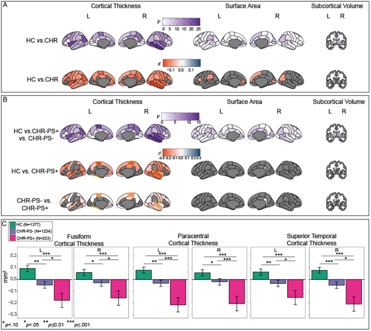
Compared to HC, the CHR group had smaller global neuroimaging measures: estimated ICV (d=-0.13, CI=-0.2 to -0.06), mean CT (d=-0.18, CI=-0.25 to -0.11) and total SA (d=-0.15, CI=-0.22 to -0.08). Fifty-three additional regions exhibited a significant effect of group (q<0.05, eTable 8). The largest group effects were observed for widespread lower CT in CHR vs. HC (42/68 comparisons, d range=-0.09 to -0.17; Figure 1A for overview; eTable 8 and eFigure 1 for details). Few subcortical (3/16) and SA (8/68) group differences were observed (d range=- 0.09 to -0.16). No group-by-sex interactions were detected (all q>0.05).

Figure 1.
  • Download figure
  • Open in new tab
Figure 1.

Effect sizes for mega-analysis of group and conversion status. A. Overview of effect sizes for HC vs. CHR. The top row reflects the results of the overall generalized linear model. A deeper purple color indicates a greater effect of group (HC vs. CHR) in this region. We observed the greatest effects of group in cortical thickness measures. The second row indicates the pairwise effect sizes for HC vs. CHR, in regions that were statistically significant (q<0.05) in the overall comparison (top row of A.). Regions that were not statistically significant in the overall comparison are in gray. In comparison to HC, CHR exhibited lower cortical thickness across the cortex. Red color indicates that HC has a larger value in comparison to CHR for this region. B. Overview of effect sizes for HC vs. CHR-PS+ vs. CHR-PS-. The top row reflects the results of the overall generalized linear model. A deeper purple color indicates a greater effect of conversion status (HC vs. CHR-PS+ vs. CHR-PS-) in this region. The second and third rows indicate the pairwise effect sizes for HC vs. CHR-PS+ and CHR-PS- vs. CHR-PS+, respectively. Pairwise comparisons are presented in regions that were statistically significant (q<0.05) in the overall comparison (top row of B.). Regions that were not statistically significant in the overall comparison are in gray. Regions that CHR-PS+ had lower cortical thickness in comparison to HC and CHR-PS-, CHR-PS+ are highlighted in yellow. C. Bar graphs for regions in which CHR-PS+ (pink) had lower CT in comparison to CHR-PS- (purple) and HC (green).

Group difference magnitudes remained similar when controlling for antipsychotic and psychotropic medication use (eTable 9). 91% of group differences remained statistically significant (q<0.05) in >90% of jackknife resampling iterations (eFigure 2), providing evidence that site effects were not driving results. Equivalence testing revealed that effect sizes for group differences were likely not meaningfully greater than our a priori-defined smallest effect size of interest in 3/56 measures (eFigure 1, eTable 10).

Figure 2.
  • Download figure
  • Open in new tab
Figure 2.

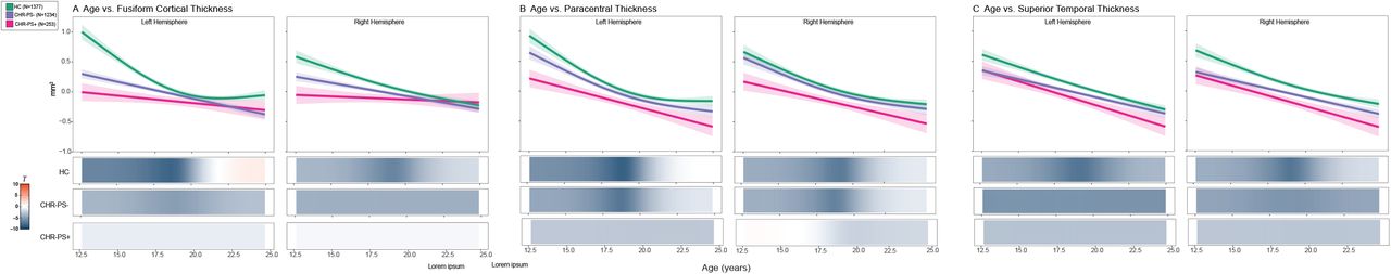
Age effects of regions that exhibited an effect of conversion status. HC are in green, CHR-PS+ are pink, and CHR-PS-are purple. Each graph has a line of best fit for the effect of age on the respective neuroimaging measures. Shading around the line indicates the standard error. The bars underneath the age plots reflect the derivative of the slope, i.e., the rate of change taking place at a particular age, scaled as a pseudo t-statistic, based on the posterior simulation. Age effects are plotted for A. fusiform cortical thickness, B. paracentral cortical thickness, and C. superior temporal cortical thickness.

Mega- and meta-analyses of neuroimaging data prior to neuroCombat harmonization provided similar results (Spearman’s ρ>0.93; eTable 11). No sMRI measures were uniquely sensitive to psychosis-risk syndrome (see Supplement and eTables 12-15 forresults).

Thinner paracentral, fusiform, and superior temporal CT are associated with psychosis conversion

Forty-eight structural neuroimaging measures (NCT=37) exhibited a significant overall effect of psychosis conversion status (q<0.05, Figure 1B; eFigure 3 and eTable 16). The three groups differed from each other on four structural neuroimaging measures: in comparison to HC and CHR-PS-, CHR-PS+ exhibited thinner cortex in bilateral paracentral, right superior temporal, and left fusiform regions (Figure 1C, average d of four sMRI measures=-0.22). CHR-PS+ and CHR-PS- (vs. HC) exhibited thinner cortex in the left superior temporal and right fusiform regions;similar trends were observed for CHR-PS+ vs. CHR-PS-differences (p<0.08; Figure 1C). Results remained stable when length of follow-up period was included as a covariate.

Figure 3.
  • Download figure
  • Open in new tab
Figure 3.

Cortical thickness alterations in CHR-PS+, schizophrenia, and 22q.11DS A. Magnitude of cortical thickness alterations for the three groups. Whole-brain cortical thickness effect sizes were not reported for SZ and 22q.11DS in original publications; values shown are the hemispheres with the smallest effect size. Colored bars represent standard error. B. Spearman’s rank correlations between cortical thickness alterations in CHR-PS+ and individuals with schizophrenia (top) and 22q.11 DS with psychosis (bottom).

Using minimal-effects testing, we observed that effect sizes for bilateral paracentral (L Z=-2.43, p=0.02; R Z=-1.86 p=0.06), right superior temporal (Z=-2.29, p=0.02) and left fusiform (Z=-2.00, p=0.05) in CHR-PS+ vs. HC were all significantly greater than 0.15, at least at trend level, underscoring the presence of notable group differences.

In the remaining follow-up comparisons, CHR-PS+ and CHR-PS-differed from HC at p<0.05, but there were no significant differences between CHR-PS+ and CHR-PS- (29/48 comparisons; eTable 16). We observed no conversion status-by-sex interactions (all q>0.05).

When we controlled for antipsychotic or psychotropic medication exposure, most overall psychosis conversion effects remained statistically significant (eTable 17). sMRI measures that differentiated the three groups showed an overall conversion effect at q<0.05 in all jackknife iterations, ruling out site effects as a potential confound (eFigure 2). No pairwise comparisons for structural neuroimaging measures that showed an overall conversion effect at q<0.05 were statistically equivalent to our predefined smallest effect size of interest (eTable 18). There were no statistically significant psychosis-risk syndrome-by-conversion status interactions (eTable 19).

CHR-PS+ fail to show expected age-associations with fusiform and paracentral CT

There were no significant group-by-age interactions in the 56 neuroimaging measures that differed between CHR and HC (q>0.05, eTable 20). In contrast, 2/4 sMRI measures in which all groups differed (HC vs. CHR-PS+ vs. CHR-PS-) displayed a statistically significant conversion status-by-age interaction.

In left fusiform, age-CT associations differed between CHR-PS+ vs. HC (F=9.8, p=4.9e-05, q=5.9e-04) and CHR-PS- vs. HC (F=8.7, p=1.5e-04, q=9.1e-04, Figure 2A, left). Between ages 12-16, HC showed a stronger negative association between age and CT, compared to CHR-PS+ and CHR-PS-, suggesting a reduced rate of CT change in CHR-PS+ and CHR-PS-. Though the age-by-conversion status interaction was not statistically significant, a similar pattern emerged for the right fusiform CT (Figure 2A, right).

Age-effects in the left paracentral CT differed between CHR-PS+ vs. HC (F=5.9, p=4.9e-03, q=0.02, Figure 2B, left). Between 12-15.8 years of age, HC showed a stronger negative association between age and CT in comparison to CHR-PS+. Age-CT associations did not differ between CHR-PS- vs. HC (F=0.2, p=0.69). This pattern of results was not observed for the right paracentral CT (Figure 2B, right).

We found no significant age-by-conversion status interactions for the superior temporal CT (Figure 2C); all three groups showed negative CT-age associations.

CT aberrations in CHR-PS+ resemble the pattern observed in SZ and 22q11DS with a psychotic disorder diagnosis, but not MDD

Figure 3A provides a visual overview of CT alterations in CHR-PS+ relative to SZ and individuals with 22q11DS and a psychotic disorder. The pattern of baseline CT alterations in CHR-PS+ (CHR-PS+ vs. HC effect sizes) correlated significantly with that observed in SZ (ρ=0.35, ppermute=0.004, Figure 3B top) and individuals with 22q11DS and psychosis (ρ=0.43, ppermute=0.001, Figure 3B bottom). CT alterations in CHR did not correlate with those observed in MDD (CHR-PS+ ρ=-0.03) and slopes for CHR-PS+/SZ and CHR-PS+/MDD correlations significantly differed (Steiger’s Z=2.06, ppermute=0.008). No significant correlations were observed for SA (SZ ρ=-0.03; 22q.11DS ρ=-0.06, eFigure 5). Subcortical volume alterations in CHR-PS+ correlated with those observed in SZ (ρ=0.54, p=0.03) and a similar non-significant trend was observed for 22q11DS and psychosis (ρ=0.46, p=0.07). Associations for CHR-PS- (vs. HC) effect sizes were similar to those reported here (see Supplement).

4. DISCUSSION

We conducted the largest multisite neuroimaging investigation to date in CHR participants, examining baseline structural neuroimaging measures associated with later transition to psychosis. We found widespread thinner cortex in CHR, consistent with previously-reported CT alterations in individuals with an established psychotic disorder. Compared to CHR-PS- and HC, at baseline, CHR-PS+ exhibited thinner cortex in bilateral paracentral, right fusiform and left superior temporal regions, with effect sizes significantly greater than what we considered to be meaningful a priori. Our results were robust to effects of medication exposure, sex, site, and length of follow-up period. Findings from this international effort suggest that conversion to psychosis amongst those at high risk is associated with thinner cortex at baseline.

We identified widespread regional thinner cortex in CHR compared to HC. Thinner cortex has been observed in SZ, as well as other psychiatric disorders69,79,93–95. Importantly, the overall pattern of thinner cortex in CHR resembled that observed in SZ and individuals with 22q11DS and a psychotic disorder, but not in MDD. For CHR-PS+, correlations with SZ CT alterations were significantly greater than the relationship observed with MDD CT alterations. Taken together, our results suggest that the overall constellation of reported CT alterations resembles the regional pattern of alterations observed in SZ and genetic disorders associated with psychosis, and thus argues in favour of psychosis-specific thinner cortex in CHR

We also observed thinner cortex specific to psychosis conversion in paracentral, superior temporal and fusiform CT; CHR-PS+ exhibit significantly lower CT than CHR-PS- and HC in these regions. Lower baseline CT and/or volume in these regions has previously been reported in CHR-PS+96,97 (data not used here). Furthermore, longitudinal CT decreases in these regions have been associated with transition to psychosis in CHR17,98,99. The magnitude of altered CT in CHR-PS+ in paracentral, superior temporal and fusiform regions was highly consistent with findings in SZ70,100–102 and lower fusiform and paracentral CT has been observed in non-clinical voice hearers103. Thus, the observed pattern of CT alterations in CHR-PS+ is consilient with morphometric disruptions across the wider psychosis spectrum.

Consistent with previous CHR studies examining baseline neuroimaging associations with later conversion to psychosis96,104, we did not observe widespread subcortical volume or SA alterations associated with later psychosis transition. Taken together, these results suggest that CT reductions may be among the most widespread, robust, and specific morphometric changes associated with psychosis risk and conversion, compared to SA or subcortical volume.

The neuroanatomic pattern of group differences and age-associated disruptions observed in CHR may provide important insights into mechanisms underlying increased risk for psychosis. Pre-clinical models 105,106 and recent genome-wide association studies107 suggest that genetic variants associated with SZ are linked to the regulation of neural progenitor cells during fetal development, while genetic markers associated with CT were associated with regulatory processes in adulthood. Thus, CT alterations may be the end result of maladaptive maturation-related mechanisms that occur during post-fetal development, including proliferation, synaptic pruning and/or myelination108–111. Thinner CT, particularly in early adolescence112,113 (Figure 2), could reflect abnormal synaptic plasticity or pruning, which have both been implicated in in vitro SZ models114. Although excessive synaptic pruning is one plausible explanation for thinner cortex associated with psychosis transition, recent evidence suggests that intracortical myelination and/or expression of myelin-related genes may be mechanisms driving cortical thinning115,116. To better understand neurobiological mechanisms underlying psychosis transition in CHR, investigations of concomitant measures of cortical thickness, macroscale white matter tracts, and intracortical myelination are necessary.

Even if CT reductions in CHR were robust, effect sizes for between-group differences were nevertheless small-to-moderate and accounted for ∼1% of the variance in CHR-PS+ vs. CHR-PS-comparisons. The subtle nature of these morphometric alterations underscores the importance of adequate statistical power, achievable only through large-scale multi-site collaborations. Consistent with recent work showing that SZ polygenic risk scores only improved differentiation of CHR-PS+ and HC (not CHR-PS+ from CHR-PS-)117, we anticipate that baseline, univariate sMRI metrics will to have a similar impact on psychosis risk prediction algorithms. Given the logistic and financial challenges that MRI brings, the use of MRI metrics in isolation may not be feasible or useful for psychosis risk prediction. A viable solution may be to adopt sequential assessment frameworks, as recently implemented34. Alternatively, sMRI alterations may be a better predictor of general psychopathology, and would be better suited for transdiagnostic risk prediction models118,119.

Limitations

One limitation common to multi-site studies is that data were collected from multiple scanners, although leave-one-out analyses suggest that site effects were not prominent. Secondly, this initial study focused on baseline cross-sectional data, and did not investigate progressive sMRI changes (trajectories) associated with psychosis conversion, as identified in prior work17,19,38,97–99,120–123. This is a future goal of the ENIGMA CHR Working Group, now that feasibility of this collaboration has been established.

Conclusions and Future Directions

In the largest study of brain abnormalities in CHR to date, we found robust evidence for a subtle, widespread pattern of CT alterations, consistent with observations in psychosis. The specificity of these alterations to CT - as well as age-associated deviations in regions sensitive to psychosis conversion - may point to abnormal development processes. These findings also point to age ranges (i.e., early adolescence) when morphometric abnormalities in CHR might be greatest.

Data Availability

All code used in this project is publicly available here: https://osf.io/p9ntr/ Due to individual site IRB restrictions, data is not publicly available. Please contact the ENIGMA CHR Working Group co-chairs if you are interested in joining and participating in the working group. Then you will be able to submit secondary data proposals.

https://osf.io/p9ntr/

Table and Figure Legends

Table 1. Demographic descriptives for every site in HC and CHR, and CHR-PS+ and CHR-PS- The Ns for CHR-PS+ and CHR-PS- do not always sum to the sample CHR N because most sites lose individuals to follow-up (CHR-UNK).

Acknowledgements

BHE (Bjørn H. Ebdrup) is part of the Advisory Board of Eli Lilly Denmark A/S, Janssen-Cilag, Lundbeck Pharma A/S, and Takeda Pharmaceutical Company Ltd; and has received lecture fees from Bristol-Myers Squibb, Otsuka Pharma Scandinavia AB, Eli Lilly Company, Boehringer Ingelheim Danmark A/S, and Lundbeck Pharma A/S. DH (Dennis Hernaus) has received financial compensation as a consultant for P1vital Produicts Ltd. F-RM (Francisco Reyes-Madrigal) has received compensation as a speaker for Janssen (Johnson & Johnson). All reported activities were unrelated to the work presented in this manuscript.

Footnotes

  • We corrected the name for Montserrat Dolz

References

  1. 1.↵
    Miller TJ, McGlashan TH, Rosen JL, et al. Prodromal assessment with the structured interview for prodromal syndromes and the scale of prodromal symptoms: predictive validity, interrater reliability, and training to reliability. Schizophr Bull. 2003;29(4):703.
    OpenUrlCrossRefPubMedWeb of Science
  2. 2.↵
    Yung AR, McGorry PD, McFarlane CA, Jackson HJ, Patton GC, Rakkar A. Monitoring and care of young people at incipient risk of psychosis. Schizophr Bull. 1996;22(2):283–303. doi:10.1093/schbul/22.2.283
    OpenUrlCrossRefPubMedWeb of Science
  3. 3.↵
    Yung AR, Phillips LJ, McGorry PD, et al. Prediction of psychosis. A step towards indicated prevention of schizophrenia. Br J Psychiatry Suppl. 1998;172(33):14–20.
    OpenUrlPubMedWeb of Science
  4. 4.↵
    Fusar-Poli P, Bonoldi I, Yung AR, et al. Predicting psychosis: meta-analysis of transition outcomes in individuals at high clinical risk. Arch Gen Psychiatry. 2012;69(3):220–229. doi:10.1001/archgenpsychiatry.2011.1472
    OpenUrlCrossRefPubMedWeb of Science
  5. 5.↵
    Simon AE, Umbricht D. High remission rates from an initial ultra-high risk state for psychosis. Schizophr Res. 2010;116(2-3):168–172. doi:10.1016/j.schres.2009.10.001
    OpenUrlCrossRefPubMedWeb of Science
  6. 6.
    Ruhrmann S, Schultze-Lutter F, Salokangas RKR, et al. Prediction of psychosis in adolescents and young adults at high risk: results from the prospective European prediction of psychosis study. Arch Gen Psychiatry. 2010;67(3):241–251. doi:10.1001/archgenpsychiatry.2009.206
    OpenUrlCrossRefPubMedWeb of Science
  7. 7.
    Cannon TD, Cadenhead K, Cornblatt B, et al. Prediction of Psychosis in Youth at High Clinical Risk: A Multisite Longitudinal Study in North America. Arch Gen Psychiatry. 2008;65(1):28. doi:10.1001/archgenpsychiatry.2007.3
    OpenUrlCrossRefPubMedWeb of Science
  8. 8.
    Yung AR, Phillips LJ, Yuen HP, et al. Psychosis prediction: 12-month follow up of a high-risk (“prodromal”) group. Schizophr Res. 2003;60(1):21–32. doi:10.1016/s0920-9964(02)00167-6
    OpenUrlCrossRefPubMedWeb of Science
  9. 9.
    Cornblatt BA, Lencz T, Smith CW, Correll CU, Auther AM, Nakayama E. The schizophrenia prodrome revisited: a neurodevelopmental perspective. Schizophr Bull. 2003;29(4):633–651. doi:10.1093/oxfordjournals.schbul.a007036
    OpenUrlCrossRefPubMedWeb of Science
  10. 10.
    Zhang T, Li H, Tang Y, et al. Validating the Predictive Accuracy of the NAPLS-2 Psychosis Risk Calculator in a Clinical High-Risk Sample from the SHARP (Shanghai At Risk for Psychosis) Program. Am J Psychiatry. 2018;175(9):906–908. doi:10.1176/appi.ajp.2018.18010036
    OpenUrlCrossRef
  11. 11.↵
    Tor J, Dolz M, Sintes A, et al. Clinical high risk for psychosis in children and adolescents: a systematic review. Eur Child Adolesc Psychiatry. 2018;27(6):683–700. doi:10.1007/s00787-017-1046-3
    OpenUrlCrossRef
  12. 12.
    Hartmann JA, Yuen HP, McGorry PD, et al. Declining transition rates to psychotic disorder in “ultra-high risk” clients: Investigation of a dilution effect. Schizophr Res. 2016;170(1):130–136. doi:10.1016/j.schres.2015.11.026
    OpenUrlCrossRefPubMed
  13. 13.↵
    Fusar-Poli P, Palombini E, Davies C, et al. Why transition risk to psychosis is not declining at the OASIS ultra high risk service: The hidden role of stable pretest risk enrichment. Schizophr Res. 2018;192:385–390. doi:10.1016/j.schres.2017.06.015
    OpenUrlCrossRef
  14. 14.↵
    Bakker G, Caan MWA, Vingerhoets WAM, et al. Cortical Morphology Differences in Subjects at Increased Vulnerability for Developing a Psychotic Disorder: A Comparison between Subjects with Ultra-High Risk and 22q11.2 Deletion Syndrome. PLOS ONE. 2016;11(11):e0159928. doi:10.1371/journal.pone.0159928
    OpenUrlCrossRef
  15. 15.↵
    Zikidi K, Gajwani R, Gross J, et al. Grey-matter abnormalities in clinical high-risk participants for psychosis. Schizophr Res. Published online November 15, 2019. doi:10.1016/j.schres.2019.08.034
    OpenUrlCrossRef
  16. 16.
    Velakoulis D, Wood SJ, Wong MTH, et al. Hippocampal and Amygdala Volumes According to Psychosis Stage and Diagnosis: A Magnetic Resonance Imaging Study of Chronic Schizophrenia, First-Episode Psychosis, and Ultra–High-Risk Individuals. Arch Gen Psychiatry. 2006;63(2):139. doi:10.1001/archpsyc.63.2.139
    OpenUrlCrossRefPubMedWeb of Science
  17. 17.↵
    Sun D, Phillips L, Velakoulis D, et al. Progressive brain structural changes mapped as psychosis develops in ‘at risk’ individuals. Schizophrenia Research. 2009;108(1-3):85–92. doi:10.1016/j.schres.2008.11.026
    OpenUrlCrossRefPubMedWeb of Science
  18. 18.
    Fornito A, Yung AR, Wood SJ, et al. Anatomic Abnormalities of the Anterior Cingulate Cortex Before Psychosis Onset: An MRI Study of Ultra-High-Risk Individuals. Biological Psychiatry. 2008;64(9):758–765. doi:10.1016/j.biopsych.2008.05.032
    OpenUrlCrossRefPubMedWeb of Science
  19. 19.↵
    Takahashi T, Wood SJ, Yung AR, et al. Insular cortex gray matter changes in individuals at ultra-high-risk of developing psychosis. Schizophrenia Research. 2009;111(1-3):94–102. doi:10.1016/j.schres.2009.03.024
    OpenUrlCrossRefPubMedWeb of Science
  20. 20.
    Tomyshev AS, Lebedeva IS, Akhadov TA, Omelchenko MA, Rumyantsev AO, Kaleda VG. Alterations in white matter microstructure and cortical thickness in individuals at ultra-high risk of psychosis: A multimodal tractography and surface-based morphometry study. Psychiatry Res Neuroimaging. 2019;289:26–36. doi:10.1016/j.pscychresns.2019.05.002
    OpenUrlCrossRef
  21. 21.
    Kwak YB, Kim M, Cho KIK, Lee J, Lee TY, Kwon JS. Reduced cortical thickness in subjects at clinical high risk for psychosis and clinical attributes. Aust N Z J Psychiatry. 2019;53(3):219–227. doi:10.1177/0004867418807299
    OpenUrlCrossRef
  22. 22.
    Yassin W, Nakatani H, Zhu Y, et al. Machine-learning classification using neuroimaging data in schizophrenia, autism, ultra-high risk and first-episode psychosis. Transl Psychiatry. 2020;10(1):278. doi:10.1038/s41398-020-00965-5
    OpenUrlCrossRef
  23. 23.
    Nakamura M, Takahashi T, Takayanagi Y, et al. Surface morphology of the orbitofrontal cortex in individuals at risk of psychosis: a multicenter study. Eur Arch Psychiatry Clin Neurosci. 2019;269(4):397–406. doi:10.1007/s00406-018-0890-6
    OpenUrlCrossRef
  24. 24.
    Sasabayashi D, Takayanagi Y, Takahashi T, et al. Increased Occipital Gyrification and Development of Psychotic Disorders in Individuals With an At-Risk Mental State: A Multicenter Study. Biol Psychiatry. 2017;82(10):737–745. doi:10.1016/j.biopsych.2017.05.018
    OpenUrlCrossRef
  25. 25.
    Iwashiro N, Suga M, Takano Y, et al. Localized gray matter volume reductions in the pars triangularis of the inferior frontal gyrus in individuals at clinical high-risk for psychosis and first episode for schizophrenia. Schizophr Res. 2012;137(1-3):124–131. doi:10.1016/j.schres.2012.02.024
    OpenUrlCrossRefPubMed
  26. 26.
    Nakamura K, Takahashi T, Nemoto K, et al. Gray matter changes in subjects at high risk for developing psychosis and first-episode schizophrenia: a voxel-based structural MRI study. Front Psychiatry. 2013;4:16. doi:10.3389/fpsyt.2013.00016
    OpenUrlCrossRefPubMed
  27. 27.
    Takayanagi Y, Kulason S, Sasabayashi D, et al. Reduced Thickness of the Anterior Cingulate Cortex in Individuals With an At-Risk Mental State Who Later Develop Psychosis. Schizophr Bull. 2017;43(4):907–913. doi:10.1093/schbul/sbw167
    OpenUrlCrossRef
  28. 28.
    Takahashi T, Tsugawa S, Nakajima S, et al. Thalamic and striato-pallidal volumes in schizophrenia patients and individuals at risk for psychosis: A multi-atlas segmentation study. Schizophr Res. Published online May 21, 2020. doi:10.1016/j.schres.2020.04.016
    OpenUrlCrossRef
  29. 29.
    Buechler R, Wotruba D, Michels L, et al. Cortical Volume Differences in Subjects at Risk for Psychosis are Driven by Surface Area. Schizophr Bull. Published online May 28, 2020. doi:10.1093/schbul/sbaa066
    OpenUrlCrossRef
  30. 30.
    Chung Y, Allswede D, Addington J, et al. Cortical abnormalities in youth at clinical high-risk for psychosis: Findings from the NAPLS2 cohort. Neuroimage Clin. 2019;23:101862. doi:10.1016/j.nicl.2019.101862
    OpenUrlCrossRef
  31. 31.
    Chung Y, Addington J, Bearden CE, et al. Use of Machine Learning to Determine Deviance in Neuroanatomical Maturity Associated With Future Psychosis in Youths at Clinically High Risk. JAMA Psychiatry. 2018;75(9):960–968. doi:10.1001/jamapsychiatry.2018.1543
    OpenUrlCrossRefPubMed
  32. 32.
    Koutsouleris N, Meisenzahl EM, Davatzikos C, et al. Use of neuroanatomical pattern classification to identify subjects in at-risk mental states of psychosis and predict disease transition. Arch Gen Psychiatry. 2009;66(7):700–712. doi:10.1001/archgenpsychiatry.2009.62
    OpenUrlCrossRefPubMedWeb of Science
  33. 33.
    Koutsouleris N, Riecher-Rössler A, Meisenzahl EM, et al. Detecting the psychosis prodrome across high-risk populations using neuroanatomical biomarkers. Schizophr Bull. 2015;41(2):471–482. doi:10.1093/schbul/sbu078
    OpenUrlCrossRefPubMed
  34. 34.↵
    Koutsouleris N, Kambeitz-Ilankovic L, Ruhrmann S, et al. Prediction Models of Functional Outcomes for Individuals in the Clinical High-Risk State for Psychosis or With Recent-Onset Depression: A Multimodal, Multisite Machine Learning Analysis. JAMA Psychiatry. 2018;75(11):1156–1172. doi:10.1001/jamapsychiatry.2018.2165
    OpenUrlCrossRef
  35. 35.↵
    Klauser P, Zhou J, Lim JKW, et al. Lack of Evidence for Regional Brain Volume or Cortical Thickness Abnormalities in Youths at Clinical High Risk for Psychosis: Findings From the Longitudinal Youth at Risk Study. Schizophr Bull. 2015;41(6):1285–1293. doi:10.1093/schbul/sbv012
    OpenUrlCrossRefPubMed
  36. 36.
    Ziermans TB, Durston S, Sprong M, et al. No evidence for structural brain changes in young adolescents at ultra high risk for psychosis. Schizophr Res. 2009;112(1-3):1–6. doi:10.1016/j.schres.2009.04.013
    OpenUrlCrossRefPubMedWeb of Science
  37. 37.↵
    Takahashi T, Yücel M, Yung AR, et al. Adhesio interthalamica in individuals at high-risk for developing psychosis and patients with psychotic disorders. Prog Neuropsychopharmacol Biol Psychiatry. 2008;32(7):1708–1714. doi:10.1016/j.pnpbp.2008.07.007
    OpenUrlCrossRefPubMed
  38. 38.↵
    Cannon TD, Chung Y, He G, et al. Progressive reduction in cortical thickness as psychosis develops: a multisite longitudinal neuroimaging study of youth at elevated clinical risk. Biol Psychiatry. 2015;77(2):147–157. doi:10.1016/j.biopsych.2014.05.023
    OpenUrlCrossRefPubMedWeb of Science
  39. 39.↵
    Sakuma A, Obara C, Katsura M, et al. No regional gray matter volume reduction observed in young Japanese people at ultra-high risk for psychosis: A voxel-based morphometry study. Asian J Psychiatr. 2018;37:167–171. doi:10.1016/j.ajp.2018.09.009
    OpenUrlCrossRef
  40. 40.↵
    Fusar-Poli P, Borgwardt S, Crescini A, et al. Neuroanatomy of vulnerability to psychosis: a voxel-based meta-analysis. Neurosci Biobehav Rev. 2011;35(5):1175–1185. doi:10.1016/j.neubiorev.2010.12.005
    OpenUrlCrossRefPubMed
  41. 41.↵
    Mechelli A, Riecher-Rössler A, Meisenzahl EM, et al. Neuroanatomical abnormalities that predate the onset of psychosis: a multicenter study. Arch Gen Psychiatry. 2011;68(5):489–495. doi:10.1001/archgenpsychiatry.2011.42
    OpenUrlCrossRefPubMedWeb of Science
  42. 42.↵
    Farris MS, Devoe DJ, Addington J. Attrition rates in trials for adolescents and young adults at clinical high-risk for psychosis: A systematic review and meta-analysis. Early Interv Psychiatry. 2020;14(5):515–527. doi:10.1111/eip.12864
    OpenUrlCrossRef
  43. 43.
    Schlosser DA, Jacobson S, Chen Q, et al. Recovery from an at-risk state: clinical and functional outcomes of putatively prodromal youth who do not develop psychosis. Schizophr Bull. 2012;38(6):1225–1233. doi:10.1093/schbul/sbr098
    OpenUrlCrossRefPubMedWeb of Science
  44. 44.↵
    Rutigliano G, Valmaggia L, Landi P, et al. Persistence or recurrence of non-psychotic comorbid mental disorders associated with 6-year poor functional outcomes in patients at ultra high risk for psychosis. J Affect Disord. 2016;203:101–110. doi:10.1016/j.jad.2016.05.053
    OpenUrlCrossRef
  45. 45.↵
    Simon AE, Borgwardt S, Riecher-Rössler A, Velthorst E, de Haan L, Fusar-Poli P. Moving beyond transition outcomes: meta-analysis of remission rates in individuals at high clinical risk for psychosis. Psychiatry Res. 2013;209(3):266–272. doi:10.1016/j.psychres.2013.03.004
    OpenUrlCrossRefPubMed
  46. 46.↵
    Button KS, Ioannidis JPA, Mokrysz C, et al. Power failure: why small sample size undermines the reliability of neuroscience. Nat Rev Neurosci. 2013;14(5):365–376. doi:10.1038/nrn3475
    OpenUrlCrossRefPubMed
  47. 47.↵
    Consideration of Sample Size in Neuroscience Studies. J Neurosci. 2020;40(21):4076–4077. doi:10.1523/JNEUROSCI.0866-20.2020
    OpenUrlFREE Full Text
  48. 48.↵
    Addington J, Cadenhead KS, Cornblatt BA, et al. North American Prodrome Longitudinal Study (NAPLS 2): Overview and recruitment. Schizophrenia Research. 2012;142(1):77–82. doi:10.1016/j.schres.2012.09.012
    OpenUrlCrossRefPubMed
  49. 49.
    Addington J, Liu L, Brummitt K, et al. North American Prodrome Longitudinal Study (NAPLS 3): Methods and baseline description. Schizophrenia Research. Published online April 18, 2020. doi:10.1016/j.schres.2020.04.010
    OpenUrlCrossRef
  50. 50.↵
    Tognin S, van Hell HH, Merritt K, et al. Towards Precision Medicine in Psychosis: Benefits and Challenges of Multimodal Multicenter Studies-PSYSCAN: Translating Neuroimaging Findings From Research into Clinical Practice. Schizophr Bull. 2020;46(2):432–441. doi:10.1093/schbul/sbz067
    OpenUrlCrossRefPubMed
  51. 51.↵
    Jones P, Rodgers B, Murray R, Marmot M. Child development risk factors for adult schizophrenia in the British 1946 birth cohort. Lancet. 1994;344(8934):1398–1402. doi:10.1016/s0140-6736(94)90569-x
    OpenUrlCrossRefPubMedWeb of Science
  52. 52.↵
    Lauronen E, Miettunen J, Veijola J, Karhu M, Jones PB, Isohanni M. Outcome and its predictors in schizophrenia within the Northern Finland 1966 Birth Cohort. Eur Psychiatry. 2007;22(2):129–136. doi:10.1016/j.eurpsy.2006.07.001
    OpenUrlCrossRefPubMed
  53. 53.↵
    Giedd JN, Blumenthal J, Jeffries NO, et al. Brain development during childhood and adolescence: a longitudinal MRI study. Nature Neuroscience. 1999;2(10):861–863. doi:10.1038/13158
    OpenUrlCrossRefPubMedWeb of Science
  54. 54.
    Raznahan A, Shaw P, Lalonde F, et al. How Does Your Cortex Grow? Journal of Neuroscience. 2011;31(19):7174–7177.
    OpenUrlAbstract/FREE Full Text
  55. 55.
    Sowell ER. Longitudinal Mapping of Cortical Thickness and Brain Growth in Normal Children. Journal of Neuroscience. 2004;24(38):8223–8231.
    OpenUrlAbstract/FREE Full Text
  56. 56.
    Shaw P, Lerch J, Greenstein D, et al. Longitudinal mapping of cortical thickness and clinical outcome in children and adolescents with attention-deficit/hyperactivity disorder. Arch Gen Psychiatry. 2006;63(5):540–549. doi:10.1001/archpsyc.63.5.540
    OpenUrlCrossRefPubMedWeb of Science
  57. 57.
    van Soelen Ilc, Brouwer RM, van Baal Gcm, et al. Genetic influences on thinning of the cerebral cortex during development. Neuroimage. 2012;59(4):3871–3880. doi:10.1016/j.neuroimage.2011.11.044
    OpenUrlCrossRefPubMedWeb of Science
  58. 58.
    Vijayakumar N, Allen NB, Youssef G, et al. Brain development during adolescence: A mixed-longitudinal investigation of cortical thickness, surface area, and volume. Hum Brain Mapp. 2016;37(6):2027–2038. doi:10.1002/hbm.23154
    OpenUrlCrossRefPubMed
  59. 59.
    Tamnes CK, Herting MM, Goddings A-L, et al. Development of the Cerebral Cortex across Adolescence: A Multisample Study of Inter-Related Longitudinal Changes in Cortical Volume, Surface Area, and Thickness. J Neurosci. 2017;37(12):3402–3412. doi:10.1523/JNEUROSCI.3302-16.2017
    OpenUrlAbstract/FREE Full Text
  60. 60.
    Mills KL, Goddings A-L, Herting MM, et al. Structural brain development between childhood and adulthood: Convergence across four longitudinal samples. Neuroimage. 2016;141:273–281.
    OpenUrlCrossRefPubMed
  61. 61.
    Mills KL, Goddings A-L, Clasen LS, Giedd JN, Blakemore S-J. The developmental mismatch in structural brain maturation during adolescence. Dev Neurosci. 2014;36(3-4):147–160. doi:10.1159/000362328
    OpenUrlCrossRefPubMed
  62. 62.
    Mills KL, Tamnes CK. Methods and considerations for longitudinal structural brain imaging analysis across development. Dev Cogn Neurosci. 2014;9:172–190. doi:10.1016/j.dcn.2014.04.004
    OpenUrlCrossRefPubMed
  63. 63.↵
    Mills KL, Lalonde F, Clasen LS, Giedd JN, Blakemore S-J. Developmental changes in the structure of the social brain in late childhood and adolescence. Soc Cogn Affect Neurosci. 2014;9(1):123–131. doi:10.1093/scan/nss113
    OpenUrlCrossRefPubMed
  64. 64.↵
    Bridgwater M, Bachman P, Tervo-Clemmens B, et al. Developmental influences on symptom expression in antipsychotic-naïve first-episode psychosis. Psychol Med. Published online October 6, 2020:1–12. doi:10.1017/S0033291720003463
    OpenUrlCrossRef
  65. 65.
    Schimmelmann BG, Michel C, Martz-Irngartinger A, Linder C, Schultze-Lutter F. Age matters in the prevalence and clinical significance of ultra-high-risk for psychosis symptoms and criteria in the general population: Findings from the BEAR and BEARS-kid studies. World Psychiatry. 2015;14(2):189–197.
    OpenUrlCrossRefPubMed
  66. 66.
    Gerstenberg M, Theodoridou A, Traber-Walker N, et al. Adolescents and adults at clinical high-risk for psychosis: age-related differences in attenuated positive symptoms syndrome prevalence and entanglement with basic symptoms. Psychol Med. 2016;46(5):1069–1078. doi:10.1017/S0033291715002627
    OpenUrlCrossRef
  67. 67.
    Schultze-Lutter F, Hubl D, Schimmelmann BG, Michel C. Age effect on prevalence of ultra-high risk for psychosis symptoms: replication in a clinical sample of an early detection of psychosis service. Eur Child Adolesc Psychiatry. 2017;26(11):1401–1405. doi:10.1007/s00787-017-0994-y
    OpenUrlCrossRef
  68. 68.↵
    Schultze-Lutter F, Schimmelmann BG, Flückiger R, Michel C. Effects of age and sex on clinical high-risk for psychosis in the community. World J Psychiatry. 2020;10(5):101–124. doi:10.5498/wjp.v10.i5.101
    OpenUrlCrossRef
  69. 69.↵
    van Erp Tgm, Hibar DP, Rasmussen JM, et al. Subcortical brain volume abnormalities in 2028 individuals with schizophrenia and 2540 healthy controls via the ENIGMA consortium. Molecular Psychiatry. 2016;21(4):547–553. doi:10.1038/mp.2015.63
    OpenUrlCrossRefPubMed
  70. 70.↵
    van Erp Tgm, Walton E, Hibar DP, et al. Cortical Brain Abnormalities in 4474 Individuals With Schizophrenia and 5098 Control Subjects via the Enhancing Neuro Imaging Genetics Through Meta Analysis (ENIGMA) Consortium. Biol Psychiatry. 2018;84(9):644–654. doi:10.1016/j.biopsych.2018.04.023
    OpenUrlCrossRefPubMed
  71. 71.↵
    Okada N, Fukunaga M, Yamashita F, et al. Abnormal asymmetries in subcortical brain volume in schizophrenia. Mol Psychiatry. 2016;21(10):1460–1466. doi:10.1038/mp.2015.209
    OpenUrlCrossRefPubMed
  72. 72.↵
    Ching CRK, Gutman BA, Sun D, et al. Mapping Subcortical Brain Alterations in 22q11.2 Deletion Syndrome: Effects of Deletion Size and Convergence With Idiopathic Neuropsychiatric Illness. AJP. Published online February 12, 2020:appi.ajp.2019.1. doi:10.1176/appi.ajp.2019.19060583
    OpenUrlCrossRef
  73. 73.↵
    Sun D, Ching CRK, Lin A, et al. Large-scale mapping of cortical alterations in 22q11.2 deletion syndrome: Convergence with idiopathic psychosis and effects of deletion size. Mol Psychiatry. Published online June 13, 2018. doi:10.1038/s41380-018-0078-5
    OpenUrlCrossRef
  74. 74.↵
    World Medical Association. World Medical Association Declaration of Helsinki: ethical principles for medical research involving human subjects. JAMA. 2013;310(20):2191–2194. doi:10.1001/jama.2013.281053
    OpenUrlCrossRefPubMedWeb of Science
  75. 75.↵
    Dale AM, Fischl B, Sereno MI. Cortical surface-based analysis. I. Segmentation and surface reconstruction. Neuroimage. 1999;9(2):179–194.
    OpenUrlCrossRefPubMedWeb of Science
  76. 76.
    Fischl B, Sereno MI, Dale AM. Cortical surface-based analysis. II: Inflation, flattening, and a surface-based coordinate system. Neuroimage. 1999;9(2):195–207.
    OpenUrlCrossRefPubMedWeb of Science
  77. 77.
    Fischl B, Dale AM. Measuring the thickness of the human cerebral cortex from magnetic resonance images. Proc Natl Acad Sci U S A. 2000;97(20):11050–11055. doi:10.1073/pnas.200033797
    OpenUrlAbstract/FREE Full Text
  78. 78.↵
    Fischl B, Salat DH, Busa E, et al. Whole brain segmentation: automated labeling of neuroanatomical structures in the human brain. Neuron. 2002;33(3):341–355.
    OpenUrlCrossRefPubMedWeb of Science
  79. 79.↵
    Hibar DP, Westlye LT, Doan NT, et al. Cortical abnormalities in bipolar disorder: an MRI analysis of 6503 individuals from the ENIGMA Bipolar Disorder Working Group. Mol Psychiatry. 2018;23(4):932–942. doi:10.1038/mp.2017.73
    OpenUrlCrossRefPubMed
  80. 80.
    van Rooij D, Anagnostou E, Arango C, et al. Cortical and Subcortical Brain Morphometry Differences Between Patients With Autism Spectrum Disorder and Healthy Individuals Across the Lifespan: Results From the ENIGMA ASD Working Group. Am J Psychiatry. 2018;175(4):359–369. doi:10.1176/appi.ajp.2017.17010100
    OpenUrlCrossRefPubMed
  81. 81.
    Hoogman M, Bralten J, Hibar DP, et al. Subcortical brain volume differences in participants with attention deficit hyperactivity disorder in children and adults: a cross-sectional mega-analysis. Lancet Psychiatry. 2017;4(4):310–319.
    OpenUrl
  82. 82.↵
    Boedhoe PSW, Schmaal L, Abe Y, et al. Cortical Abnormalities Associated With Pediatric and Adult Obsessive-Compulsive Disorder: Findings From the ENIGMA Obsessive-Compulsive Disorder Working Group. Am J Psychiatry. 2018;175(5):453–462. doi:10.1176/appi.ajp.2017.17050485
    OpenUrlCrossRef
  83. 83.↵
    Radua J, Vieta E, Shinohara R, et al. Increased power by harmonizing structural MRI site differences with the ComBat batch adjustment method in ENIGMA. Neuroimage. 2020;218:116956. doi:10.1016/j.neuroimage.2020.116956
    OpenUrlCrossRef
  84. 84.↵
    Johnson WE, Li C, Rabinovic A. Adjusting batch effects in microarray expression data using empirical Bayes methods. Biostatistics. 2007;8(1):118–127. doi:10.1093/biostatistics/kxj037
    OpenUrlCrossRefPubMedWeb of Science
  85. 85.↵
    Benjamini Y, Hochberg Y. Controlling the False Discovery Rate: A Practical and Powerful Approach to Multiple Testing. Journal of the Royal Statistical Society: Series B (Methodological). 1995;57(1):289–300. doi:https://doi.org/10.1111/j.2517-6161.1995.tb02031.x
    OpenUrlCrossRefPubMedWeb of Science
  86. 86.↵
    Efron B, Stein C. The Jackknife Estimate of Variance. Ann Statist. 1981;9(3):586–596. doi:10.1214/aos/1176345462
    OpenUrlCrossRef
  87. 87.↵
    Efron B. The Jackknife, the Bootstrap, and Other Resampling Plans. Society for Industrial and Applied Mathematics (SIAM, 3600 Market Street, Floor 6, Philadelphia, PA 19104); 1982.
  88. 88.↵
    Lakens D. Equivalence Tests: A Practical Primer for t Tests, Correlations, and Meta-Analyses. Soc Psychol Personal Sci. 2017;8(4):355–362. doi:10.1177/1948550617697177
    OpenUrlCrossRefPubMed
  89. 89.↵
    Hastie T, Tibshirani R. Generalized Additive Models. Statist Sci. 1986;1(3):297–310. doi:10.1214/ss/1177013604
    OpenUrlCrossRefPubMed
  90. 90.↵
    Hastie T, Tibshirani R. Generalized Additive Models. CRC Press; 1990.
  91. 91.↵
    Jalbrzikowski M, Larsen B, Hallquist MN, Foran W, Calabro F, Luna B. Development of White Matter Microstructure and Intrinsic Functional Connectivity Between the Amygdala and Ventromedial Prefrontal Cortex: Associations With Anxiety and Depression. Biol Psychiatry. 2017;82(7):511–521. doi:10.1016/j.biopsych.2017.01.008
    OpenUrlCrossRefPubMed
  92. 92.↵
    Marek S, Hwang K, Foran W, Hallquist MN, Luna B. The Contribution of Network Organization and Integration to the Development of Cognitive Control. PLoS Biol. 2015;13(12):e1002328. doi:10.1371/journal.pbio.1002328
    OpenUrlCrossRefPubMed
  93. 93.↵
    Schmaal L, Veltman DJ, van Erp Tgm, et al. Subcortical brain alterations in major depressive disorder: findings from the ENIGMA Major Depressive Disorder working group. Mol Psychiatry. 2016;21(6):806–812. doi:10.1038/mp.2015.69
    OpenUrlCrossRefPubMed
  94. 94.
    Rimol LM, Nesvåg R, Hagler DJ, et al. Cortical volume, surface area, and thickness in schizophrenia and bipolar disorder. Biol Psychiatry. 2012;71(6):552–560. doi:10.1016/j.biopsych.2011.11.026
    OpenUrlCrossRefPubMedWeb of Science
  95. 95.↵
    Writing Committee for the Attention-Deficit/Hyperactivity Disorder, Autism Spectrum Disorder, Bipolar Disorder, et al. Virtual Histology of Cortical Thickness and Shared Neurobiology in 6 Psychiatric Disorders. JAMA Psychiatry. Published online August 26, 2020. doi:10.1001/jamapsychiatry.2020.2694
    OpenUrlCrossRef
  96. 96.↵
    Del Re EC, Stone WS, Bouix S, et al. Baseline Cortical Thickness Reductions in Clinical High Risk for Psychosis: Brain Regions Associated with Conversion to Psychosis Versus Non-Conversion as Assessed at One-Year Follow-Up in the Shanghai-At-Risk-for-Psychosis (SHARP) Study. Schizophr Bull. Published online September 14, 2020. doi:10.1093/schbul/sbaa127
    OpenUrlCrossRef
  97. 97.↵
    Borgwardt SJ, McGuire PK, Aston J, et al. Structural brain abnormalities in individuals with an at-risk mental state who later develop psychosis. Br J Psychiatry Suppl. 2007;51:s69–75. doi:10.1192/bjp.191.51.s69
    OpenUrlCrossRefPubMed
  98. 98.↵
    Pantelis C, Velakoulis D, Mcgorry PD, et al. Neuroanatomical abnormalities before and after onset of psychosis: a cross-sectional and longitudinal MRI comparison. Lance t. 2003;361(9354):281–288.
    OpenUrl
  99. 99.↵
    Takahashi T, Wood SJ, Yung AR, et al. Progressive gray matter reduction of the superior temporal gyrus during transition to psychosis. Arch Gen Psychiatry. 2009;66(4):366–376. doi:10.1001/archgenpsychiatry.2009.12
    OpenUrlCrossRefPubMedWeb of Science
  100. 100.↵
    Lee CU, Shenton ME, Salisbury DF, et al. Fusiform gyrus volume reduction in first-episode schizophrenia: a magnetic resonance imaging study. Arch Gen Psychiatry. 2002;59(9):775–781. doi:10.1001/archpsyc.59.9.775
    OpenUrlCrossRefPubMedWeb of Science
  101. 101.
    Onitsuka T, Shenton ME, Kasai K, et al. Fusiform gyrus volume reduction and facial recognition in chronic schizophrenia. Arch Gen Psychiatry. 2003;60(4):349–355. doi:10.1001/archpsyc.60.4.349
    OpenUrlCrossRefPubMedWeb of Science
  102. 102.↵
    Takahashi T, Suzuki M, Zhou S-Y, et al. Temporal lobe gray matter in schizophrenia spectrum: a volumetric MRI study of the fusiform gyrus, parahippocampal gyrus, and middle and inferior temporal gyri. Schizophr Res. 2006;87(1-3):116–126. doi:10.1016/j.schres.2006.04.023
    OpenUrlCrossRefPubMedWeb of Science
  103. 103.↵
    van Lutterveld R, van den Heuvel MP, Diederen KMJ, et al. Cortical thickness in individuals with non-clinical and clinical psychotic symptoms. Brain. 2014;137(Pt 10):2664–2669. doi:10.1093/brain/awu167
    OpenUrlCrossRefPubMed
  104. 104.↵
    Sasabayashi D, Takayanagi Y, Takahashi T, et al. Subcortical Brain Volume Abnormalities in Individuals With an At-risk Mental State. Schizophr Bull. 2020;46(4):834–845. doi:10.1093/schbul/sbaa011
    OpenUrlCrossRef
  105. 105.↵
    Pontious A, Kowalczyk T, Englund C, Hevner RF. Role of intermediate progenitor cells in cerebral cortex development. Dev Neurosci. 2008;30(1-3):24–32. doi:10.1159/000109848
    OpenUrlCrossRefPubMedWeb of Science
  106. 106.↵
    Rakic P. Specification of cerebral cortical areas. Science. 1988;241(4862):170–176.
    OpenUrlAbstract/FREE Full Text
  107. 107.↵
    Grasby KL, Jahanshad N, Painter JN, et al. The genetic architecture of the human cerebral cortex. Science. 2020;367(6484). doi:10.1126/science.aay6690
    OpenUrlAbstract/FREE Full Text
  108. 108.↵
    Huttenlocher PR. Synaptic density in human frontal cortex - developmental changes and effects of aging. Brain Res. 1979;163(2):195–205. doi:10.1016/0006-8993(79)90349-4
    OpenUrlCrossRefPubMedWeb of Science
  109. 109.
    Rakic P, Bourgeois JP, Eckenhoff MF, Zecevic N, Goldman-Rakic PS. Concurrent overproduction of synapses in diverse regions of the primate cerebral cortex. Science. 1986;232(4747):232–235. doi:10.1126/science.3952506
    OpenUrlAbstract/FREE Full Text
  110. 110.
    Petanjek Z, Judas M, Kostović I, Uylings HBM. Lifespan alterations of basal dendritic trees of pyramidal neurons in the human prefrontal cortex: a layer-specific pattern. Cereb Cortex. 2008;18(4):915–929. doi:10.1093/cercor/bhm124
    OpenUrlCrossRefPubMedWeb of Science
  111. 111.↵
    Natu VS, Gomez J, Barnett M, et al. Apparent thinning of human visual cortex during childhood is associated with myelination. Proc Natl Acad Sci U S A. 2019;116(41):20750–20759. doi:10.1073/pnas.1904931116
    OpenUrlAbstract/FREE Full Text
  112. 112.↵
    Pantelis C, Yücel M, Wood SJ, et al. Structural brain imaging evidence for multiple pathological processes at different stages of brain development in schizophrenia. Schizophr Bull. 2005;31(3):672–696. doi:10.1093/schbul/sbi034
    OpenUrlCrossRefPubMedWeb of Science
  113. 113.↵
    Sun D, Stuart GW, Jenkinson M, et al. Brain surface contraction mapped in first-episode schizophrenia: a longitudinal magnetic resonance imaging study. Mol Psychiatry. 2009;14(10):976–986. doi:10.1038/mp.2008.34
    OpenUrlCrossRefPubMedWeb of Science
  114. 114.↵
    Sellgren CM, Gracias J, Watmuff B, et al. Increased synapse elimination by microglia in schizophrenia patient-derived models of synaptic pruning. Nat Neurosci. 2019;22(3):374–385. doi:10.1038/s41593-018-0334-7
    OpenUrlCrossRefPubMed
  115. 115.↵
    Whitaker KJ, Vértes PE, Romero-Garcia R, et al. Adolescence is associated with genomically patterned consolidation of the hubs of the human brain connectome. Proc Natl Acad Sci U S A. 2016;113(32):9105–9110. doi:10.1073/pnas.1601745113
    OpenUrlAbstract/FREE Full Text
  116. 116.↵
    Parker N, Patel Y, Jackowski AP, et al. Assessment of Neurobiological Mechanisms of Cortical Thinning During Childhood and Adolescence and Their Implications for Psychiatric Disorders. JAMA Psychiatry. Published online June 17, 2020. doi:10.1001/jamapsychiatry.2020.1495
    OpenUrlCrossRef
  117. 117.↵
    Perkins DO, Olde Loohuis L, Barbee J, et al. Polygenic Risk Score Contribution to Psychosis Prediction in a Target Population of Persons at Clinical High Risk. Am J Psychiatry. 2020;177(2):155–163. doi:10.1176/appi.ajp.2019.18060721
    OpenUrlCrossRef
  118. 118.↵
    1. Baune BT
    Mei C, Nelson B, Hartmann J, Spooner R, McGorry PD. Chapter 4 - Transdiagnostic early intervention, prevention, and prediction in psychiatry. In: Baune BT, ed. Personalized Psychiatry. Academic Press; 2020:27–37. doi:10.1016/B978-0-12-813176-3.00004-3
    OpenUrlCrossRef
  119. 119.↵
    McGorry PD, Hartmann JA, Spooner R, Nelson B. Beyond the “at risk mental state” concept: transitioning to transdiagnostic psychiatry. World Psychiatry. 2018;17(2):133–142. doi:10.1002/wps.20514
    OpenUrlCrossRef
  120. 120.↵
    Dazzan P, Soulsby B, Mechelli A, et al. Volumetric abnormalities predating the onset of schizophrenia and affective psychoses: an MRI study in subjects at ultrahigh risk of psychosis. Schizophr Bull. 2012;38(5):1083–1091. doi:10.1093/schbul/sbr035
    OpenUrlCrossRefPubMedWeb of Science
  121. 121.
    Ziermans TB, Schothorst PF, Schnack HG, et al. Progressive structural brain changes during development of psychosis. Schizophr Bull. 2012;38(3):519–530. doi:10.1093/schbul/sbq113
    OpenUrlCrossRefPubMedWeb of Science
  122. 122.
    Walter A, Studerus E, Smieskova R, et al. Hippocampal volume in subjects at high risk of psychosis: a longitudinal MRI study. Schizophr Res. 2012;142(1-3):217–222. doi:10.1016/j.schres.2012.10.013
    OpenUrlCrossRefPubMed
  123. 123.↵
    Valli I, Marquand AF, Mechelli A, et al. Identifying Individuals at High Risk of Psychosis: Predictive Utility of Support Vector Machine using Structural and Functional MRI Data. Front Psychiatry. 2016;7:52. doi:10.3389/fpsyt.2016.00052
    OpenUrlCrossRef
Back to top
PreviousNext
Posted January 21, 2021.
Download PDF

Supplementary Material

Data/Code
Email

Thank you for your interest in spreading the word about medRxiv.

NOTE: Your email address is requested solely to identify you as the sender of this article.

Enter multiple addresses on separate lines or separate them with commas.
Thinner cortex is associated with psychosis onset in individuals at Clinical High Risk for Developing Psychosis: An ENIGMA Working Group mega-analysis
(Your Name) has forwarded a page to you from medRxiv
(Your Name) thought you would like to see this page from the medRxiv website.
CAPTCHA
This question is for testing whether or not you are a human visitor and to prevent automated spam submissions.
Share
Thinner cortex is associated with psychosis onset in individuals at Clinical High Risk for Developing Psychosis: An ENIGMA Working Group mega-analysis
Maria Jalbrzikowski, Rebecca A. Hayes, Stephen J. Wood, Dorte Nordholm, Juan H. Zhou, Paolo Fusar-Poli, Peter J. Uhlhaas, Tsutomu Takahashi, Gisela Sugranyes, Yoo Bin Kwak, Daniel H. Mathalon, Naoyuki Katagiri, Christine I. Hooker, Lukasz Smigielski, Tiziano Colibazzi, Esther Via, Jinsong Tang, Shinsuke Koike, Paul E. Rasser, Chantal Michel, Irina Lebedeva, Wenche ten Velden Hegelstad, Camilo de la Fuente-Sandoval, James A. Waltz, Romina R.M. Mizrahi, Cheryl Corcoran, Franz Resch, Christian K. Tamnes, Shalaila S. Haas, Imke L.J. Lemmers-Jansen, Ingrid Agartz, Paul Allen, Ole A. Andreassen, Kimberley Atkinson, Peter Bachman, Inmaculada Baeza, Helen Baldwin, Cali F. Bartholomeusz, Kolbjørn S. Brønnick, Sabrina Catalano, Michael W.L. Chee, Xiaogang Chen, Kang Ik K. Cho, Rebecca E. Cooper, Vanessa L. Cropley, Montserrat Dolz, Bjørn H. Ebdrup, Adriana Fortea, Louise B. Glenthøj, Birte Y. Glenthøj, Lieuwe de Haan, Holly K. Hamilton, Mathew A. Harris, Kristen M. Haut, Ying He, Karsten Heekeren, Andreas Heinz, Daniela Hubl, Wu Jeong Hwang, Michael Kaess, Kiyoto Kasai, Minah Kim, Jochen Kindler, Mallory J. Klaunig, Tina D. Kristensen, Jun Soo Kwon, Stephen M. Lawrie, Jimmy Lee, Pablo León-Ortiz, Ashleigh Lin, Rachel L. Loewy, Xiaoqian Ma, Daniel H. Mathalon, Patrick McGorry, Philip McGuire, Masafumi Mizuno, Paul Møller, Toma Moncada-Habib, Daniel Munoz-Samons, Barnaby Nelson, Takahiro Nemoto, Merete Nordentoft, Maria A. Omelchenko, Ketil Oppedal, Lijun Ouyang, Christos Pantelis, Jose C. Pariente, Jayachandra Raghava, Francisco Reyes-Madrigal, Brian J. Roach, Jan I. Røssberg, Wulf Rössler, Dean F. Salisbury, Daiki Sasabayashi, Ulrich Schall, Jason Schiffman, Florian Schlagenhauf, Andre Schmidt, Mikkel E. Sørensen, Michio Suzuki, Anastasia Theodoridou, Alexander S. Tomyshev, Jordina Tor, Tor G Vaernes, Dennis Velakoulis, Gloria D. Venegoni, Sophia Vinogradov, Christina Wenneberg, Lars T. Westlye, Hidenori Yamasue, Liu Yuan, Alison R. Yung, Thérèse A.M.J. van Amelsvoort, Jessica A. Turner, Theo G.M. van Erp, Paul M. Thompson, Dennis Hernaus, Koppel, Borgwardt
medRxiv 2021.01.05.20248768; doi: https://doi.org/10.1101/2021.01.05.20248768
Twitter logo Facebook logo LinkedIn logo Mendeley logo
Citation Tools
Thinner cortex is associated with psychosis onset in individuals at Clinical High Risk for Developing Psychosis: An ENIGMA Working Group mega-analysis
Maria Jalbrzikowski, Rebecca A. Hayes, Stephen J. Wood, Dorte Nordholm, Juan H. Zhou, Paolo Fusar-Poli, Peter J. Uhlhaas, Tsutomu Takahashi, Gisela Sugranyes, Yoo Bin Kwak, Daniel H. Mathalon, Naoyuki Katagiri, Christine I. Hooker, Lukasz Smigielski, Tiziano Colibazzi, Esther Via, Jinsong Tang, Shinsuke Koike, Paul E. Rasser, Chantal Michel, Irina Lebedeva, Wenche ten Velden Hegelstad, Camilo de la Fuente-Sandoval, James A. Waltz, Romina R.M. Mizrahi, Cheryl Corcoran, Franz Resch, Christian K. Tamnes, Shalaila S. Haas, Imke L.J. Lemmers-Jansen, Ingrid Agartz, Paul Allen, Ole A. Andreassen, Kimberley Atkinson, Peter Bachman, Inmaculada Baeza, Helen Baldwin, Cali F. Bartholomeusz, Kolbjørn S. Brønnick, Sabrina Catalano, Michael W.L. Chee, Xiaogang Chen, Kang Ik K. Cho, Rebecca E. Cooper, Vanessa L. Cropley, Montserrat Dolz, Bjørn H. Ebdrup, Adriana Fortea, Louise B. Glenthøj, Birte Y. Glenthøj, Lieuwe de Haan, Holly K. Hamilton, Mathew A. Harris, Kristen M. Haut, Ying He, Karsten Heekeren, Andreas Heinz, Daniela Hubl, Wu Jeong Hwang, Michael Kaess, Kiyoto Kasai, Minah Kim, Jochen Kindler, Mallory J. Klaunig, Tina D. Kristensen, Jun Soo Kwon, Stephen M. Lawrie, Jimmy Lee, Pablo León-Ortiz, Ashleigh Lin, Rachel L. Loewy, Xiaoqian Ma, Daniel H. Mathalon, Patrick McGorry, Philip McGuire, Masafumi Mizuno, Paul Møller, Toma Moncada-Habib, Daniel Munoz-Samons, Barnaby Nelson, Takahiro Nemoto, Merete Nordentoft, Maria A. Omelchenko, Ketil Oppedal, Lijun Ouyang, Christos Pantelis, Jose C. Pariente, Jayachandra Raghava, Francisco Reyes-Madrigal, Brian J. Roach, Jan I. Røssberg, Wulf Rössler, Dean F. Salisbury, Daiki Sasabayashi, Ulrich Schall, Jason Schiffman, Florian Schlagenhauf, Andre Schmidt, Mikkel E. Sørensen, Michio Suzuki, Anastasia Theodoridou, Alexander S. Tomyshev, Jordina Tor, Tor G Vaernes, Dennis Velakoulis, Gloria D. Venegoni, Sophia Vinogradov, Christina Wenneberg, Lars T. Westlye, Hidenori Yamasue, Liu Yuan, Alison R. Yung, Thérèse A.M.J. van Amelsvoort, Jessica A. Turner, Theo G.M. van Erp, Paul M. Thompson, Dennis Hernaus, Koppel, Borgwardt
medRxiv 2021.01.05.20248768; doi: https://doi.org/10.1101/2021.01.05.20248768

Citation Manager Formats

  • BibTeX
  • Bookends
  • EasyBib
  • EndNote (tagged)
  • EndNote 8 (xml)
  • Medlars
  • Mendeley
  • Papers
  • RefWorks Tagged
  • Ref Manager
  • RIS
  • Zotero
  • Tweet Widget
  • Facebook Like
  • Google Plus One

Subject Area

  • Psychiatry and Clinical Psychology
Subject Areas
All Articles
  • Addiction Medicine (349)
  • Allergy and Immunology (668)
  • Allergy and Immunology (668)
  • Anesthesia (181)
  • Cardiovascular Medicine (2648)
  • Dentistry and Oral Medicine (316)
  • Dermatology (223)
  • Emergency Medicine (399)
  • Endocrinology (including Diabetes Mellitus and Metabolic Disease) (942)
  • Epidemiology (12228)
  • Forensic Medicine (10)
  • Gastroenterology (759)
  • Genetic and Genomic Medicine (4103)
  • Geriatric Medicine (387)
  • Health Economics (680)
  • Health Informatics (2657)
  • Health Policy (1005)
  • Health Systems and Quality Improvement (985)
  • Hematology (363)
  • HIV/AIDS (851)
  • Infectious Diseases (except HIV/AIDS) (13695)
  • Intensive Care and Critical Care Medicine (797)
  • Medical Education (399)
  • Medical Ethics (109)
  • Nephrology (436)
  • Neurology (3882)
  • Nursing (209)
  • Nutrition (577)
  • Obstetrics and Gynecology (739)
  • Occupational and Environmental Health (695)
  • Oncology (2030)
  • Ophthalmology (585)
  • Orthopedics (240)
  • Otolaryngology (306)
  • Pain Medicine (250)
  • Palliative Medicine (75)
  • Pathology (473)
  • Pediatrics (1115)
  • Pharmacology and Therapeutics (466)
  • Primary Care Research (452)
  • Psychiatry and Clinical Psychology (3432)
  • Public and Global Health (6527)
  • Radiology and Imaging (1403)
  • Rehabilitation Medicine and Physical Therapy (814)
  • Respiratory Medicine (871)
  • Rheumatology (409)
  • Sexual and Reproductive Health (410)
  • Sports Medicine (342)
  • Surgery (448)
  • Toxicology (53)
  • Transplantation (185)
  • Urology (165)