medRxiv
  • Home
  • About
  • Submit
  • ALERTS / RSS
Advanced Search

Salivary detection of COVID-19. Clinical performance of oral sponge sampling for SARS-CoV-2 testing

View ORCID ProfileCharles Hugo Marquette, View ORCID ProfileJacques Boutros, View ORCID ProfileJonathan Benzaquen, View ORCID ProfileMarius Ilié, Mickelina Labaky, Didier Benchetrit, Thibaut Lavrut, View ORCID ProfileSylvie Leroy, Richard Chemla, View ORCID ProfileMichel Carles, Virginie Tanga, Charlotte Maniel, Olivier Bordone, Maryline Allégra, Virginie Lespinet, Julien Fayada, Jennifer Griffonnet, Véronique Hofman, View ORCID ProfilePaul Hofman
doi: https://doi.org/10.1101/2021.02.17.21251556
Charles Hugo Marquette
1Université Côte d’Azur, Centre Hospitalier Universitaire de Nice, FHU OncoAge, Department of Pulmonary Medicine and Thoracic Oncology, Nice, France
2Université Côte d’Azur, CNRS, INSERM, Institute of Research on Cancer and Aging (IR- CAN), Nice, France
  • Find this author on Google Scholar
  • Find this author on PubMed
  • Search for this author on this site
  • ORCID record for Charles Hugo Marquette
  • For correspondence: marquette.c{at}chu-nice.fr
Jacques Boutros
1Université Côte d’Azur, Centre Hospitalier Universitaire de Nice, FHU OncoAge, Department of Pulmonary Medicine and Thoracic Oncology, Nice, France
  • Find this author on Google Scholar
  • Find this author on PubMed
  • Search for this author on this site
  • ORCID record for Jacques Boutros
Jonathan Benzaquen
1Université Côte d’Azur, Centre Hospitalier Universitaire de Nice, FHU OncoAge, Department of Pulmonary Medicine and Thoracic Oncology, Nice, France
2Université Côte d’Azur, CNRS, INSERM, Institute of Research on Cancer and Aging (IR- CAN), Nice, France
  • Find this author on Google Scholar
  • Find this author on PubMed
  • Search for this author on this site
  • ORCID record for Jonathan Benzaquen
Marius Ilié
2Université Côte d’Azur, CNRS, INSERM, Institute of Research on Cancer and Aging (IR- CAN), Nice, France
3Université Côte d’Azur, FHU OncoAge, Centre Hospitalier Universitaire de Nice, Laboratory of Clinical and Experimental Pathology, Biobank (BB-0033-00025), Nice, France
  • Find this author on Google Scholar
  • Find this author on PubMed
  • Search for this author on this site
  • ORCID record for Marius Ilié
Mickelina Labaky
4SYNLAB Barla, 41 boulevard Louis Braille, Nice, France
  • Find this author on Google Scholar
  • Find this author on PubMed
  • Search for this author on this site
Didier Benchetrit
4SYNLAB Barla, 41 boulevard Louis Braille, Nice, France
  • Find this author on Google Scholar
  • Find this author on PubMed
  • Search for this author on this site
Thibaut Lavrut
5Université Côte d’Azur, Centre Hospitalier Universitaire de Nice, Laboratoire de Virologie, CHU de Nice, Nice, France
  • Find this author on Google Scholar
  • Find this author on PubMed
  • Search for this author on this site
Sylvie Leroy
1Université Côte d’Azur, Centre Hospitalier Universitaire de Nice, FHU OncoAge, Department of Pulmonary Medicine and Thoracic Oncology, Nice, France
6CNRS UMR 7275 - Institut de Pharmacologie Moléculaire et Cellulaire - Université Côte d’Azur, Nice, France
  • Find this author on Google Scholar
  • Find this author on PubMed
  • Search for this author on this site
  • ORCID record for Sylvie Leroy
Richard Chemla
7Ville de Nice, Nice, France
  • Find this author on Google Scholar
  • Find this author on PubMed
  • Search for this author on this site
Michel Carles
8Université Côte d’Azur, Centre Hospitalier Universitaire de Nice, Department of Infectious Diseases, Nice, France
  • Find this author on Google Scholar
  • Find this author on PubMed
  • Search for this author on this site
  • ORCID record for Michel Carles
Virginie Tanga
3Université Côte d’Azur, FHU OncoAge, Centre Hospitalier Universitaire de Nice, Laboratory of Clinical and Experimental Pathology, Biobank (BB-0033-00025), Nice, France
  • Find this author on Google Scholar
  • Find this author on PubMed
  • Search for this author on this site
Charlotte Maniel
1Université Côte d’Azur, Centre Hospitalier Universitaire de Nice, FHU OncoAge, Department of Pulmonary Medicine and Thoracic Oncology, Nice, France
  • Find this author on Google Scholar
  • Find this author on PubMed
  • Search for this author on this site
Olivier Bordone
3Université Côte d’Azur, FHU OncoAge, Centre Hospitalier Universitaire de Nice, Laboratory of Clinical and Experimental Pathology, Biobank (BB-0033-00025), Nice, France
  • Find this author on Google Scholar
  • Find this author on PubMed
  • Search for this author on this site
Maryline Allégra
3Université Côte d’Azur, FHU OncoAge, Centre Hospitalier Universitaire de Nice, Laboratory of Clinical and Experimental Pathology, Biobank (BB-0033-00025), Nice, France
  • Find this author on Google Scholar
  • Find this author on PubMed
  • Search for this author on this site
Virginie Lespinet
3Université Côte d’Azur, FHU OncoAge, Centre Hospitalier Universitaire de Nice, Laboratory of Clinical and Experimental Pathology, Biobank (BB-0033-00025), Nice, France
  • Find this author on Google Scholar
  • Find this author on PubMed
  • Search for this author on this site
Julien Fayada
3Université Côte d’Azur, FHU OncoAge, Centre Hospitalier Universitaire de Nice, Laboratory of Clinical and Experimental Pathology, Biobank (BB-0033-00025), Nice, France
  • Find this author on Google Scholar
  • Find this author on PubMed
  • Search for this author on this site
Jennifer Griffonnet
1Université Côte d’Azur, Centre Hospitalier Universitaire de Nice, FHU OncoAge, Department of Pulmonary Medicine and Thoracic Oncology, Nice, France
  • Find this author on Google Scholar
  • Find this author on PubMed
  • Search for this author on this site
Véronique Hofman
2Université Côte d’Azur, CNRS, INSERM, Institute of Research on Cancer and Aging (IR- CAN), Nice, France
3Université Côte d’Azur, FHU OncoAge, Centre Hospitalier Universitaire de Nice, Laboratory of Clinical and Experimental Pathology, Biobank (BB-0033-00025), Nice, France
  • Find this author on Google Scholar
  • Find this author on PubMed
  • Search for this author on this site
Paul Hofman
2Université Côte d’Azur, CNRS, INSERM, Institute of Research on Cancer and Aging (IR- CAN), Nice, France
3Université Côte d’Azur, FHU OncoAge, Centre Hospitalier Universitaire de Nice, Laboratory of Clinical and Experimental Pathology, Biobank (BB-0033-00025), Nice, France
  • Find this author on Google Scholar
  • Find this author on PubMed
  • Search for this author on this site
  • ORCID record for Paul Hofman
  • Abstract
  • Full Text
  • Info/History
  • Metrics
  • Data/Code
  • Preview PDF
Loading

ABSTRACT

Background The current diagnostic standard for coronavirus 2019 disease (COVID-19) is reverse transcriptase-polymerase chain reaction (RT-PCR) testing with naso-pharyngeal (NP) swabs. The invasiveness and need for trained personnel make the NP technique unsuited for repeated community-based mass screening. We developed a technique to collect saliva in a simple and easy way with the sponges that are usually used for tamponade of epistaxis. This study was carried out to validate the clinical performance of oral sponge (OS) sampling for SARS-CoV-2 testing.

Methods Over a period of 22 weeks, we collected prospectively 409 paired NP and OS samples from consecutive subjects presenting to a public community-based free screening center. Subjects were referred by their attending physician because of recent COVID-19 symptoms (n=147) or by the contact tracing staff of the French public health insurance since they were considered as close contacts of a laboratory-confirmed COVID-19 case (n=262).

Results In symptomatic subjects, RT-PCR SARS-CoV-2 testing with OS showed a 96.5% (95%CI: 89.6-94.8) concordance with NP testing, and, a 93.3% [95%CI: 89.1-97.3] sensitivity. In close contacts the NP-OS concordance (93.8% [95%CI: 90.9-96.7]) and OS sensitivity (71.9% [95%CI: 66.5-77.3]) were slightly lower.

Conclusion These results strongly suggest that OS testing is a straightforward, low-cost and high-throughput sampling method that can be used for frequent RT-PCR testing of COVID-19 patients and mass screening of populations.

Summary of the “take home” message OS sampling for SARS-CoV2 RT-PCR is an easy to perform, straightforward self-administered sampling technique, which has a sensitivity of up to 93.3% in symptomatic patients and 71% in close contact subjects.

Introduction

To date, reverse transcriptase-polymerase chain reaction (RT-PCR) testing of naso-pharyn-geal swab specimens (NP) is the gold standard for diagnosis of coronavirus 2019 disease (COVID-19) [1–4]. While its specificity is 100%, its sensitivity is 80-90% during the first 10 days of the disease of a COVID-19 positive subject [5, 6] and depends on the operator and on the moment the sampling is performed during the course of the infection. RT-PCR on NP, known as being of “high analytical sensitivity", is particularly well suited for symptomatic patients. However, in the context of a pandemic, when mass screening or repeated testing is required, the use of NP for RT-PCR is not appropriate [7] since its acceptability is debated and its implementation requires time and dedicated trained staff.

Antigen-detecting rapid diagnostic tests (Ag-RDT) are quick, simple, inexpensive and allow the decentralization of testing of symptomatic people at the point of care. As for a conventional RT-PCR test, they are performed with NP and thus need to be done by a trained operator [8]. The sensitivity varies considerably (60 to 95%) [8–10] and the invasiveness makes Ag-RDT poorly suited for repeated testing or mass screening.

Sampling of saliva offers a promising alternative to NP [11]. Various pathophysiological pathways explain the presence of the SARS-CoV-2 in saliva of patients with COVID-19 [12–17]. Tests with saliva involve amplification of viral RNA by RT-PCR or by Reverse Transcriptase Loop-Mediated Isothermal Amplification (RT-LAMP). These tests are non-invasive, easily re-peatable and can be performed with or even without assistance, leading in the latter case to a lower risk of contamination of nursing staff. RT-PCR on saliva samples has been approved in Japan since June 2020 and in the USA since October 2020. Following the publication of an umbrella review [18] including ten meta-analyses [11, 19–27] as well as trials meeting prede-fined quality criteria, the French “Haute Autorité de Santé” [28] retained very recently the indication of RT-PCR on saliva in the following three situations: 1 / on symptomatic patients as a second-line alternative to NP when NP is difficult or impossible (deviation of the nasal septum, very young patients, patients with psychiatric disorders, etc.); 2 / on contact persons as a second-line alternative to NP for whom NP is difficult or impossible; 3 / in the first instance as part of targeted mass screening, particularly if it is repeated regularly such as in schools, universities, for staff in health care establishments, in nursing homes, etc … From a practical point of view, saliva sampling should be performed more than 30 minutes after the last drink, food, cigarette/e-cigarette, tooth brushing or mouth rinsing [28]. Saliva spit in a dry and sterile bottle is preferred; otherwise, saliva is collected using a dedicated system. Based on our research on the respiratory epithelium and on COVID-19 [29–33] we aimed to investigate the potential use of the RT-PCR detection of SARS-COV-2 on oral sponge samples (OS) as an alternative to NP for symptomatic subjects and close contacts.

Methods

Study design and participants

The present study was conducted on a prospective cohort of consecutive volunteers at the Nice-Côte d’Azur Metropolis community-based COVID-19 center (Nice, France), accessible for free screening to the general population.

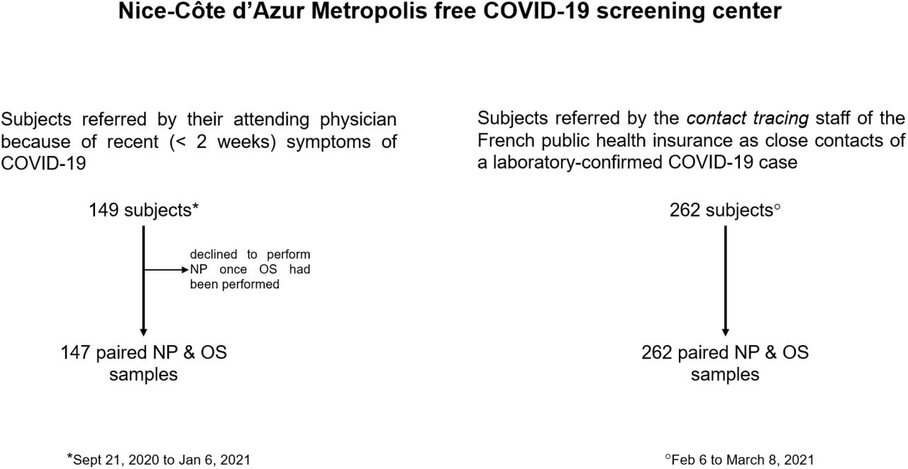
During the first part of the study (Sept 21, 2020 to Jan 6, 2021) we enrolled subjects referred by their attending physician because of recent (≤ 2 weeks) symptoms of COVID-19 [34]. During the second part of the study (Feb 6 to March 8, 2021) we enrolled subjects referred by the contact tracing staff of the French public health insurance [35], since they were considered as close contacts of a laboratory-confirmed COVID-19 case (figure 1).

Figure 1:
  • Download figure
  • Open in new tab
Figure 1:

Trial profile

Procedures

Specimen Collection

After signing an informed consent to participate, all volunteers were interviewed and underwent paired sampling: NP and OS in a random order. Disposable NP nylon swabs (type A-04. Jiangsu Han-Heng Medical technology Co., Ltd. Changzou, Jiangsu, China) were eluted into a vial containing viral transport medium (appendix 1) and transported within 4 hours to the Synlab Barla laboratory (Nice, France). Small hydroxylated polyvinyl acetate (PVA) sponges (Merocel® Standard Dressing, réf 400400, Medtronic), usually used for tamponade of epistaxis, were placed in the mouth by the subjects themselves and taken out after one minute and inserted into a sterile collection tube transported within 4 hours to the Idylla platform (Biocartis, Mechelen, Belgium) of the Laboratory of Clinical and Experimental Pathology (LPCE), (Hôpital Pasteur 1, Nice). No instructions were given regarding the need to fast, to have chewed gum or smoked a cigarette/e-cigarette.

Detection of SARS-CoV-2 RNA on NP samples

The Synlab Barla laboratory is a private biological analysis laboratory that is under contract with the Nice-Côte d’Azur Metropolis for COVID screening tests. This laboratory used RT-PCR to detect SARS-CoV-2 RNA on NP during the first part of the study. For logistical reasons, this laboratory switched to the Transcription Mediated Amplification (TMA) technique to detect SARS-CoV-2 RNA during the second part of the study. According to the guidelines of the French society for Microbiology [36] for RT-PCR, the result was considered positive when the Ct (cycle threshold) value for the N and/or on Orf1b genes was equal or less than 36. For TMA the result was considered as positive when the RLU was above 850.

Analysis of OS samples

We described previously RT-PCR for detection of SARS-CoV-2 RNA on the Idylla platform and showed a 100% positive and negative agreement between this technique and standard-of-care RT-PCR tests [33]. Upon arrival of the sample, 200μl of the OS eluate were pipetted with a sterile tip (D. Dutscher, reference 134000), placed individually into Idylla SARS-CoV-2 test cartridges (Biocartis, reference A1043/6) and underwent fully automated nucleic acid testing including extraction, amplification, and detection in a single-use cartridge. The duration of the run took 90 minutes (+/-10 min). The residual volume OS eluate was immediately aliquoted and stored at −80°C in the Nice COVID-19 Biobank, which is S96-900 certified by AFNOR [30]. According to the manufacturer, the Idylla SARS-CoV-2 test provides a qualitative result for the presence or absence of SARS-CoV-2 RNA with a corresponding quality status. The Biocartis SARS-CoV-2 test included 2 genes (N, Orf1b) covered by 5 PCR targets (2 N targets and 3 Orf1b targets). A positive result required at least 2 amplified N targets [by setting a quantification cycle (Cq) of 41.9] and/or at least one or more amplified Orf1b targets. As Orf1b is highly specific no threshold was required for this gene.

External validation

All OS eluates were sent to the Synlab Barla laboratory and underwent RT-PCR using the ORF1ab and N genes Da An Gene DA0992-Detection Kit for 2019-nCoV (Da An Gene Co., Ltd. Sun Yat-sen University, Guangzhou, Guangdong, China). During the first part of the study this external validation was performed on the OS eluates stored at −80°C in the Nice COVID-19 biobank) [30]. During the second part of the study, external validation was carried out in the Synlab Barla laboratory on fresh samples transferred from the Nice COVID-19 biobank.

The different analyses were processed in a double-blind way: the results of the reference standard test were unavailable to the readers of the index test and vice versa.

Outcomes

The primary outcome was the clinical performance of OS compared to NP for SARS- CoV-2 screening in symptomatic and close contact subjects. For each group (symptomatic vs close contacts) we extracted the number of individuals positive for SARS- CoV-2 in both NP and OS (a), those positive only with NP (b), those positive only with OS (c) and (d) those negative with both NO and OS (Table 1). From these data, we calculated the concordance of the two types of sample (a+d)/(a+b+c+d). We also calculated the sensitivity of the test on each type of sample. The estimation of the sensitivity of a test requires a reference diagnosis. Since NP sampling has been shown to produce false negatives by RT-PCR, sensitivities for NP and OS are defined here respectively as (a+b)/(a+b+c) and (a+c)/(a+b+c), considering as a true positive any individual with a positive result on one or the other sample. This definition of a positive individual is in agreement with the US-CDC and the ECDC directives on SARS-CoV-2 testing [37, 38].

View this table:
  • View inline
  • View popup
  • Download powerpoint
Table 1a:

Clinical performance of OS compared to NP for SARS- CoV-2 screening in symptomatic subjects (Idylla platform).

View this table:
  • View inline
  • View popup
  • Download powerpoint
Table 1b:

Clinical performance of OS compared to NP for SARS- CoV-2 screening of close contact subjects. External validation at the Synlab Barla laboratory platform

Secondary outcomes included the description of the studied population and the comparison between Ct values measured with NP and OS, an approximate proxy of viral loads. To calculate the sample size in the absence of data regarding the sensitivity of OS in symptomatic subjects, we calculated the sensitivity of OS once the first 40 paired (NP/OS) samples were obtained. This sensitivity was 85%. Given this estimate and a desired lower bound of the 95% confidence interval for sensitivity of at least 80%, 144 volunteers were needed to complete the study in symptomatic subjects. To deal with the dropout risk we decided to include 149 volunteers.

Statistics

Continuous variables are presented as means (± SD), and categorical variables as numbers and percentages. Baseline characteristics between patients with and without COVID-19 were compared using the Student’s t-test or Wilcoxon - Mann Whitney for quantitative variables based on the normality of the distribution of parameters or using the Chi-Square test for qualitative variables.

Ethics and regulatory authorizations

The promoter of the study was the Center Hospitalier Universitaire de Nice. The agreement for the study of the Institutional review board Sud Méditerranée V was obtained on April 22, 2020 (registration # 20.04014.35208). SHAM liability insurance (n° 159087). The study is registered in ClinicalTrial.gov (NCT04418206).

Role of the funding sources

The organizations that supported this study played no role in its design, patient selection, data collection, analysis or interpretation, report writing, or decision to submit the document for publication. Authors had full access to all data and responsibility of submission for publication.

Results

Four hundred and eleven subjects underwent COVID-19 diagnostic testing over the 22 weeks of the study, 149 during the first part of the study conducted on symptomatic subjects and 262 during the second part of the study conducted on close contacts. Two symptomatic subjects declined to perform NP once OS had been performed and were excluded from subsequent analyses. One had a positive OS RT-PCR, and the other was negative. The study therefore covers the remaining 409 participants (figure 1).

The 147 symptomatic subjects were predominantly women 86/147 (58.5%). Their mean age was 40 ± 15 years. The interval between symptom onset and testing was 3.6 ± 2.6 days and most participants (107/147 [72.8%]) were sampled at the early stage of the disease, i.e., within 4 days of symptom onset. Of these symptomatic subjects, 59 (40.1%) had a positive RT-PCR result with one or both of the sampling techniques and were thus diagnosed COVID-19. No clinical symptoms distinguished between RT-PCR positive and negative subjects (figure 2), with the exception of anosmia and dysgeusia, which were more frequent in RT-PCR positive subjects (42% vs. 10%, p <0.001 and 38 vs. 16%, p = 0.004) and sore throat, which was significantly more common in RT-PCR negative subjects (36 vs. 10%, p = 0.001).

Figure 2:
  • Download figure
  • Open in new tab
Figure 2:

Self-reported symptoms (%)

Figure 3:
  • Download figure
  • Open in new tab
Figure 3:

Viral load of NP and OS as indirectly assessed by the Ct for the Orf1b and N genes

For these symptomatic subjects, the Ct values for the Orf 1b and N genes with OS showed a more than 3-point increase compared with NP; the magnitude of this difference indicates a more than 10-fold lower quantity of viral genetic material in OS samples [39].

The 262 close contacts with a laboratory-confirmed COVID-19 case were predominantly women 145/262 (55.3%). The mean age was 41 ± 15 years. The contact was identified in the household (n=41; 15.6%), in the family (n=56; 21.4%); in the close environment (n=40; 15.3%), at work (n=65; 24.8%), or was not known by the subject (n=60; 22.9%). Of these close contact subjects, 57 (21.7%) had a positive RT-PCR result with one or both of the sampling techniques. Differences in viral load between the NP and the OS could not be assessed in close contact subjects since viral RNA detection with NP relied on TMA and not on RT-PCR in this part of the study.

In the symptomatic subjects, the NP-OS concordance was 96.5% (95%CI: 93.5 – 99.5) and the OS sensitivity was 93.2% (95%CI: 89.1 – 97.3) with the Idylla platform. With the Synlab Barla platform, the NP-OS concordance was 89.0% (95%CI: 83.9 – 94.1) and the OS sensitivity was 76.3% (95%CI: 69.4 – 83.2) (table 1).

In the close contact subjects, the NP-OS concordance was 93.8% (95%CI: 90.9 – 96.7) and the OS sensitivity was 71.9% (95%CI: 66.5 – 77.3) with the Idylla platform. With the Synlab barla platform, the NP-OS concordance was 94.2% (95%CI: 91.4 – 97.0) and the OS sensitivity was 73.7% (95%CI: 68.4 – 79.0) (table 1).

Discussion

In this study, RT-PCR SARS-CoV-2 testing with OS showed a high level of concordance (with NP testing), 96.5% (95%CI: 89.6-94.8) and sensitivity, 93.3 (95%CI: 89.1-97.3) for symptomatic subjects and a slightly lower level of concordance, 93.8% (95%CI: 90.9-96.7) and sensitivity, 71.9% (95%CI: 66.5-77.3) in close contacts of a laboratory-confirmed COVID-19 case. These results obtained in ambulatory subjects presenting to a public community-based screening center, strongly suggest that OS, a straightforward, low-cost and high-throughput sampling material can be used for frequent RT-PCR testing of COVID-19 patients and mass screening of populations.

During the early phase of the COVID-19 pandemic, many molecular tests, immunoassays and sampling methods were rapidly developed and validated using archived biological samples of known virological status, albeit many still await clinical validation [40]. To evaluate the clinical performance of OS, compared to NP, the present study was carried out according to the standards recommended by the French “Haute Autorité de Santé” [8]; i.e., a prospective comparative clinical study relating to a series of individuals of unknown COVID-19 status, recruited consecutively; a salivary test including at least two molecular targets; and by the US and European CDC directives; i.e. definition of a positive individual when comparing two diagnostic SARS-Cov-2 testing methods [37,38].

In published studies that compared salivary and NP testing, symptomatic subjects were the dominant inclusion profile (85%), especially in the majority of short series of hospitalized patients [18]. Only 6 cohorts of equal or larger size than ours reported comparative test results obtained in a community-based settings [41–46].

Given the particularly high incidence of COVID-19 in the Nice metropolitan area throughout our study period (incidence between 330 and 720/100,000), our rate of positivity (21-41%) was higher than the rates observed in these 6 cohorts (1.5% to 10.7%). Despite this difference, our NP-OS concordance and the sensitivity of RT-PCR on NP and on OS are in agreement with the results reported for these cohorts; and with the notion of no association between salivary and NP test performance and viral prevalence [19].

RT-PCR with OS performed at the Synlab Barla platform showed a significantly lower sensitivity than Idylla platform in symptomatic subjects. This discrepancy may be due to 2 factors: the Idylla platform uses 3 Orf1 and 2 N targets while the Synlab Barla platform uses only one Orf1 and one N target; preservation at −80°C for several weeks may have altered the nucleic acids. The discrepancy disappeared in the second part of the study in which the external validation of OS testing at the Synlab Barla platform was done on fresh samples in real time.

The significant difference in viral load observed between samples of nasopharyngeal and buccal origin may be due to several parameters, notably endogenous to the laboratory [(i.e. total volume of sample collection buffer/medium, sample preparation method (heat, lysis methods)] and laboratory reagent volumes used in each step of the RT-PCR process. A similar difference has been observed by others [2, 47–49] and explains why the diagnostic yield of NP is considered to be higher than that of throat swabs [48, 49]. Thus, this difference most likely has a pathophysiological basis, related to the fact that the mouth squamous cells act as a viral reservoir to a lesser degree than the nasopharynx ciliated cells [31, 32, 50]. As a consequence, the interpretative cut-offs for NP Ct [36] hardly apply to saliva samples.

The OS sampling method is well standardized. It does not need sialagogic drugs, nor clearing the throat or spitting effort, nor particular constraints such as early morning saliva sampling before tooth brushing and breakfast, avoiding eating, drinking, gum chewing, smoking, or vaping. [51–53]. It does not need dedicated trained nursing staff and its acceptability makes it possible to consider repeating the test even in institutionalized elderly people or in children.

The question as to whether the 72% sensitivity we observed in contact subjects is good enough to identify SARS-CoV-2 infected people in the setting of mass screening has been debated by Mina et al. in a landmark position paper [7]. In this paper they distinguished between a low frequency tests with a high analytical sensitivity and high frequency tests with low analytical sensitivity. RT-PCR with NP samples belongs to the former category and is well suited to diagnosis of COVID-19 in symptomatic subjects. Whereas the OS sampling method described herein belongs to the latter category and would be more effective in identifying, isolating, and thus filtering out currently infected persons, including those who are asymptomatic.

We industrialized the OS process in the form of an “all-in-one” kit simplifying extraction of the biological liquid (figure 4) and presently intend to clinically validate the performance of this sampling method with the pooling technique [54] for mass screening.

Figure 4:
  • Download figure
  • Open in new tab
Figure 4:

All-in-on saliva sampling technique with a PVA sponge and a flexible, double-capped plastic tube.

Data Availability

all data referred to in this manuscript can be available after contacting the corresponding author

Funding

This work was supported by Conseil Départemental 06, Ville de Nice, Métropole Nice Côte d’Azur, Fonds de Dotation AVENI, and from private donators.

Acknowledgements

Special thanks to E. Faidhi, N. Fridlyand, A. Rauscher, E. Maris, the Lauro family and to the many private donators for their generous contribution.

1 APPENDIX PROCEDURES

Detection of SARS-CoV-2 RNA with naso-pharyngeal swab specimens

During the first study period (September 21, 2020 to January 6, 2021; symptomatic patients), naso-pharyngeal (NP) swab specimens were analyzed by RT-PCR using the ORF1ab and N genes Da An Gene DA0992-Detection Kit for 2019-nCoV (Da An Gene Co., Ltd. Sun Yat-sen University, Guangzhou, Guangdong, China). NP swabs were eluted into a vial containing 400 µl of viral RNA extraction buffer (RNA/DNA purification kit, Da An Gene, ref DA0940); and 5 μl was then processed on an AGS 4800 Thermocycler (Hangzhou, Zhejiang, China). According to the guidelines of the French Society for Microbiology [reference 30 Avis du 25 septembre 2020 de la Société Française de Microbiologie (SFM) relatif à l’interprétation de la valeur de Ct] the result was considered as positive when the Ct (cycle threshold) value for the N and/or Orf1b genes was equal or less than 36.

During the second study period (February 6 to March 6, 2021; close contacts) NP samples were analyses with the Aptima® SARS-CoV-2 assay (Hologic, Inc. 10210 Genetic Center Drive San Diego, CA 92121 USA), which combines Transcription Mediated Amplification (TMA), and Dual Kinetic Assay (DKA) that amplifies and detects two conserved regions of the ORF1ab gene. NP swabs were eluted into a vial containing 710 µl of medium (Hologic Specimen Lysis Tubes, ref PDD-06554) before being tested on the Panther system. Assay results were determined with a cut-off based on the total Relative Light Units (RLU). According to the guidelines of the French Society for Microbiology the results were considered as positive when the RLU was above 850.

Detection of SARS-CoV-2 RNA with oral sponge (OS) specimens at the Synlab Barla laboratory

25 μl of the OS eluates transferred fresh from the Nice COVID-19 biobank were put into a vial containing 400 µl of viral RNA extraction buffer (RNA/DNA purification kit, Da An Gene, ref DA0940); and 5 μl was then processed on an AGS 4800 Thermocycler (Hangzhou, Zhejiang, China).

Footnotes

  • Background: The current diagnostic standard for coronavirus 2019 disease (COVID-19) is reverse transcriptase-polymerase chain reaction (RT-PCR) testing with naso-pharyngeal (NP) swabs. The invasiveness and need for trained personnel make the NP technique unsuited for repeated community-based mass screening. We developed a technique to collect saliva in a simple and easy way with the sponges that are usually used for tamponade of epistaxis. This study was carried out to validate the clinical performance of oral sponge (OS) sampling for SARS-CoV-2 testing. Methods: Over a period of 22 weeks, we collected prospectively 409 paired NP and OS samples from consecutive subjects presenting to a public community-based free screening center. Subjects were referred by their attending physician because of recent COVID-19 symptoms (n=147) or by the contact tracing staff of the French public health insurance since they were considered as close contacts of a laboratory-confirmed COVID-19 case (n=262). Results: In symptomatic subjects, RT-PCR SARS-CoV-2 testing with OS showed a 96.5% (95%CI: 89.6-94.8) concordance with NP testing, and, a 93.3% [95%CI: 89.1-97.3] sensitivity. In close contacts the NP-OS concordance (93.8% [95%CI: 90.9-96.7]) and OS sensitivity (71.9% [95%CI: 66.5-77.3]) were slightly lower. Conclusion: These results strongly suggest that OS testing is a straightforward, low-cost and high-throughput sampling method that can be used for frequent RT-PCR testing of COVID-19 patients and mass screening of populations.

References

  1. 1.↵
    Hanson KE, Caliendo AM, Arias CA, Englund JA, Lee MJ, Patel R, Alayli AE, Kalot MA, Falck-Ytter Y, Morgan RL, Murad MH, Sultan S, Bhimraj A, Mustafa RA. Infectious Diseases Society of America Guidelines on the Diagnosis of COVID-19. : 72.
  2. 2.↵
    Zou L, Ruan F, Huang M, Liang L, Huang H, Hong Z, Yu J, Kang M, Song Y, Xia J, Guo Q, Song T, He J, Yen H-L, Peiris M, Wu J. SARS-CoV-2 Viral Load in Upper Respiratory Specimens of Infected Patients. New England Journal of Medicine Massachusetts Medical Society; 2020; 382: 1177–1179.
    OpenUrl
  3. 3.
    Sullivan PS, Sailey C, Guest JL, Guarner J, Kelley C, Siegler AJ, Valentine-Graves M, Gravens L, Del Rio C, Sanchez TH. Detection of SARS-CoV-2 RNA and Antibodies in Diverse Samples: Protocol to Validate the Sufficiency of Provider-Observed, Home-Collected Blood, Saliva, and Oropharyngeal Samples. JMIR Public Health Surveill 2020; 6: e19054.
    OpenUrlCrossRef
  4. 4.↵
    Pondaven-Letourmy S, Alvin F, Boumghit Y, Simon F. How to perform a nasopharyngeal swab in adults and children in the COVID-19 era. Eur Ann Otorhinolaryngol Head Neck Dis 2020; 137: 325–327.
    OpenUrl
  5. 5.↵
    Kucirka LM, Lauer SA, Laeyendecker O, Boon D, Lessler J. Variation in False-Negative Rate of Reverse Transcriptase Polymerase Chain Reaction–Based SARS-CoV-2 Tests by Time Since Exposure. Ann Intern Med American College of Physicians; 2020; 173: 262–267.
    OpenUrl
  6. 6.↵
    Arevalo-Rodriguez I, Buitrago-Garcia D, Simancas-Racines D, Zambrano-Achig P, Campo RD, Ciapponi A, Sued O, Martinez-García L, Rutjes AW, Low N, Bossuyt PM, Perez-Molina JA, Zamora J. False-negative results of initial RT-PCR assays for COVID-19: A systematic review. PLOS ONE Public Library of Science; 2020; 15: e0242958.
    OpenUrl
  7. 7.↵
    Mina MJ, Parker R, Larremore DB. Rethinking Covid-19 Test Sensitivity — A Strategy for Containment. New England Journal of Medicine Massachusetts Medical Society; 2020; 383: e120.
    OpenUrl
  8. 8.↵
    Haute Autorité de Santé. Revue rapide sur les tests de détection antigénique du virus SARS-CoV-2. 8 octobre 2020 (https://www.has-sante.fr/upload/docs/application/pdf/2020-10/synthese_tests_antigeniques_vd.pdf).
  9. 9.
    World Health Organization. (2020). Antigen-detection in the diagnosis of SARS-CoV-2 infection using rapid immunoassays: interim guidance, 11 September 2020. World Health Organization. https://apps.who.int/iris/handle/10665/334253. License: CC BY-NC-SA 3.0 IGO [Internet]. [cited 2021 Jan 22]. Available from: https://www.who.int/publications-detail-redirect/antigen-detection-in-the-diagnosis-of-sars-cov-2infection-using-rapid-immunoassays.
  10. 10.↵
    Évaluation de la performance diagnostique de neuf tests rapides antigéniques COVID-19 [Internet]. [cited 2021 Jan 22].Available from: https://www.aphp.fr/contenu/evaluation-de-la-performance-diagnostique-de-neuf-tests-rapides-antigeniques-covid-19.
  11. 11.↵
    Butler-Laporte G, Lawandi A, Schiller I, Yao M, Dendukuri N, McDonald EG, Lee TC. Comparison of Saliva and Nasopharyngeal Swab Nucleic Acid Amplification Testing for Detection of SARS-CoV-2: A Systematic Review and Meta-analysis. JAMA Intern Med 2021; 181: 353–360.
    OpenUrl
  12. 12.↵
    To KK-W, Tsang OT-Y, Leung W-S, Tam AR, Wu T-C, Lung DC, Yip CC-Y, Cai J-P, Chan JM-C, Chik TS-H, Lau DP-L, Choi CY-C, Chen L-L, Chan W-M, Chan K-H, Ip JD, Ng AC-K, Poon RW-S, Luo C-T, Cheng VC-C, Chan JF-W, Hung IF-N, Chen Z, Chen H, Yuen K-Y. Temporal profiles of viral load in posterior oropharyngeal saliva samples and serum antibody responses during infection by SARS-CoV-2: an observational cohort study. The Lancet Infectious Diseases Elsevier; 2020; 20: 565–574.
    OpenUrlCrossRefPubMed
  13. 13.
    Liu L, Wei Q, Alvarez X, Wang H, Du Y, Zhu H, Jiang H, Zhou J, Lam P, Zhang L, Lackner A, Qin C, Chen Z. Epithelial cells lining salivary gland ducts are early target cells of severe acute respiratory syndrome coronavirus infection in the upper respiratory tracts of rhesus macaques. J Virol 2011; 85: 4025–4030.
    OpenUrlAbstract/FREE Full Text
  14. 14.
    Lu B, Huang Y, Huang L, Li B, Zheng Z, Chen Z, Chen J, Hu Q, Wang H. Effect of mucosal and systemic immunization with virus-like particles of severe acute respiratory syndrome coronavirus in mice. Immunology 2010; 130: 254–261.
    OpenUrlCrossRefPubMed
  15. 15.
    Silva-Boghossian CM, Colombo APV, Tanaka M, Rayo C, Xiao Y, Siqueira WL. Quantitative Proteomic Analysis of Gingival Crevicular Fluid in Different Periodontal Conditions. PLOS ONE Public Library of Science; 2013; 8: e75898.
    OpenUrl
  16. 16.
    Xu H, Zhong L, Deng J, Peng J, Dan H, Zeng X, Li T, Chen Q. High expression of ACE2 receptor of 2019-nCoV on the epithelial cells of oral mucosa. International Journal of Oral Science Nature Publishing Group; 2020; 12: 1–5.
    OpenUrl
  17. 17.↵
    Dawes C, Wong DTW. Role of Saliva and Salivary Diagnostics in the Advancement of Oral Health. J Dent Res SAGE Publications Inc; 2019; 98: 133–141.
    OpenUrl
  18. 18.↵
    Haute Autorité de Santé. Méta-analyse de l’intérêt diagnostique des tests RT-PCR salivaires de détection du SARS-CoV-2. https://www.has-sante.fr/jcms/p_3237008/fr/meta-analyse-de-l-interet-diagnostique-des-tests-rt-pcr-salivaires-de-detection-du-sars-cov-2 (accessed Apr. 10, 2021)]
  19. 19.↵
    Ibrahimi N, Delaunay-Moisan A, Hill C, Teuff GL, Rupprecht J-F, Thuret J-Y, Chaltiel D, Potier M-C. Screening for SARS-CoV-2 by RT-PCR: saliva or nasopharyngeal swab? Systematic review and meta-analysis. medRxiv Cold Spring Harbor Laboratory Press; 2021; : 2021.02.10.21251508.
  20. 20.
    Moreira VM, Mascarenhas P, Machado V, Botelho J, Mendes JJ, Taveira N, Almeida MG. Diagnosis of SARS-Cov-2 Infection by RT-PCR Using Specimens Other Than Naso-and Oropharyngeal Swabs: A Systematic Review and Meta-Analysis. Diagnostics Multidisciplinary Digital Publishing Institute; 2021; 11: 363.
    OpenUrl
  21. 21.
    Bastos ML, Perlman-Arrow S, Menzies D, Campbell JR. The Sensitivity and Costs of Testing for SARS-CoV-2 Infection With Saliva Versus Nasopharyngeal Swabs?: A Systematic Review and Meta-analysis. Ann Intern Med 2021;.
  22. 22.
    Lee RA, Herigon JC, Benedetti A, Pollock NR, Denkinger CM. Performance of Saliva, Oropharyngeal Swabs, and Nasal Swabs for SARS-CoV-2 Molecular Detection: A Systematic Review and Meta-analysis. J Clin Microbiol 2021;.
  23. 23.
    Kivelä JM, Jarva H, Lappalainen M, Kurkela S. Saliva-based testing for diagnosis of SARS-CoV-2 infection: A meta-analysis. J Med Virol 2021; 93: 1256–1258.
    OpenUrl
  24. 24.
    Meng M, Zhou X, Zhang Q, Zou J. The positive rate of saliva for the detection of 2019-nCoV and possible factors related to the sensitivity results. J Med Virol 2020;.
  25. 25.
    Peeters E, Singh SKDA, Vandesompele J, Mestdagh P, Hutse V, Arbyn M. Rapid systematic review of the sensitivity of SARS-CoV-2 molecular testing on saliva compared to nasopharyngeal swabs. medRxiv Cold Spring Harbor Laboratory Press; 2020; : 2020.08.05.20168716.
  26. 26.
    Riccò M, Ranzieri S, Peruzzi S, Valente M, Marchesi F, Balzarini F, Bragazzi NL, Signorelli C. RT-qPCR assays based on saliva rather than on nasopharyngeal swabs are possible but should be interpreted with caution: results from a systematic review and meta-analysis.: Acta Bio Med 2020; 91: e2020025–e2020025.
    OpenUrl
  27. 27.↵
    Czumbel LM, Kiss S, Farkas N, Mandel I, Hegyi A, Nagy Á, Szakács Z, Hegyi P, Steward MC, Varga G. Saliva as a Candidate for COVID-19 Diagnostic Testing: A Meta-Analysis. Front Med (Lausanne) 2020; : 465–465.
  28. 28.↵
    Avis n° 2021.0007/AC/SEAP du 10 février 2021 du collège de la HAS relatif aux modifications des conditions d’inscription sur la LAP mentionnée à l’article L. 162-1-7 du CSS, à la détection du génome du virus SARS-CoV-2 par technique de transcription inverse suivie d’une amplification (RT-PCR) sur prélèvement salivaire [Internet]. Haute Autorité de Santé [cited 2021 Apr 7].Available from: https://www.has-sante.fr/jcms/p_3236975/fr/avis-n-2021-0007/ac/seap-du-10-fevrier-2021-du-college-de-la-has-relatif-aux-modifications-des-conditions-d-inscription-sur-la-lap-mentionnee-a-l-article-l-162-1-7-du-css-a-la-detection-du-genome-du-virus-sars-cov-2-par-technique-de-transcription-inverse-suivie-d-une-amplification-rt-pcr-sur-prelevement-salivaire.
  29. 29.↵
    Deprez M, Zaragosi L-E, Truchi M, Becavin C, Ruiz García S, Arguel M-J, Plaisant M, Magnone V, Lebrigand K, Abelanet S, Brau F, Paquet A, Pe’er D, Marquette C-H, Leroy S, Barbry P. A Single-Cell Atlas of the Human Healthy Airways. Am J Respir Crit Care Med 2020; 202: 1636–1645.
    OpenUrl
  30. 30.↵
    Tanga V, Leroy S, Fayada J, Hamila M, Allegra M, Messaoudi Z, Bonnetaud C, Lespinet V, Bordone O, Washetine K, Griffonnet J, Maniel C, Philibert L, Selva E, Benzaquen J, Ilie M, Long E, Lassalle S, Lantéri E, Marquette C-H, Hofman V, Hofman P. Establishment of a Collection of Blood-Derived Products from COVID-19 Patients for Translational Research: Experience of the LPCE Biobank (Nice, France). Biopreservation and Biobanking Mary Ann Liebert, Inc., publishers; 2020; 18: 517–524.
    OpenUrl
  31. 31.↵
    Sungnak W, Huang N, Bécavin C, Berg M, Queen R, Litvinukova M, Talavera-López C, Maatz H, Reichart D, Sampaziotis F, Worlock KB, Yoshida M, Barnes JL. SARS-CoV-2 entry factors are highly expressed in nasal epithelial cells together with innate immune genes. Nature Medicine Nature Publishing Group; 2020; 26: 681–687.
    OpenUrl
  32. 32.↵
    Ziegler CGK, Allon SJ, Nyquist SK, Mbano IM, Miao VN, Tzouanas CN, Cao Y, Yousif AS, Bals J, Hauser BM, Feldman J, Muus C, Wadsworth MH, Kazer SW, Hughes TK, Doran B, Gatter GJ, Vukovic M, Taliaferro F, Mead BE, Guo Z, Wang JP, Gras D, Plaisant M, Ansari M, Angelidis I, Adler H, Sucre JMS, Taylor CJ, Lin B, et al. SARS-CoV-2 Receptor ACE2 Is an Interferon-Stimulated Gene in Human Airway Epithelial Cells and Is Detected in Specific Cell Subsets across Tissues. Cell 2020; 181: 1016-1035.e19.
    OpenUrlCrossRefPubMed
  33. 33.↵
    Hofman P, Boutros J, Benchetrit D, Benzaquen J, Leroy S, Tanga V. A rapid near-patient RT-PCR test for suspected COVID-19. A study of the diagnostic accuracy. Annals of Translational Medicine 2021; ATM-21-690 accepted for publication on the 21st of march 2021.
  34. 34.↵
    Lechien JR, Chiesa-Estomba CM, Place S, Van Laethem Y, Cabaraux P, Mat Q, Huet K, Plzak J, Horoi M, Hans S, Rosaria Barillari M, Cammaroto G, Fakhry N, Martiny D, Ayad T, Jouffe L, Hopkins C, Saussez S, COVID-19 Task Force of YO-IFOS. Clinical and epidemiological characteristics of 1420 European patients with mild-to-moderate coronavirus disease 2019. J Intern Med 2020; 288: 335–344.
    OpenUrlCrossRefPubMed
  35. 35.↵
    «?Contact tracing»?: arrêter les chaînes de transmission [Internet]. [cited 2021 Apr 13].Available from: https://www.ameli.fr/assure/covid-19/tester-alerter-proteger-comprendre-la-strategie-pour-stopper-lepidemie/contact-tracing-arreter-les-chaines-de-transmission.
  36. 36.↵
    Avis du 25 septembre 2020 de la Société Française de Microbiologie (SFM) relatif à l’interprétation de la valeur de Ct (estimation de la charge virale) obtenue en cas de RT-PCR SARS-CoV-2 positive sur les prélèvements cliniques réalisés à des fins diagnostiques ou de dépistage. Version 3 [Internet]. 2020 [cited 2021 Jan 22].Available from: https://www.sfm-microbiologie.org/wp-content/uploads/2020/10/Avis-SFM-valeur-Ct-excre%CC%81tion-virale-_-Version-Finale-07102020-V3.pdf.
  37. 37.↵
    CDC. Healthcare Workers [Internet]. Centers for Disease Control and Prevention 2020 [cited 2021 Apr 12].Available from: https://www.cdc.gov/coronavirus/2019-ncov/hcp/testing-overview.html.
  38. 38.↵
    Diagnostic testing and screening for SARS-CoV-2 [Internet]. European Centre for Disease Prevention and Control [cited 2021 Apr 12].Available from: https://www.ecdc.europa.eu/en/covid-19/latest-evidence/diagnostic-testing.
  39. 39.↵
    Understanding cycle threshold (Ct) in SARS-CoV-2 RT-PCR. [Internet] Public Health England. Available from: https://assets.publishing.service.gov.uk/government/uploads/system/uploads/attachment_data/file/926410/Understanding_Cycle_ThresholdCtin_SARS-CoV-2_RT-PCR_.pdf. : 12.
  40. 40.↵
    Vandenberg O, Martiny D, Rochas O, van Belkum A, Kozlakidis Z. Considerations for diagnostic COVID-19 tests. Nat Rev Microbiol 2020; : 1–13.
  41. 41.↵
    Caulley L, Corsten M, Eapen L, Whelan J, Angel JB, Antonation K, Bastien N, Poliquin G, Johnson-Obaseki S. Salivary Detection of COVID-19. Ann Intern Med American College of Physicians; 2020; 174: 131–133.
    OpenUrl
  42. 42.
    Barat B, Das S, Giorgi VD, Henderson DK, Kopka S, Lau AF, Miller T, Moriarty T, Palmore TN, Sawney S, Spalding C, Tanjutco P, Wortmann G, Zelazny AM, Frank KM. Pooled Saliva Specimens for SARS-CoV-2 Testing. Journal of Clinical Microbiology [Internet] American Society for Microbiology Journals; 2021 [cited 2021 Apr 12]; 59 Available from: https://jcm.asm.org/content/59/3/e02486-20.
  43. 43.
    Kandel C, Zheng J, McCready J, Serbanescu MA, Racher H, Desaulnier M, Powis JE, Vojdani K, Finlay L, Sheldrake E, Vermeiren C, Katz K, McGeer A, Kozak R, Goneau LW. Detection of SARS-CoV-2 from Saliva as Compared to Nasopharyngeal Swabs in Outpatients. Viruses Multidisciplinary Digital Publishing Institute; 2020; 12: 1314.
    OpenUrl
  44. 44.
    Senok A, Alsuwaidi H, Atrah Y, Ayedi OA, Zahid JA, Han A, Marzooqi AA, Heialy SA, Altrabulsi B, AbdelWareth L, Idaghdour Y, Ali R, Loney T, Alsheikh-Ali A. <p>Saliva as an Alternative Specimen for Molecular COVID-19 Testing in Community Settings and Population-Based Screening</p>. IDR Dove Press; 2020; 13: 3393–3399.
    OpenUrl
  45. 45.
    Vogels CBF, Watkins AE, Harden CA, Brackney DE, Shafer J, Wang J, Caraballo C, Kalinich CC, Ott IM, Fauver JR, Kudo E, Lu P, Venkataraman A, Tokuyama M, Moore AJ, Muenker MC, Casanovas-Massana A, Fournier J, Bermejo S, Campbell M, Datta R, Nelson A, Anastasio K, Askenase MH, Batsu M, Bickerton S, Brower K, Bucklin ML, Cahill S, Cao Y, et al. SalivaDirect: A simplified and flexible platform to enhance SARS-CoV-2 testing capacity. Med 2021; 2: 263–280.e6.
    OpenUrl
  46. 46.↵
    Yokota I, Shane PY, Okada K, Unoki Y, Yang Y, Inao T, Sakamaki K, Iwasaki S, Hayasaka K, Sugita J, Nishida M, Fujisawa S, Teshima T. Mass screening of asymptomatic persons for SARS-CoV-2 using saliva. Clinical Infectious Diseases [Internet] 2020 [cited 2021 Apr 12]; Available from: https://doi.org/10.1093/cid/ciaa1388.
  47. 47.↵
    Vlek ALM, Wesselius TS, Achterberg R, Thijsen SFT. Combined throat/nasal swab sampling for SARS-CoV-2 is equivalent to nasopharyngeal sampling. Eur J Clin Microbiol Infect Dis 2021; 40: 193–195.
    OpenUrl
  48. 48.↵
    Wang X, Tan L, Wang X, Liu W, Lu Y, Cheng L, Sun Z. Comparison of nasopharyngeal and oropharyngeal swabs for SARS-CoV-2 detection in 353 patients received tests with both specimens simultaneously. International Journal of Infectious Diseases 2020; 94: 107–109.
    OpenUrlCrossRefPubMed
  49. 49.↵
    Patel MR, Carroll D, Ussery E, Whitham H, Elkins CA, Noble-Wang J, Rasheed JK, Lu X, Lindstrom S, Bowen V, Waller J, Armstrong G, Gerber S, Brooks JT. Performance of Oropharyngeal Swab Testing Compared With Nasopharyngeal Swab Testing for Diagnosis of Coronavirus Disease 2019—United States, January 2020–February 2020. Clin Infect Dis [Internet] 2020 [cited 2021 Feb 8]; Available from: https://www.ncbi.nlm.nih.gov/pmc/articles/PMC7337670/.
  50. 50.↵
    Muus C, Luecken MD, Eraslan G, Sikkema L, Waghray A, Heimberg G, Kobayashi Y, Vaishnav ED, Subramanian A, Smillie C, Jagadeesh KA, Duong ET, Fiskin E, Triglia ET, Ansari M, Cai P, Lin B, Buchanan J, Chen S, Shu J, Haber AL, Chung H, Montoro DT, Adams T, Aliee H, Allon SJ, Andrusivova Z, Angelidis I, Ashenberg O, Bassler K, et al. Single-cell meta-analysis of SARS-CoV-2 entry genes across tissues and demographics. Nature Medicine Nature Publishing Group; 2021; 27: 546–559.
    OpenUrl
  51. 51.↵
    L’Helgouach N, Champigneux P, Schneider FS, Molina L, Espeut J, Alali M, Baptiste J, Cardeur L, Dubuc B, Foulongne V, Galtier F, Makinson A, Marin G, Picot M-C, Prieux-Lejeune A, Quenot M, Robles FC, Salvetat N, Vetter D, Reynes J, Molina F. EasyCOV?: LAMP based rapid detection of SARS-CoV-2 in saliva. medRxiv Cold Spring Harbor Laboratory Press; 2020; : 2020.05.30.20117291.
  52. 52.
    Lai CKC, Chen Z, Lui G, Ling L, Li T, Wong MCS, Ng RWY, Tso EYK, Ho T, Fung KSC, Ng ST, Wong BKC, Boon SS, Hui DSC, Chan PKS. Prospective Study Comparing Deep Throat Saliva With Other Respiratory Tract Specimens in the Diagnosis of Novel Coronavirus Disease 2019. The Journal of Infectious Diseases 2020; 222: 1612–1619.
    OpenUrl
  53. 53.↵
    Chen JH-K, Yip CC-Y, Poon RW-S, Chan K-H, Cheng VC-C, Hung IF-N, Chan JF-W, Yuen K-Y, To KK-W. Evaluating the use of posterior oropharyngeal saliva in a point-of-care assay for the detection of SARS-CoV-2. Emerging Microbes & Infections 2020; 9: 1356–1359.
    OpenUrl
  54. 54.↵
    Watkins AE, Fenichel EP, Weinberger DM, Vogels CBF, Brackney DE, Casanovas-Massana A, Campbell M, Fournier J, Bermejo S, Datta R, Dela Cruz CS, Farhadian SF, Iwasaki A, Ko AI, Grubaugh ND, Wyllie AL, Yale IMPACT Research Team2. Increased SARS-CoV-2 Testing Capacity with Pooled Saliva Samples. Emerg Infect Dis 2021; 27.
Back to top
PreviousNext
Posted April 16, 2021.
Download PDF
Data/Code
Email

Thank you for your interest in spreading the word about medRxiv.

NOTE: Your email address is requested solely to identify you as the sender of this article.

Enter multiple addresses on separate lines or separate them with commas.
Salivary detection of COVID-19. Clinical performance of oral sponge sampling for SARS-CoV-2 testing
(Your Name) has forwarded a page to you from medRxiv
(Your Name) thought you would like to see this page from the medRxiv website.
CAPTCHA
This question is for testing whether or not you are a human visitor and to prevent automated spam submissions.
Share
Salivary detection of COVID-19. Clinical performance of oral sponge sampling for SARS-CoV-2 testing
Charles Hugo Marquette, Jacques Boutros, Jonathan Benzaquen, Marius Ilié, Mickelina Labaky, Didier Benchetrit, Thibaut Lavrut, Sylvie Leroy, Richard Chemla, Michel Carles, Virginie Tanga, Charlotte Maniel, Olivier Bordone, Maryline Allégra, Virginie Lespinet, Julien Fayada, Jennifer Griffonnet, Véronique Hofman, Paul Hofman
medRxiv 2021.02.17.21251556; doi: https://doi.org/10.1101/2021.02.17.21251556
Twitter logo Facebook logo LinkedIn logo Mendeley logo
Citation Tools
Salivary detection of COVID-19. Clinical performance of oral sponge sampling for SARS-CoV-2 testing
Charles Hugo Marquette, Jacques Boutros, Jonathan Benzaquen, Marius Ilié, Mickelina Labaky, Didier Benchetrit, Thibaut Lavrut, Sylvie Leroy, Richard Chemla, Michel Carles, Virginie Tanga, Charlotte Maniel, Olivier Bordone, Maryline Allégra, Virginie Lespinet, Julien Fayada, Jennifer Griffonnet, Véronique Hofman, Paul Hofman
medRxiv 2021.02.17.21251556; doi: https://doi.org/10.1101/2021.02.17.21251556

Citation Manager Formats

  • BibTeX
  • Bookends
  • EasyBib
  • EndNote (tagged)
  • EndNote 8 (xml)
  • Medlars
  • Mendeley
  • Papers
  • RefWorks Tagged
  • Ref Manager
  • RIS
  • Zotero
  • Tweet Widget
  • Facebook Like
  • Google Plus One

Subject Area

  • Infectious Diseases (except HIV/AIDS)
Subject Areas
All Articles
  • Addiction Medicine (349)
  • Allergy and Immunology (668)
  • Allergy and Immunology (668)
  • Anesthesia (181)
  • Cardiovascular Medicine (2648)
  • Dentistry and Oral Medicine (316)
  • Dermatology (223)
  • Emergency Medicine (399)
  • Endocrinology (including Diabetes Mellitus and Metabolic Disease) (942)
  • Epidemiology (12228)
  • Forensic Medicine (10)
  • Gastroenterology (759)
  • Genetic and Genomic Medicine (4103)
  • Geriatric Medicine (387)
  • Health Economics (680)
  • Health Informatics (2657)
  • Health Policy (1005)
  • Health Systems and Quality Improvement (985)
  • Hematology (363)
  • HIV/AIDS (851)
  • Infectious Diseases (except HIV/AIDS) (13695)
  • Intensive Care and Critical Care Medicine (797)
  • Medical Education (399)
  • Medical Ethics (109)
  • Nephrology (436)
  • Neurology (3882)
  • Nursing (209)
  • Nutrition (577)
  • Obstetrics and Gynecology (739)
  • Occupational and Environmental Health (695)
  • Oncology (2030)
  • Ophthalmology (585)
  • Orthopedics (240)
  • Otolaryngology (306)
  • Pain Medicine (250)
  • Palliative Medicine (75)
  • Pathology (473)
  • Pediatrics (1115)
  • Pharmacology and Therapeutics (466)
  • Primary Care Research (452)
  • Psychiatry and Clinical Psychology (3432)
  • Public and Global Health (6527)
  • Radiology and Imaging (1403)
  • Rehabilitation Medicine and Physical Therapy (814)
  • Respiratory Medicine (871)
  • Rheumatology (409)
  • Sexual and Reproductive Health (410)
  • Sports Medicine (342)
  • Surgery (448)
  • Toxicology (53)
  • Transplantation (185)
  • Urology (165)