medRxiv
  • Home
  • About
  • Submit
  • ALERTS / RSS
Advanced Search

Analysis of Whole Genome Sequencing in a Cohort of Individuals with PHACE Syndrome Suggests Dysregulation of RAS/PI3K Signaling

View ORCID ProfileElizabeth S. Partan, View ORCID ProfileFrancine Blei, View ORCID ProfileSarah L. Chamlin, View ORCID ProfileOlivia M. T. Davies, View ORCID ProfileBeth A. Drolet, View ORCID ProfileIlona J. Frieden, View ORCID ProfileIoannis Karakikes, View ORCID ProfileChien-Wei Lin, View ORCID ProfileAnthony J. Mancini, View ORCID ProfileDenise Metry, View ORCID ProfileAnthony Oro, View ORCID ProfileNicole S. Stefanko, View ORCID ProfileLaksshman Sundaram, View ORCID ProfileMonika Tutaj, View ORCID ProfileAlexander E. Urban, View ORCID ProfileKevin C. Wang, View ORCID ProfileXiaowei Zhu, View ORCID ProfileNara Sobreira, View ORCID ProfileDawn H. Siegel
doi: https://doi.org/10.1101/2021.08.05.21261553
Elizabeth S. Partan
1McKusick-Nathans Department of Genetic Medicine, Johns Hopkins Medicine, Baltimore, MD
2Predoctoral Training Program in Human Genetics, Johns Hopkins School of Medicine, Baltimore, MD
  • Find this author on Google Scholar
  • Find this author on PubMed
  • Search for this author on this site
  • ORCID record for Elizabeth S. Partan
Francine Blei
3Department of Pediatrics / Vascular Anomalies Program, NYU Langone Health, Grossman School of Medicine, New York, NY
  • Find this author on Google Scholar
  • Find this author on PubMed
  • Search for this author on this site
  • ORCID record for Francine Blei
Sarah L. Chamlin
4Department of Dermatology, Northwestern University Feinberg School of Medicine, Chicago, IL
5Department of Pediatrics, Northwestern University Feinberg School of Medicine, Chicago, IL
6Division of Dermatology, Ann & Robert H. Lurie Children’s Hospital of Chicago, Chicago, IL
  • Find this author on Google Scholar
  • Find this author on PubMed
  • Search for this author on this site
  • ORCID record for Sarah L. Chamlin
Olivia M. T. Davies
7Medical College of Wisconsin, Milwaukee, WI
  • Find this author on Google Scholar
  • Find this author on PubMed
  • Search for this author on this site
  • ORCID record for Olivia M. T. Davies
Beth A. Drolet
8Department of Dermatology, University of Wisconsin School of Medicine and Public Health, Madison, WI
  • Find this author on Google Scholar
  • Find this author on PubMed
  • Search for this author on this site
  • ORCID record for Beth A. Drolet
Ilona J. Frieden
9Department of Dermatology, University of California San Francisco, San Francisco, CA
10Department of Pediatrics, University of California San Francisco, San Francisco, CA
  • Find this author on Google Scholar
  • Find this author on PubMed
  • Search for this author on this site
  • ORCID record for Ilona J. Frieden
Ioannis Karakikes
11Department of Cardiothoracic Surgery, Stanford University, Palo Alto, CA
  • Find this author on Google Scholar
  • Find this author on PubMed
  • Search for this author on this site
  • ORCID record for Ioannis Karakikes
Chien-Wei Lin
12Division of Biostatistics, Institute for Health and Equity, Medical College of Wisconsin, Milwaukee, WI
  • Find this author on Google Scholar
  • Find this author on PubMed
  • Search for this author on this site
  • ORCID record for Chien-Wei Lin
Anthony J. Mancini
4Department of Dermatology, Northwestern University Feinberg School of Medicine, Chicago, IL
5Department of Pediatrics, Northwestern University Feinberg School of Medicine, Chicago, IL
6Division of Dermatology, Ann & Robert H. Lurie Children’s Hospital of Chicago, Chicago, IL
  • Find this author on Google Scholar
  • Find this author on PubMed
  • Search for this author on this site
  • ORCID record for Anthony J. Mancini
Denise Metry
13Department of Dermatology, Baylor College of Medicine, Houston, TX
  • Find this author on Google Scholar
  • Find this author on PubMed
  • Search for this author on this site
  • ORCID record for Denise Metry
Anthony Oro
14Department of Dermatology, Stanford University, Palo Alto, CA
  • Find this author on Google Scholar
  • Find this author on PubMed
  • Search for this author on this site
  • ORCID record for Anthony Oro
Nicole S. Stefanko
4Department of Dermatology, Northwestern University Feinberg School of Medicine, Chicago, IL
  • Find this author on Google Scholar
  • Find this author on PubMed
  • Search for this author on this site
  • ORCID record for Nicole S. Stefanko
Laksshman Sundaram
15Department of Computer Science, Stanford School of Engineering, Palo Alto, CA
  • Find this author on Google Scholar
  • Find this author on PubMed
  • Search for this author on this site
  • ORCID record for Laksshman Sundaram
Monika Tutaj
16Department of Biomedical Engineering, Medical College of Wisconsin, Milwaukee, WI
  • Find this author on Google Scholar
  • Find this author on PubMed
  • Search for this author on this site
  • ORCID record for Monika Tutaj
Alexander E. Urban
17Department of Psychiatry and Behavioral Sciences, Stanford University, Palo Alto, CA
18Department of Genetics, Stanford University, Palo Alto, CA
  • Find this author on Google Scholar
  • Find this author on PubMed
  • Search for this author on this site
  • ORCID record for Alexander E. Urban
Kevin C. Wang
14Department of Dermatology, Stanford University, Palo Alto, CA
19Veterans Affairs Healthcare System, Palo Alto, CA
  • Find this author on Google Scholar
  • Find this author on PubMed
  • Search for this author on this site
  • ORCID record for Kevin C. Wang
Xiaowei Zhu
17Department of Psychiatry and Behavioral Sciences, Stanford University, Palo Alto, CA
18Department of Genetics, Stanford University, Palo Alto, CA
  • Find this author on Google Scholar
  • Find this author on PubMed
  • Search for this author on this site
  • ORCID record for Xiaowei Zhu
Nara Sobreira
1McKusick-Nathans Department of Genetic Medicine, Johns Hopkins Medicine, Baltimore, MD
20Department of Pediatrics, Johns Hopkins Medicine, Baltimore, MD
  • Find this author on Google Scholar
  • Find this author on PubMed
  • Search for this author on this site
  • ORCID record for Nara Sobreira
Dawn H. Siegel
21Department of Dermatology, Medical College of Wisconsin, Milwaukee, WI
  • Find this author on Google Scholar
  • Find this author on PubMed
  • Search for this author on this site
  • ORCID record for Dawn H. Siegel
  • For correspondence: dsiegel4{at}stanford.edu
  • Abstract
  • Full Text
  • Info/History
  • Metrics
  • Supplementary material
  • Data/Code
  • Preview PDF
Loading

ABSTRACT

The acronym PHACE stands for the co-occurrence of posterior brain fossa malformations, hemangiomas, arterial anomalies, cardiac defects, and eye abnormalities. The majority of patients have a segmental hemangioma and at least one developmental structural anomaly. The etiology and pathogenesis are unknown. Here we discuss the candidate causative genes identified in a de novo analysis of whole genome sequencing of germline samples from 98 unrelated trios in which the probands had PHACE, all sequenced as part of the Gabriella Miller Kids First Pediatric Research Program. A g:Profiler pathway analysis of the genes with rare, de novo variants suggested dysregulation of the RAS/MAPK and PI3K/AKT pathways that regulate cell growth, migration, and angiogenesis. These findings, along with the developmental anomalies and the vascular birthmark, support including PHACE within the RASopathy family of syndromes.

INTRODUCTION

The RASopathies represent a collection of rare genetic conditions caused by mutations in individual genes within the RAS/MAP kinase (MAPK) pathway. RAS/MAPK signaling is upstream of many cellular pathways; therefore, depending on the individual mutation and affected tissue, single-gene perturbations can cause numerous syndromes, including Cardio-Facio-Cutaneous (CFC), Costello (CS), Legius (LS), Neurofibromatosis type 1 (NF1), Noonan (NS), Noonan-like (NS with Multiple Lentigines, NSML; NS with Loose Anagen Hair, NSLH) and Capillary Malformation-Arteriovenous Malformation (CM-AVM) (Grant et al. 2018; Stevenson et al. 2016). These syndromes share many clinical features including distinct facial features, developmental delays, cardiac defects, growth delays, and structural brain anomalies. While these individual syndromes are rare, as a group the RASopathies are among the most common genetic conditions in the world. The vast majority of mosaic neurocutaneous syndromes have been shown to be caused by somatic variants in RAS pathway genes, including the association of HRAS and KRAS variants with sebaceous nevus syndrome and NRAS variants with neurocutaneous melanosis (Chacon□macho et al. 2019; Kinsler et al. 2013).

The PHACE (OMIM 606519) acronym represents the sporadic vascular syndrome exhibiting the co-occurrence of posterior brain fossa malformations, hemangiomas, arterial anomalies, cardiac defects, and eye abnormalities (Frieden et al. 1996). The majority of patients have a segmental infantile hemangioma and at least one other feature (Garzon et al. 2016; Metry et al. 2009). Dysplasia and stenosis of the arteries of the head and neck is the second most common feature (occurring in >85%), followed by structural brain anomalies (including hypoplastic cerebellum), cardiovascular anomalies (including aortic coarctation or ventricular septal defect), and eye anomalies (Metry et al. 2009; Siegel 2017). The significant overlap in developmental anomaly phenotypes between PHACE and RASopathy patients suggests a common mechanistic underpinning of sporadic PHACE and germline RASopathy variants (Ruggieri et al. 2020).

The etiology and pathogenesis of PHACE are unknown, although consideration of the variety of body systems affected suggests that the age of onset is between 3 and 12 weeks of gestation during early vasculogenesis (Frieden et al. 1996; Metry et al. 2006; Siegel 2017). There is a 6:1 female:male preponderance, but no significant difference in the severity of the phenotype between males and females (Metry et al. 2009; Wan et al. 2017). PHACE is a sporadic disorder; there are no reported familial cases and at least four female patients with PHACE have given birth to clinically unaffected children (Martel et al. 2015; Stefanko et al. 2019). PHACE is believed to have a genetic cause because there is no evidence of a common exposure during gestation or early infancy for children diagnosed with PHACE (Wan et al. 2017). Autosomal dominant inheritance caused by de novo, germline pathogenic variants or mosaicism caused by somatic mosaic pathogenic variants have been suggested as possible modes of inheritance (Mitchell et al. 2012; Siegel 2017).

Here we present the de novo analysis of whole genome sequencing (WGS) of germline samples from 98 unrelated trios in which the probands had PHACE, all sequenced as part of the Gabriella Miller Kids First project (X01HL140519-01). Additionally, we analyzed the DECIPHER database (Firth et al. 2009) for genes affected by copy number variants (CNVs) identified in individuals with the search terms “hemangioma” and “facial hemangioma” and used this analysis to prioritize genes with de novo variants identified among our cohort of 98 patients. Among the rare, de novo variants, we prioritized variants in 10 genes in 18 patients; six of these genes were in the RAS pathway. Our results support including PHACE within the RASopathy family of syndromes.

RESULTS

Germline whole genome sequencing and de novo analysis pipeline

We identified a total of 16,107 de novo single nucleotide variants (SNVs) in our cohort of 98 patients; on average, this is 164 SNVs per patient. Our analysis found 15,752 rare (minor allele frequency [MAF] < 1%), de novo variants, including 219 coding variants (119 missense/nonsense/stoploss/splicing and 100 synonymous) and 15,533 noncoding variants. No de novo variant was found in more than two patients, and no gene had nonsynonymous, de novo coding variants in more than one patient.

Variant prioritization

Among the rare, de novo variants, we prioritized 18 candidate SNVs in 10 genes in 18 patients, and two indels in these same genes (Supplemental Table 1; see Methods for a description of prioritization metrics). Briefly, variants were prioritized if they were rare or novel (population frequency < 1% or 0), affected a gene known to cause a phenotype similar to PHACE in humans or mice, or affected genes disrupted by CNVs in a previous study of PHACE (Siegel et al. 2013).

Genes associated with vascular human phenotypes

Using OMIM annotation, we identified a missense, heterozygous STAMBP–p.Ile301Asn variant in Patient 16. Missense and frameshift homozygous or compound heterozygous variants in this gene cause autosomal recessive microcephaly-capillary malformation syndrome (OMIM 614261), which is characterized by capillary malformation, microcephaly, optic atrophy, cleft palate, tapered fingers, and seizures. STAMBP–p.Ile301Asn is not found in gnomAD and is predicted to be pathogenic by 18 in silico algorithms. Additionally, 88% of non-VUS missense variants in this gene are pathogenic. Patient 16 was heterozygous for the STAMBP–p.Ile301Asn variant and a second candidate variant in this gene was not identified despite careful analysis of the WGS data including a search for deletions or duplications.

Genes associated with vascular mouse phenotypes

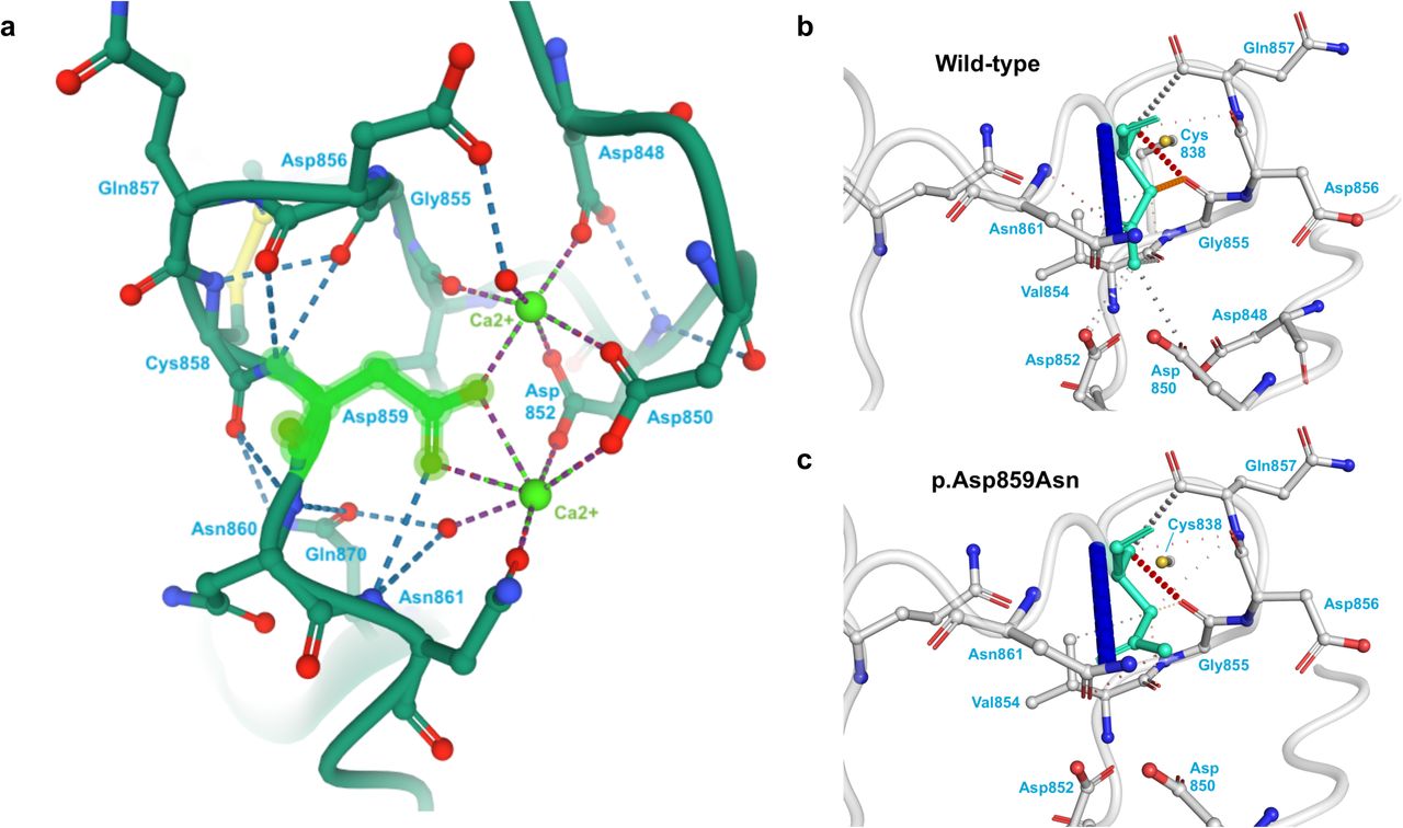
We then identified coding and noncoding variants in six genes associated with lethal knockout mouse models due to vascular abnormalities. Two variants, RASA3–p.Val85Met in Patient 4 and THBS2–p.Asp859Asn in Patient 14, were exonic. The RASA3– p.Val85Met variant is predicted to be pathogenic by 11 algorithms (CADD, DEOGEN2, EIGEN, FATHMM-MKL, FATHMM-XF, LRT, Mutation assessor, MutationTaster, PrimateAI, SIFT, and SIFT4G) and to affect splicing. The THBS2–p.Asp859Asn variant is found in the C-terminal type 3 repeats and likely disrupts intraprotein interactions and calcium binding (Figure 1); this variant is predicted to be pathogenic by 19 algorithms (BayesDel-addAF, BayesDel-noAF, CADD, DEOGEN2, EIGEN, EIGEN PC, FATHMM, FATHMM-MKL, FATHMM-XF, LIST-S2, MVP, MetaLR, MetaSVM, Mutation assessor, MutationTaster, PROVEAN, REVEL, SIFT, and SIFT4G). The other variants were noncoding and included two intronic SNVs in BCAS3, three intronic SNVs in DLC1, one intronic SNV in GLRX3, one intronic SNV in PIK3CA, and one intronic SNV in THBS2 (see Supplemental Table 1). Most of the identified variants are novel. Variants in BCAS3, DLC1, GLRX3, and PIK3CA are predicted to affect splicing. Variants in BCAS3, DLC1, PIK3CA, and THBS2 are predicted to be pathogenic by at least one algorithm. The BCAS3 SNV in intron 23 is predicted to fall within a binding site for GATA1 and the THBS2 intronic SNV is predicted to fall in a binding site for IKZF1. All three variants in DLC1 and the variants in GLRX3 and PIK3CA are predicted to be in open chromatin in human vascular endothelial cells (HUVECs) by Encode and therefore accessible to proteins regulating gene expression: four are predicted to be transcribed, and one (DLC1 intron 5) is predicted to be in an enhancer.

Figure 1:
  • Download figure
  • Open in new tab
Figure 1: Location and consequences of THBS2–p.Asp859Asn variant

A) The location and interactions of p.Asp859 in the THBS2 protein structure 1YO8 in PDB. The p.Asp859 amino acid is highlighted in green. The surrounding amino acids that participate in hydrogen binding with p.Asp859 and/or coordination with the calcium ions are labeled. B-C) The wild-type (B) and mutant (C) structure predictions from DynaMut are given. The affected amino acid is highlighted in teal. Red dashes indicate hydrogen bonds, and orange dashes indicate weak hydrogen bonds. The p.Asp859Asn variant disrupts many of the interactions of p.Asp859 with its neighboring amino acids.

Genes previously implicated in CNV analysis

We next looked for variants in genes affected by CNVs identified in a cohort of patients with PHACE described by Siegel et al. (2013) among our cohort. This analysis identified the aforementioned intronic variant in PIK3CA again. We also prioritized two intronic variants in AFF2, an intronic variant in EPHA3, and four intronic variants in EXOC4. All four genes were affected by a CNV in a single patient in the prior study. The EPHA3 intronic variant is predicted to be pathogenic by EIGEN (0.58), to affect splicing, and to fall within binding sites for CTCF, TCF12, and RAD21. All four EXOC4 variants are predicted to affect splicing, and the EXOC4 variant in intron 17 is predicted to be pathogenic by EIGEN (0.05) and GWAVA (0.5) and to fall within binding sites for GATA2 and GATA3.

Indels in prioritized genes

Finally, we investigated our cohort for rare, de novo indels affecting any of the 12 prioritized genes and identified two more variants: a 2-bp intronic deletion in BCAS3 in Patient 20 and an intronic 8-bp insertion in THBS2 in Patient 12, just upstream of the previously identified intronic SNV in THBS2 in the same patient.

DECIPHER analyses

Next, we analyzed published CNVs using DECIPHER to identify previously reported individuals with a phenotypic description containing the term “hemangioma” and CNVs containing our prioritized genes. This analysis identified five individuals in DECIPHER with deletions affecting THBS2; a single individual each with a deletion affecting BCAS3, GLRX3, and RASA3; and a single individual each with a duplication affecting DLC1 and PIK3CA.

We expanded our DECIPHER analysis to identify individuals with “facial hemangioma” and the genes disrupted by their CNVs. Next, we searched for rare, de novo variants in our cohort in the genes disrupted in two or more DECIPHER individuals. THBS2 was only disrupted by deletions in DECIPHER. Another two genes were each disrupted by two CNVs, one deletion and one duplication, in DECIPHER individuals with facial hemangioma. ALDH3A1 has a novel p.Val373Met variant in our Patient 8; this SNV is in the first codon of exon eight, is predicted to affect splicing by Human Splicing Finder (HSF), and is predicted to be pathogenic by CADD (23.3) and FATHMM-MKL (0.48). ATP7B has a p.Thr482Ala variant in our Patient 10; this SNV is predicted to affect splicing by HSF.

We performed an enrichment analysis for the DECIPHER CNVs disrupting ALDH3A1, ATP7B, and THBS2. Only deletions disrupting THBS2 were significantly more common among individuals with facial hemangioma (FET, p=3.7e-2, OR=7.17) or hemangioma (FET, p=3.9e-3, OR=5.24) in DECIPHER than among the individuals without these features in DECIPHER, respectively.

Pathway analysis

Genes containing rare, de novo, non-intergenic SNVs (n=4,320 genes) were analyzed through g:Profiler to look for common pathways disrupted in our cohort (Table 1). The five gene ontology (GO) terms with the most significant p-values were all related to development and morphogenesis. The most significant term, nervous system development (adjusted p=6.22e-24), included our candidate genes AFF2, DLC1, EPHA3, PIK3CA, and THBS2. We also observed an enrichment for cell adhesion (adjusted p=1.34e-10, including BCAS3, DLC1, EPHA3, PIK3CA, and THBS2), which is critical for angiogenesis. The three KEGG terms with the most significant p-values implicated RAP signaling (adjusted p=1.61e-6, including PIK3CA), focal adhesion (adjusted p=2.43e-6, including PIK3CA and THBS2), and phospholipase D signaling (adjusted p=8.13e-6, including PIK3CA).

View this table:
  • View inline
  • View popup
Table 1: g:Profiler pathway analysis of genes with rare, de novo variants

Expression analysis

We explored the expression of the candidate genes RASA3, AFF2, DLC1, EPHA3, PIK3CA, and THBS2 across diverse cell types in the human developing heart (Figure 2) by incorporating chromatin accessibility (scATAC-seq) (Ameen & Sundaram et. al., unpublished) and gene expression (scRNA-seq) datasets (Asp et al. 2019; Miao et al. 2020; Suryawanshi et al. 2020). We observed that AFF2, EPHA3, PIK3CA, and THBS2 are co-expressed in the vasculature (pre-smooth muscle cells and vascular smooth muscle cells) in the fetal heart, whilst AFF2, EPHA3, and PIK3CA are co-expressed in the endothelium. Furthermore, RASA3, DLC1, and THBS2 are co-expressed in pericytes, the mural cells surrounding blood vessels and embedded within the basement membrane of the vasculature and adjacent to endothelial cells (Armulik et al. 2005), and RASA3 and THBS2 are co-expressed in perivascular fibroblasts, which may serve as pericyte precursors (Rajan et al. 2020). Together, these data suggest that the expression of the candidate genes is enriched in endothelial cells and mural cells (pericytes and vascular smooth muscle cells) in the blood vessel wall.

Figure 2:
  • Download figure
  • Open in new tab
Figure 2: Expression of candidate genes in the developing heart at single cell level

A-F) Clustering of single cells by Uniform Manifold Approximation and Projection (UMAP) based on the differentially accessibility of open chromatin regions (scATAC-seq). Gene expression levels of RASA3 (A), DLC1 (B), AFF2 (C), EPHA3 (D), PIK3CA (E), and THBS2 (F) imputed from gene accessibility scores (scATAC-seq) and RNA expression (scRNA-seq). Annotation shows the expression level of each gene in the corresponding cell type cluster.

DISCUSSION

Our whole genome sequencing analyses support a disruption in RAS pathway signaling as the primary pathogenic mechanism behind PHACE. The RAS pathway genes identified in our analysis are associated with abnormal angiogenesis, including changes to cytoskeletal organization, cell migration, and adhesion, in relevant knockout mouse models. DLC1 binds RASA1 (a paralog of RASA3) at focal adhesions, and Dlc1 knockout causes embryonic lethal placenta vascular malformations (Durkin et al. 2005; Yang et al. 2009). EPHA3 regulates cytoskeletal organization, cell-cell adhesion, and cell migration in angiogenesis, and Epha3 knockout causes perinatal lethal cardiac defects (Clifford et al. 2008; Vaidya et al. 2003; Vearing et al. 2005). PIK3CA regulates cell migration and adhesion and has been associated with vascular anomalies, and Pik3ca knockout causes embryonic lethal vascular defects and hemorrhage (Bi et al. 1999; Graupera et al. 2008; Lelievre et al. 2005). RASA3 regulates cell-cell and cell-matrix adhesion and cell migration, and Rasa3 knockout causes embryonic lethal hemorrhage following vascular lumenization defects (Iwashita et al. 2007; Molina-Ortiz et al. 2018). THBS2 binds heparin in the extracellular matrix (ECM), mediates cell-matrix interactions, and inhibits cell adhesion and angiogenesis, and Thbs2 knockout causes increased vascularity and premature death (Bornstein et al. 2000; Kyriakides et al. 1998; Noh et al. 2003; Streit et al. 1999; Yang et al. 2000).

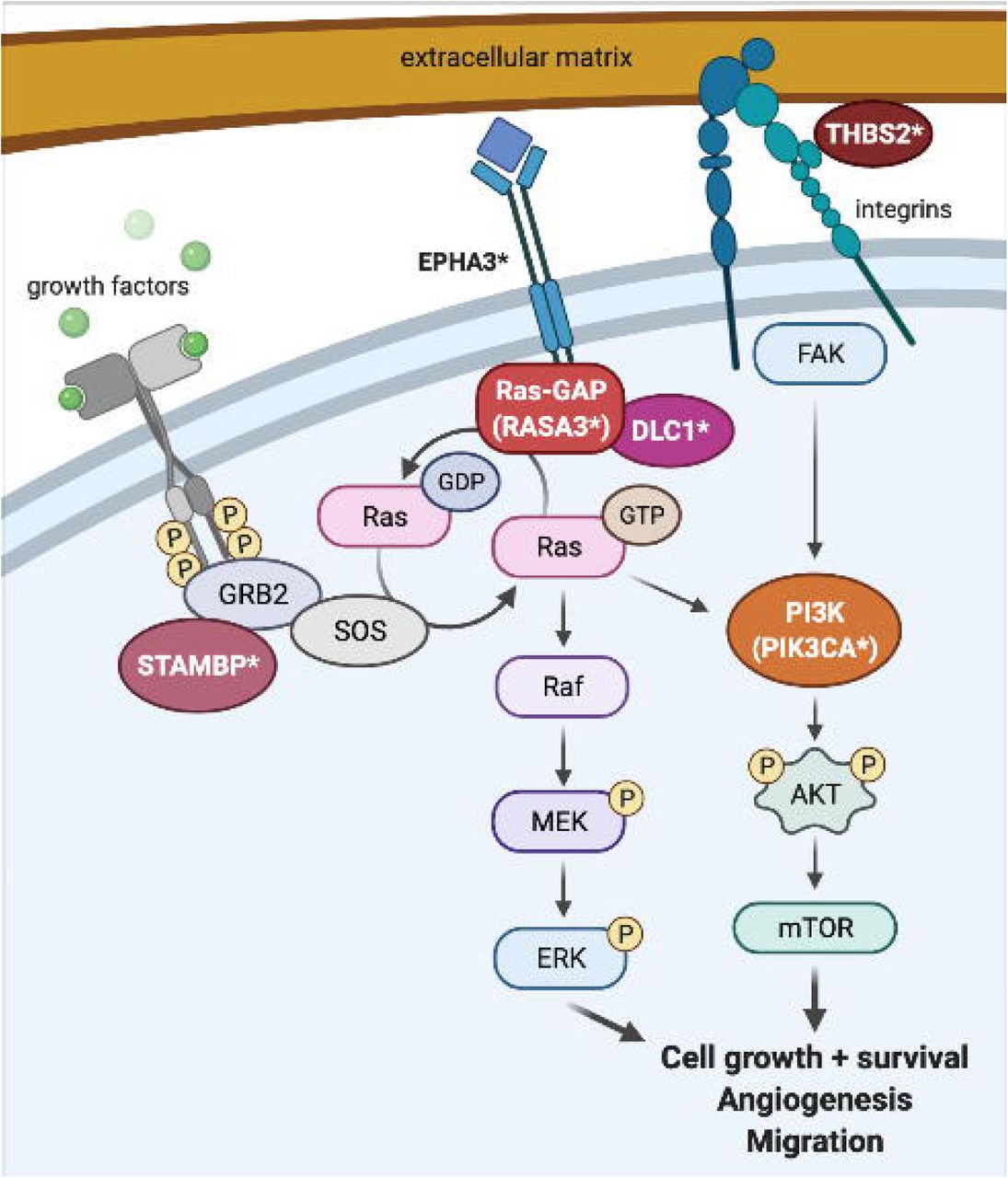
The g:Profiler pathway analysis of the genes with rare, de novo SNVs in these patients also implicated these same candidate genes. KEGG pathway analysis identified RAP signaling, focal adhesion, and phospholipase D (PLD) signaling as the most significantly affected pathways. RASA3 downregulates RAP1 GTPase signaling (Battram et al. 2017; Molina-Ortiz et al. 2018). BCAS3, DLC1, PIK3CA, and THBS2 all participate in the regulation of focal adhesion (Durkin et al. 2005; Elzie and Murphy-Ullrich 2004; Jain et al. 2012; Matsuoka et al. 2012; Yang et al. 2009). PLD is required for actin cytoskeleton remodeling in cell migration and angiogenesis, participates in RAS activation, and is activated by both RAS/MAPK/ERK and PI3K/AKT/mTOR signaling (Bruntz et al. 2014). RASA3 downregulates RAS signaling through MEK/ERK and also binds PI3K and regulates cell migration though PI3K-dependent integrin signaling (Battram et al. 2017; Castellano and Downward 2011). PIK3CA forms part of the PI3K complex and regulates endothelial cell migration during vascular development (Graupera et al. 2008). STAMBP binds GRB2 to regulate both the RAS/MAPK and PI3K/AKT/mTOR pathways (McDonell et al. 2013; Tsang et al. 2006). THBS2 signals through FAK activation of the MAPK/ERK and PI3K/AKT pathways to stimulate actin reorganization, decrease cell adhesion, and increase migration (Goicoechea et al. 2000; Greenwood et al. 1998; Orr et al. 2002). DLC1 and EPHA3 both bind RASA1, a close paralog of RASA3 (Haupaix et al. 2013; Yang et al. 2009); the EPHA3 intronic variant in particular is also in a binding site for TCF12, which downregulates ERK signaling (Yi et al. 2017). DLC1 is also phosphorylated by AKT, which abrogates its tumor suppression activity (Ko et al. 2010). The identification of variants in these genes among the probands with PHACE suggests that defects in matricellular signaling affecting endothelial cell adhesion and migration through the RAS and PI3K pathways may contribute to the pathogenesis of PHACE (Figure 3).

Figure 3:
  • Download figure
  • Open in new tab
Figure 3: Diagram of proposed RAS signaling pathway

RAS is activated downstream of growth factor binding to cellular receptors, which signal through GRB2/SOS/STAMBP, and deactivated by RAS-GAP proteins (RASA1, RASA3), which interact with EPHA3 and DLC1. Activated RAS-GTP signals through the MEK/ERK and PI3K/AKT/mTOR pathways to upregulate cell growth, cell migration, and angiogenesis. PI3K is also activated through extracellular matrix (ECM) signaling: THBS2 binds to the ECM and to integrins, which link the cell to the ECM, and THBS2 signals through focal adhesion kinase (FAK) to PI3K. Protein names denoted with an asterisk (DLC1, EPHA3, PIK3CA, RASA3, STAMBP, THBS2) were found to have de novo variants in our cohort. Image created using BioRender.

Our analysis of rare, de novo germline variants combined with the DECIPHER analysis of CNVs found in individuals with hemangioma identified THBS2 as a strong candidate causative gene for PHACE. THBS2 had missense and intronic variants in our cohort and was deleted in five individuals with hemangioma in DECIPHER; an enrichment analysis showed that deletions disrupting THBS2 were significantly more common among individuals with hemangioma than in individuals without this feature in DECIPHER. While no human phenotype has been associated with this gene, knockout of this gene in a mouse causes blood vessel overgrowth and premature death (Bornstein et al. 2000; Kyriakides et al. 1998; Yang et al. 2000). THBS2 is highly expressed in arterial tissues (aorta, coronary, and tibial) described in GTEx and in the developing vasculature in our scRNA analysis. THBS2 interacts with the ECM, mediates cell-matrix interactions, inhibits cell adhesion, inhibits angiogenesis, and acts as a tumor suppressor (Bornstein et al. 2000; Kyriakides et al. 1998; Noh et al. 2003; Streit et al. 1999; Yang et al. 2000). The THBS2 residue p.Asp859, which had a missense variant in our cohort, is found in the C-terminal type 3 repeats and participates in calcium binding (Figure 1). This calcium binding is necessary for correct folding of the C terminus, which itself must correctly trimerize in order to support the integrin-binding and cell motility functions of the THBS2 protein (Anilkumar et al. 2002; Carlson et al. 2008; Kvansakul et al. 2004). Therefore, a variant in this position could affect the protein function by disrupting protein folding and trimerization.

Our analysis identified RASA3 as a second strong candidate causative gene for PHACE. RASA3 had a missense variant in our cohort and was deleted in one DECIPHER individual with capillary hemangioma. Germline pathogenic variants in a related gene, RASA1, are known to cause capillary malformation–arteriovenous malformation (Eerola et al. 2003). No human phenotype has been associated with RASA3, but knockout of this gene in a mouse causes hemorrhage and embryonic lethality due to vascular abnormalities (Iwashita et al. 2007; Molina-Ortiz et al. 2018). RASA3 also regulates angiogenesis via the RAS/RAP pathway (Molina-Ortiz et al. 2018), which was implicated in our pathway analysis, and was highly expressed in pericytes in our scRNA analysis.

We also identified seven noncoding variants in four RAS pathway genes (DLC1, EPHA3, PIK3CA, and THBS2). These variants were predicted to affect splicing, functional DNA, transcription factor binding, and/or open chromatin in vascular cells (Table 2). The genes DLC1 and PIK3CA are highly intolerant to loss-of-function coding mutations (probability of loss-of-function intolerance scores > 0.9) (Lek et al. 2016) and have enhanced expression in PHACE-relevant tissues: DLC1 in neurons and retinal amacrine cells and PIK3CA in aorta and coronary artery (Genotype-Tissue Expression (GTEx) Project 2021). Single cell RNA expression analyses also showed that our candidate RAS pathway genes have high expression in neural crest, smooth muscle, and endothelial cells in the developing heart (Figure 2). The predicted disruption of expression and/or splicing of these genes by our identified noncoding variants may therefore contribute to other PHACE features. For instance, an intronic SNV, rs1905014-T, in DLC1 is significantly associated with optic disc size (Han et al. 2019). However, the effects of the noncoding variants on splicing and/or functional DNA need to be investigated further.

View this table:
  • View inline
  • View popup
Table 2: Candidate germline de novo variants in RAS pathway genes

In conclusion, we have found strong candidate coding and noncoding variants in genes in the RAS/MAPK and PI3K/AKT pathways in individual patients with PHACE. These variants, paired with the vascular birthmark (infantile hemangioma) and the structural brain, heart, arterial, and eye anomalies in PHACE, support the inclusion of PHACE as a RASopathy syndrome. It is striking that the majority of these variants were predicted to have effects that may lead to altered gene expression during development. However, because of the complex cellular interactions between vascular cell types, further in vivo studies are necessary to confirm pathogenicity of the variants identified and to understand their effect on the pathways that are likely disrupted by these variants.

SUBJECTS AND METHODS

Subjects

Patients with PHACE and their parents were recruited and sequenced as part of the Gabriella Miller Kids First Pediatric Research Program (X01HL140519-01). The study is approved by the Medical College of Wisconsin IRB #PRO00034867 and patients gave written, informed consent. Of the 98 patients discussed here, 19 were male, and 85 were white (Table 3). The average age of the cohort was 5.8 years at the time of enrollment. Ninety patients had facial hemangioma.

View this table:
  • View inline
  • View popup
  • Download powerpoint
Table 3: Patient demographics

Germline whole genome sequencing and de novo analysis pipeline

WGS was performed on germline DNA from 98 unrelated patients with PHACE and their parents. WGS libraries were sequenced on the Illumina NovaSeq 6000 platform using 150-bp paired ends and the NovaSeq 6000 S4 Reagent Kit (Illumina, San Diego, California). Alignment was performed using DRAGEN (v.SW: 01.011.269.3.2.8) (Ma et al. 2015); data conversion from CRAM to BAM files with samtools (v.1.9); data pre-processing with Picard RevertSam, MarkDuplicates, SortSam, and MergeBamAlignment (v2.18.2) (Broad Institute 2019) and base quality recalibration with GATK BQSR (v.4.0.3.0). Germline variant calling and single-sample gVCF generation were performed using GATK HaplotypeCaller (v.3.5-0-g36282e4). Genotype analysis of trios/families, de novo variant discovery, variant quality recalibration, joint variant calling on all samples, and recalculation of genotype call accuracy on the population level were performed with GATK tools (Van der Auwera et al. 2013): HaplotypeCaller (v.3.5-0-g36282e4), VariantAnnotator (v.3.8-0-ge9d806836), VQSR, CalculateGenotypePosteriors, and VariantFiltration (v.4.0.12.0). All outputs were released to the DRC Portal and dbGaP, where the files can be accessed and downloaded (github.com/kids-first/kf-alignment-workflow).

High confidence de novo SNVs and indels were called for variants present only in the proband with GQ scores ≥ 20 for all trio members. Resulting variants were lifted to GRCh37/hg19 using UCSC’s liftOver tool and annotated using Annovar (v2013_09_11) (Wang et al. 2010). The liftover was necessary because the TraP and GWAVA annotations (see Variant prioritization) are not available under hg38.

Variant prioritization

Variants were subset to include SNVs with gnomAD (Karczewski et al. 2020) minor allele frequency (MAF) < 1% (hereafter defined as rare). Next, we prioritized SNVs that:

  • 1) were not present in gnomAD (novel);

  • 2) affected a gene already known to cause a phenotype overlapping that of PHACE in OMIM (omim.org) or GWAS annotation (Buniello et al. 2019);

  • 3) affected a gene where the mouse model phenotype (informatics.jax.org) overlapped that of PHACE;

  • 4) affected genes disrupted by CNVs described by Siegel et al. (2013); or

  • 5) affected genes with coding variants in two or more patients.

Prioritized coding variants were further evaluated for pathogenicity prediction scores using VarSome (Kopanos et al. 2019).

Prioritized noncoding variants were further evaluated for scores designed to predict effects on splicing [TraP (Gelfman et al. 2017), Human Splicing Finder (HSF) (Desmet et al. 2009)]; functional DNA [CADD (Rentzsch et al. 2019), DANN (Quang et al. 2015), GWAVA (Ritchie et al. 2014), EIGEN (Ionita-Laza et al. 2016), FATHMM (Shihab et al. 2015)]; transcription factor binding sites (TFBS) (Euskirchen et al. 2007); and open chromatin in vascular cells in Encode (Ernst et al. 2011).

Finally, we investigated the de novo indels identified in the genes prioritized using metrics 1-5.

DECIPHER analyses

We first selected the pathogenic or likely pathogenic deletions and duplications described in DECIPHER (Firth et al. 2009) in individuals with hemangioma. We compared the genes affected by these CNVs with the genes prioritized in our cohort (described above) to identify other individuals in DECIPHER with hemangiomas and CNVs affecting our prioritized genes.

We then selected all pathogenic or likely pathogenic CNVs described in DECIPHER in individuals with facial hemangioma. The “facial hemangioma” feature was chosen to represent PHACE because it is the most common manifestation of this syndrome. We identified 47 DECIPHER individuals with facial hemangioma, with a total of 55 CNVs (17 gains, 38 losses) disrupting 5,703 genes; 1,191 of these genes were disrupted in at least two DECIPHER individuals. This subset of 1,191 genes was compared to the list of rare, de novo variants identified in our cohort of patients with PHACE to prioritize further candidates.

An enrichment analysis was performed for each gene identified by the second DECIPHER analysis. The DECIPHER pathogenic or likely pathogenic deletions disrupting the gene of interest in individuals with facial hemangioma were classified as “cases”. The DECIPHER pathogenic or likely pathogenic deletions disrupting the same gene in individuals without facial hemangioma were classified as “controls”. For each gene we built a contingency table comparing the number of cases to controls and performed a Fisher’s exact test (FET) using a cutoff of p-value < 0.05 for statistical significance. For significant genes we also calculated an odds ratio.

Sanger sequencing

Prioritized candidate variants were validated by Sanger sequencing. DNA from the proband and each parent was amplified using Accuprime Taq polymerase (Invitrogen; 12339-016) using standard thermal cycling conditions with annealing at 60°C and variant-specific primer sequences (Supplemental Table 2). Small aliquots of each PCR product were run on a 1% agarose gel to verify amplification, and the remainder of each PCR product was cleaned up using a mixture of exonuclease 1 (Affymetrix; 70073X) and shrimp alkaline phosphatase (Affymetrix; 78390) and sequenced using the same primers.

Protein structure analysis

The experimentally derived structure of THBS2 was obtained from RCSB PDB (Berman et al. 2000) structure 1YO8 (Carlson et al. 2005). The effect of the identified p.Asp859Asn variant in THBS2 was examined using DynaMut (Rodrigues et al. 2018), which predicts the effects of missense mutations on known PDB structures.

Pathway analysis

All 4,320 genes with rare, de novo SNVs were analyzed with gProfiler (Raudvere et al. 2019) to determine if they had similar functions or affected the same pathways. Multiple testing-adjusted p-values (using the default g:SCS method in gProfiler) were reported, and 0.05 was used as the cutoff for statistical significance.

Expression analysis

The chromatin accessibility atlas and gene expression data of the human developing heart was derived from nuclei isolated from human fetal hearts (Ameen & Sundaram et al., unpublished). Briefly, the Chromium 10X platform (Satpathy et al. 2019) was used to generate scATAC-seq data from three primary human fetal heart samples at 6, 8, and 19 weeks post-gestation. The scRNA-seq data from fetal hearts were analyzed from published studies (Asp et al. 2019; Miao et al. 2020; Suryawanshi et al. 2020) using the batch correction algorithm Harmony (Korsunsky et al. 2019), and then annotated using known cell-type-specific marker genes.

Data Availability

Datasets related to this article are accessible through the Kids First Data Resource Portal (kidsfirstdrc.org) and/or dbGaP (ncbi.nlm.nih.gov/gap) accession phs001785.v1.p1.

DATA AVAILABILITY

Datasets related to this article are accessible through the Kids First Data Resource Portal (kidsfirstdrc.org) and/or dbGaP (ncbi.nlm.nih.gov/gap) accession phs001785.v1.p1.

CONFLICT OF INTEREST

Francine Blei has received honoraria and non-drug related educational grants from Pierre Fabre. Sarah Chamlin is a speaker for Sanofi Genzyme and Regeneron Pharmaceuticals. Beth Drolet is a consultant and on the clinical advisory board for Venthera. Ilona Frieden serves on the Data Safety monitoring boards for Pfizer, is a consultant and advisory board member for Novartis, and is a consultant for Venthera. Dawn Siegel has received royalties from UpToDate, is a consultant for Arqule/Merck, and received a SID SUN mid-career investigator award.

AUTHOR CONTRIBUTIONS (CREDIT)

Elizabeth S. Partan: Conceptualization, Data curation, Investigation, Visualization, Writing – original draft, Writing – review & editing

Francine Blei: Data curation, Writing – review & editing

Sarah L. Chamlin: Data curation, Writing – review & editing

Olivia M. T. Davies: Data curation, Writing – review & editing

Beth A. Drolet: Data curation, Writing – review & editing

Ilona J. Frieden: Data curation, Writing – review & editing

Ioannis Karakikes: Writing – review & editing

Chien-Wei Lin: Formal analysis, Writing – review & editing

Anthony J. Mancini: Data curation, Writing – review & editing

Denise Metry: Data curation, Writing – review & editing

Anthony Oro: Writing – review & editing

Nicole S. Stefanko: Data curation, Writing – review & editing

Laksshman Sundaram: Investigation, Visualization

Monika Tutaj: Data curation, Formal analysis, Writing – review & editing

Alexander E. Urban: Writing – review & editing

Kevin C. Wang: Writing – review & editing

Xiaowei Zhu: Writing – review & editing

Nara Sobreira: Conceptualization, Funding acquisition, Project administration, Supervision, Writing – review & editing

Dawn H. Siegel: Conceptualization, Data curation, Funding acquisition, Project administration, Supervision, Writing – review & editing

ACKNOWLEDGMENTS

We thank all the families for their participation in this study, and the PHACE Syndrome Community and Pediatric Dermatology Research Alliance (PeDRA) for support of this project. We acknowledge the support of the Children’s Research Institute at Children’s Wisconsin and the Genomic Sciences and Precision Medicine Center at Medical College of Wisconsin.

The results analyzed and shown here are based in part upon data generated by Gabriella Miller Kids First Pediatric Research Program (Kids First) projects phs001785.v1.p1. Kids First was supported by the Common Fund of the Office of the Director of the National Institutes of Health (commonfund.nih.gov/KidsFirst). The Hudson-Alpha Institute for Biotechnology was awarded a U24HD090744-01 to sequence structural birth defect cohort samples submitted by investigators through the Kids First program (1X01HL140519-01).

This study was supported by the NICHD, NHLBI, and NIAMS under award numbers 1X01HL14519, R03HD098526, and R01AR064258, respectively. The content is solely the responsibility of the authors and does not necessarily represent the official views of the National Institutes of Health. Additional support was received from a SID SUN grant, PHACE Foundation Canada, and the Children’s Research Institute.

This study makes use of data generated by the DECIPHER community. A full list of centers who contributed to the generation of the data is available from decipher.sanger.ac.uk and via email from decipher{at}sanger.ac.uk. Funding for the project was provided by Wellcome. The individuals who carried out the original analysis and collection of the data bear no responsibility for the further analysis or interpretation of it in this paper.

Footnotes

  • Work was performed in Baltimore, MD, USA; Milwaukee, WI, USA; and Palo Alto, CA, USA

Abbreviations used

SNV
single nucleotide variant
CNV
copy number variant
ECM
extracellular matrix

REFERENCES

  1. ↵
    Anilkumar N, Annis DS, Mosher DF, Adams JC. Trimeric assembly of the C-terminal region of thrombospondin-1 or thrombospondin-2 is necessary for cell spreading and fascin spike organisation. J Cell Sci. The Company of Biologists Ltd; 2002;115(11):2357–66 Available from: https://jcs.biologists.org/content/115/11/2357
    OpenUrlAbstract/FREE Full Text
  2. ↵
    Armulik A, Abramsson A, Betsholtz C. Endothelial/pericyte interactions. Circulation Research. American Heart Association; 2005;97(6):512–23 Available from: http://www.ahajournals.org/doi/10.1161/01.RES.0000182903.16652.d7
    OpenUrl
  3. ↵
    Asp M, Giacomello S, Larsson L, Wu C, Fürth D, Qian X, et al. A spatiotemporal organ-wide gene expression and cell atlas of the developing human heart. Cell. 2019;179(7):1647–1660.e19 Available from: https://www.sciencedirect.com/science/article/pii/S0092867419312826
    OpenUrlCrossRefPubMed
  4. ↵
    Battram AM, Durrant TN, Agbani EO, Heesom KJ, Paul DS, Piatt R, et al. The phosphatidylinositol 3,4,5-trisphosphate (PI(3,4,5)P3) binder Rasa3 regulates phosphoinositide 3-kinase (PI3K)-dependent integrin α 3 outside-in signaling. J Biol Chem. 2017;292(5):1691–704
    OpenUrlAbstract/FREE Full Text
  5. ↵
    Berman HM, Westbrook J, Feng Z, Gilliland G, Bhat TN, Weissig H, et al. The Protein Data Bank. Nucleic Acids Research. 2000;28(1):235–42 Available from: https://doi.org/10.1093/nar/28.1.235
    OpenUrlCrossRefPubMedWeb of Science
  6. ↵
    Bi L, Okabe I, Bernard DJ, Wynshaw-Boris A, Nussbaum RL. Proliferative defect and embryonic lethality in mice homozygous for a deletion in the p110α subunit of phosphoinositide 3-kinase. J. Biol. Chem. American Society for Biochemistry and Molecular Biology; 1999;274(16):10963–8 Available from: http://www.jbc.org/content/274/16/10963
    OpenUrlAbstract/FREE Full Text
  7. ↵
    Bornstein P, Kyriakides TR, Yang Z, Armstrong LC, Birk DE. Thrombospondin 2 modulates collagen fibrillogenesis and angiogenesis. Journal of Investigative Dermatology Symposium Proceedings. 2000;5(1):61–6 Available from: http://www.sciencedirect.com/science/article/pii/S0022202X15528601
    OpenUrlCrossRefPubMed
  8. ↵
    Broad Institute. Picard Toolkit [Internet]. Broad Institute, GitHub Repository; 2019. Available from: http://broadinstitute.github.io/picard/
  9. ↵
    Bruntz RC, Lindsley CW, Brown HA. Phospholipase D signaling pathways and phosphatidic acid as therapeutic targets in cancer. Pharmacol Rev. 2014;66(4):1033–79 Available from: https://www.ncbi.nlm.nih.gov/pmc/articles/PMC4180337/
    OpenUrlAbstract/FREE Full Text
  10. ↵
    Buniello A, MacArthur JAL, Cerezo M, Harris LW, Hayhurst J, Malangone C, et al. The NHGRI-EBI GWAS Catalog of published genome-wide association studies, targeted arrays and summary statistics 2019. Nucleic Acids Res. 2019;47(D1):D1005–12
    OpenUrlCrossRefPubMed
  11. ↵
    Carlson CB, Bernstein DA, Annis DS, Misenheimer TM, Hannah BA, Mosher DF, et al. Structure of the calcium-rich signature domain of human thrombospondin-2. Nat Struct Mol Biol. 2005;12(10):910–4 Available from: http://www.nature.com/articles/nsmb997
    OpenUrlCrossRefPubMedWeb of Science
  12. ↵
    Carlson CB, Gunderson KA, Mosher DF. Mutations targeting intermodular interfaces or calcium binding destabilize the thrombospondin-2 signature domain. J Biol Chem. 2008;283(40):27089–99 Available from: https://www.ncbi.nlm.nih.gov/pmc/articles/PMC2555992/
    OpenUrlAbstract/FREE Full Text
  13. ↵
    Castellano E, Downward J. RAS interaction with PI3K. Genes Cancer. 2011;2(3):261–74 Available from: https://www.ncbi.nlm.nih.gov/pmc/articles/PMC3128635/
    OpenUrlCrossRefPubMed
  14. ↵
    Chacon Camacho OF, Lopez Moreno D, MoralesLJ anchez MA, Hofmann E, Pacheco Quito M, Wieland I, et al. Expansion of the phenotypic spectrum and description of molecular findings in a cohort of patients with oculocutaneous mosaic RASopathies. Mol Genet Genomic Med. 2019;7(5):e625 Available from: https://www.ncbi.nlm.nih.gov/pmc/articles/PMC6503218/
    OpenUrl
  15. ↵
    Clifford N, Smith LM, Powell J, Gattenlöhner S, Marx A, O’Connor R. The EphA3 receptor is expressed in a subset of rhabdomyosarcoma cell lines and suppresses cell adhesion and migration. Journal of Cellular Biochemistry. 2008;105(5):1250–9 Available from: http://onlinelibrary.wiley.com/doi/abs/10.1002/jcb.21926
    OpenUrlCrossRefPubMedWeb of Science
  16. ↵
    Desmet F-O, Hamroun D, Lalande M, Collod-Béroud G, Claustres M, Béroud C. Human Splicing Finder: An online bioinformatics tool to predict splicing signals. Nucleic Acids Res. 2009;37(9):e67
    OpenUrlCrossRefPubMed
  17. ↵
    Durkin ME, Avner MR, Huh C-G, Yuan B-Z, Thorgeirsson SS, Popescu NC. DLC-1, a Rho GTPase-activating protein with tumor suppressor function, is essential for embryonic development. FEBS Letters. 2005;579(5):1191–6 Available from: http://www.sciencedirect.com/science/article/pii/S001457930500089X
    OpenUrlCrossRefPubMedWeb of Science
  18. ↵
    Eerola I, Boon LM, Mulliken JB, Burrows PE, Dompmartin A, Watanabe S, et al. Capillary malformation–arteriovenous malformation, a new clinical and genetic disorder caused by RASA1 mutations. The American Journal of Human Genetics. Elsevier; 2003;73(6):1240–9 Available from: https://www.cell.com/ajhg/abstract/S0002-9297(07)63977-9
    OpenUrlCrossRefPubMedWeb of Science
  19. ↵
    Elzie CA, Murphy-Ullrich JE. The N-terminus of thrombospondin: The domain stands apart. The International Journal of Biochemistry & Cell Biology. 2004;36(6):1090–101 Available from: http://www.sciencedirect.com/science/article/pii/S1357272503004357
    OpenUrl
  20. ↵
    Ernst J, Kheradpour P, Mikkelsen TS, Shoresh N, Ward LD, Epstein CB, et al. Mapping and analysis of chromatin state dynamics in nine human cell types. Nature. 2011;473(7345):43–9 Available from: https://www.ncbi.nlm.nih.gov/pmc/articles/PMC3088773/
    OpenUrlCrossRefPubMedWeb of Science
  21. ↵
    Euskirchen GM, Rozowsky JS, Wei C-L, Lee WH, Zhang ZD, Hartman S, et al. Mapping of transcription factor binding regions in mammalian cells by ChIP: Comparison of array- and sequencing-based technologies. Genome Res. 2007;17(6):898–909
    OpenUrlAbstract/FREE Full Text
  22. ↵
    Firth HV, Richards SM, Bevan AP, Clayton S, Corpas M, Rajan D, et al. DECIPHER: Database of Chromosomal Imbalance and Phenotype in Humans Using Ensembl Resources. Am J Hum Genet. 2009;84(4):524–33 Available from: https://www.ncbi.nlm.nih.gov/pmc/articles/PMC2667985/
    OpenUrlCrossRefPubMedWeb of Science
  23. ↵
    Frieden IJ, Reese V, Cohen D. PHACE syndrome: The association of posterior fossa brain malformations, hemangiomas, arterial anomalies, coarctation of the aorta and cardiac defects, and eye abnormalities. Arch Dermatol. 1996;132(3):307–11 Available from: https://jamanetwork.com/journals/jamadermatology/fullarticle/557585
    OpenUrlCrossRefPubMedWeb of Science
  24. ↵
    Garzon MC, Epstein LG, Heyer GL, Frommelt PC, Orbach DB, Baylis AL, et al. PHACE syndrome: Consensus-derived diagnosis and care recommendations. The Journal of Pediatrics. 2016;178:24–33.e2 Available from: https://www.jpeds.com/article/S0022-3476(16)30656-4/fulltext
    OpenUrlCrossRefPubMed
  25. ↵
    Gelfman S, Wang Q, McSweeney KM, Ren Z, La Carpia F, Halvorsen M, et al. Annotating pathogenic non-coding variants in genic regions. Nature Communications. 2017;8(1):236 Available from: http://www.nature.com/articles/s41467-017-00141-2
    OpenUrl
  26. ↵
    Genotype-Tissue Expression (GTEx) Project. GTEx portal [Internet]. 2021 [cited 2021 Jun 22]. Available from: https://www.gtexportal.org/home/
  27. ↵
    Goicoechea S, Orr AW, Pallero MA, Eggleton P, Murphy-Ullrich JE. Thrombospondin mediates focal adhesion disassembly through interactions with cell surface calreticulin. J. Biol. Chem. American Society for Biochemistry and Molecular Biology; 2000;275(46):36358–68 Available from: http://www.jbc.org/content/275/46/36358
    OpenUrlAbstract/FREE Full Text
  28. ↵
    Grant AR, Cushman BJ, Cavé H, Dillon MW, Gelb BD, Gripp KW, et al. Assessing the gene–disease association of 19 genes with the RASopathies using the ClinGen gene curation framework. Hum Mutat. 2018;39(11):1485–93 Available from: https://www.ncbi.nlm.nih.gov/pmc/articles/PMC6326381/
    OpenUrlCrossRef
  29. ↵
    Graupera M, Guillermet-Guibert J, Foukas LC, Phng L-K, Cain RJ, Salpekar A, et al. Angiogenesis selectively requires the p110α isoform of PI3K to control endothelial cell migration. Nature. Nature Publishing Group; 2008;453(7195):662–6 Available from: https://www.nature.com/articles/nature06892
    OpenUrlCrossRefPubMedWeb of Science
  30. ↵
    Greenwood JA, Pallero MA, Theibert AB, Murphy-Ullrich JE. Thrombospondin signaling of focal adhesion disassembly requires activation of phosphoinositide 3-kinase. J. Biol. Chem. American Society for Biochemistry and Molecular Biology; 1998;273(3):1755–63 Available from: http://www.jbc.org/content/273/3/1755
    OpenUrlAbstract/FREE Full Text
  31. ↵
    Han X, Qassim A, An J, Marshall H, Zhou T, Ong J-S, et al. Genome-wide association analysis of 95,549 individuals identifies novel loci and genes influencing optic disc morphology. Hum Mol Genet. 2019;28(21):3680–90
    OpenUrl
  32. ↵
    Haupaix N, Stolfi A, Sirour C, Picco V, Levine M, Christiaen L, et al. p120RasGAP mediates ephrin/Eph-dependent attenuation of FGF/ERK signals during cell fate specification in ascidian embryos. Development. 2013;140(21):4347–52
    OpenUrlAbstract/FREE Full Text
  33. Hopkin RJ, Schorry E, Bofinger M, Milatovich A, Stern HJ, Jayne C, et al. New insights into the phenotypes of 6q deletions. American Journal of Medical Genetics. 1997;70(4):377–86 Available from: http://onlinelibrary.wiley.com/doi/abs/10.1002/%28SICI%291096-8628%2819970627%2970%3A4%3C377%3A%3AAID-AJMG9%3E3.0.CO%3B2-Q
    OpenUrlCrossRefPubMedWeb of Science
  34. ↵
    Ionita-Laza I, McCallum K, Xu B, Buxbaum JD. A spectral approach integrating functional genomic annotations for coding and noncoding variants. Nat. Genet. 2016;48(2):214–20
    OpenUrlCrossRefPubMed
  35. Ishii N, Owada Y, Yamada M, Miura S, Murata K, Asao H, et al. Loss of neurons in the hippocampus and cerebral cortex of AMSH-deficient mice. Mol Cell Biol. 2001;21(24):8626–37
    OpenUrlAbstract/FREE Full Text
  36. ↵
    Iwashita S, Kobayashi M, Kubo Y, Hinohara Y, Sezaki M, Nakamura K, et al. Versatile roles of R-Ras GAP in neurite formation of PC12 cells and embryonic vascular development. J. Biol. Chem. American Society for Biochemistry and Molecular Biology; 2007;282(6):3413–7 Available from: http://www.jbc.org/content/282/6/3413
    OpenUrlAbstract/FREE Full Text
  37. ↵
    Jain M, Bhat GP, VijayRaghavan K, Inamdar MS. Rudhira/BCAS3 is a cytoskeletal protein that controls Cdc42 activation and directional cell migration during angiogenesis. Experimental Cell Research. 2012;318(6):753–67 Available from: http://www.sciencedirect.com/science/article/pii/S0014482712000377
    OpenUrlPubMed
  38. ↵
    Karczewski KJ, Francioli LC, Tiao G, Cummings BB, Alföldi J, Wang Q, et al. The mutational constraint spectrum quantified from variation in 141,456 humans. Nature. Nature Publishing Group; 2020;581(7809):434–43 Available from: https://www.nature.com/articles/s41586-020-2308-7
    OpenUrlCrossRefPubMed
  39. ↵
    Kinsler VA, Thomas AC, Ishida M, Bulstrode NW, Loughlin S, Hing S, et al. Multiple congenital melanocytic nevi and neurocutaneous melanosis are caused by postzygotic mutations in codon 61 of NRAS. J Invest Dermatol. 2013;133(9):2229–36 Available from: https://www.ncbi.nlm.nih.gov/pmc/articles/PMC3678977/
    OpenUrlCrossRefPubMedWeb of Science
  40. ↵
    Ko FCF, Chan L, Tung EK, Lowe SW, Ng IO, Yam JWP. Akt phosphorylation of Deleted in Liver Cancer 1 abrogates its suppression of liver cancer tumorigenesis and metastasis. Gastroenterology. 2010;139(4):1397–1407.e6 Available from: https://www.sciencedirect.com/science/article/pii/S0016508510009601
    OpenUrlCrossRefPubMed
  41. ↵
    Kopanos C, Tsiolkas V, Kouris A, Chapple CE, Albarca Aguilera M, Meyer R, et al. VarSome: The human genomic variant search engine. Bioinformatics. 2019;35(11):1978–80 Available from: https://doi.org/10.1093/bioinformatics/bty897
    OpenUrlCrossRefPubMed
  42. ↵
    Korsunsky I, Millard N, Fan J, Slowikowski K, Zhang F, Wei K, et al. Fast, sensitive, and accurate integration of single cell data with Harmony. Nat Methods. 2019;16(12):1289–96 Available from: https://www.ncbi.nlm.nih.gov/pmc/articles/PMC6884693/
    OpenUrlPubMed
  43. ↵
    Kvansakul M, Adams JC, Hohenester E. Structure of a thrombospondin C-terminal fragment reveals a novel calcium core in the type 3 repeats. The EMBO Journal. John Wiley & Sons, Ltd; 2004;23(6):1223–33 Available from: https://www.embopress.org/doi/full/10.1038/sj.emboj.7600166
    OpenUrlAbstract/FREE Full Text
  44. ↵
    Kyriakides TR, Zhu Y-H, Smith LT, Bain SD, Yang Z, Lin MT, et al. Mice that lack thrombospondin 2 display connective tissue abnormalities that are associated with disordered collagen fibrillogenesis, an increased vascular density, and a bleeding diathesis. Journal of Cell Biology. 1998;140(2):419–30 Available from: https://doi.org/10.1083/jcb.140.2.419
    OpenUrlAbstract/FREE Full Text
  45. ↵
    Lek M, Karczewski KJ, Minikel EV, Samocha KE, Banks E, Fennell T, et al. Analysis of protein-coding genetic variation in 60,706 humans. Nature. 2016;536(7616):285–91 Available from: https://www.nature.com/articles/nature19057
    OpenUrlCrossRefPubMedWeb of Science
  46. ↵
    Lelievre E, Bourbon P-M, Duan L-J, Nussbaum RL, Fong G-H. Deficiency in the p110α subunit of PI3K results in diminished Tie2 expression and Tie2-/-–like vascular defects in mice. Blood. 2005;105(10):3935–8 Available from: https://www.ncbi.nlm.nih.gov/pmc/articles/PMC1895075/
    OpenUrlAbstract/FREE Full Text
  47. ↵
    Ma S, Jiang T, Jiang R. Differential regulation enrichment analysis via the integration of transcriptional regulatory network and gene expression data. Bioinformatics. 2015;31(4):563–71
    OpenUrlCrossRefPubMed
  48. ↵
    Martel C, Robertson R, Williams FB, Moore RC, Clark A. Anesthetic management of a parturient with PHACE syndrome for cesarean delivery. A&A Practice. 2015;5(10):176–8 Available from: https://journals.lww.com/aacr/subjects/Cardiovascular/Fulltext/2015/11150/Anesthetic_Management_of_a_Parturient_with_PHACE.3.aspx
    OpenUrl
  49. ↵
    Matsuoka T, Yashiro M, Nishioka N, Hirakawa K, Olden K, Roberts JD. PI3K/Akt signalling is required for the attachment and spreading, and growth in vivo of metastatic scirrhous gastric carcinoma. British Journal of Cancer. Nature Publishing Group; 2012;106(9):1535–42 Available from: https://www.nature.com/articles/bjc2012107
    OpenUrlCrossRefPubMed
  50. ↵
    McDonell LM, Mirzaa GM, Alcantara D, Schwartzentruber J, Carter MT, Lee LJ, et al. Mutations in STAMBP, encoding a deubiquitinating enzyme, cause Microcephaly-Capillary Malformation syndrome. Nat Genet. 2013;45(5):556–62 Available from: https://www.ncbi.nlm.nih.gov/pmc/articles/PMC4000253/
    OpenUrlCrossRefPubMed
  51. ↵
    Metry DW, Haggstrom AN, Drolet BA, Baselga E, Chamlin S, Garzon M, et al. A prospective study of PHACE syndrome in infantile hemangiomas: Demographic features, clinical findings, and complications. American Journal of Medical Genetics Part A. 2006;140A(9):975–86 Available from: https://onlinelibrary.wiley.com/doi/abs/10.1002/ajmg.a.31189
    OpenUrlPubMed
  52. ↵
    Metry DW, Heyer G, Hess C, Garzon M, Haggstrom A, Frommelt P, et al. Consensus statement on diagnostic criteria for PHACE syndrome. Pediatrics. 2009;124(5):1447–56 Available from: http://pediatrics.aappublications.org/content/124/5/1447
    OpenUrlAbstract/FREE Full Text
  53. ↵
    Miao Y, Tian L, Martin M, Paige SL, Galdos FX, Li J, et al. Intrinsic endocardial defects contribute to hypoplastic left heart syndrome. Cell Stem Cell. 2020;27(4):574–589.e8 Available from: https://www.sciencedirect.com/science/article/pii/S1934590920303532
    OpenUrlCrossRef
  54. ↵
    Mitchell S, Siegel DH, Shieh JTC, Stevenson DA, Grimmer JF, Lewis T, et al. Candidate locus analysis for PHACE syndrome. Am. J. Med. Genet. A. 2012;158A(6):1363–7
    OpenUrlCrossRefPubMed
  55. ↵
    Molina-Ortiz P, Orban T, Martin M, Habets A, Dequiedt F, Schurmans S. Rasa3 controls turnover of endothelial cell adhesion and vascular lumen integrity by a Rap1-dependent mechanism. PLOS Genetics. Public Library of Science; 2018;14(1):e1007195 Available from: https://journals.plos.org/plosgenetics/article?id=10.1371/journal.pgen.1007195
    OpenUrl
  56. ↵
    Noh Y-H, Matsuda K, Hong Y-K, Kunstfeld R, Riccardi L, Koch M, et al. An N-terminal 80 kDa recombinant fragment of human thrombospondin-2 inhibits vascular endothelial growth factor induced endothelial cell migration in vitro and tumor growth and angiogenesis in vivo. Journal of Investigative Dermatology. 2003;121(6):1536–43 Available from: http://www.sciencedirect.com/science/article/pii/S0022202X15305637
    OpenUrlCrossRefPubMedWeb of Science
  57. ↵
    Orr AW, Pallero MA, Murphy-Ullrich JE. Thrombospondin stimulates focal adhesion disassembly through Gi- and phosphoinositide 3-kinase-dependent ERK activation. Journal of Biological Chemistry. 2002;277(23):20453–60 Available from: http://www.sciencedirect.com/science/article/pii/S0021925820848881
    OpenUrlAbstract/FREE Full Text
  58. Peeden JN, Scarbrough P, Taysi K, Wilroy RS, Finley S, Luthardt F, et al. Ring chromosome 6: Variability in phenotypic expression. Am J Med Genet. 1983;16(4):563–73
    OpenUrlPubMed
  59. Petit P, Fryns JP, van den Berghe H. Partial trisomy 13 with phenotype of Patau syndrome due to maternal reciprocal translocation t(6;13)(q25;q13). Ann Genet. 1980;23(1):57–9
    OpenUrlPubMedWeb of Science
  60. ↵
    Quang D, Chen Y, Xie X. DANN: A deep learning approach for annotating the pathogenicity of genetic variants. Bioinformatics. 2015;31(5):761–3
    OpenUrlCrossRefPubMed
  61. ↵
    Rajan AM, Ma RC, Kocha KM, Zhang DJ, Huang P. Dual function of perivascular fibroblasts in vascular stabilization in zebrafish. PLOS Genetics. Public Library of Science; 2020;16(10):e1008800 Available from: https://journals.plos.org/plosgenetics/article?id=10.1371/journal.pgen.1008800
    OpenUrl
  62. ↵
    Raudvere U, Kolberg L, Kuzmin I, Arak T, Adler P, Peterson H, et al. g:Profiler: A web server for functional enrichment analysis and conversions of gene lists (2019 update). Nucleic Acids Res. Oxford Academic; 2019;47(W1):W191–8 Available from: https://academic.oup.com/nar/article/47/W1/W191/5486750
    OpenUrl
  63. ↵
    Rentzsch P, Witten D, Cooper GM, Shendure J, Kircher M. CADD: Predicting the deleteriousness of variants throughout the human genome. Nucleic Acids Res. 2019;47(D1):D886–94
    OpenUrlCrossRefPubMed
  64. ↵
    Ritchie GRS, Dunham I, Zeggini E, Flicek P. Functional annotation of noncoding sequence variants. Nat Methods. Nature Publishing Group; 2014;11(3):294–6 Available from: https://www.nature.com/articles/nmeth.2832
    OpenUrlCrossRefPubMedWeb of Science
  65. ↵
    Rodrigues CH, Pires DE, Ascher DB. DynaMut: Predicting the impact of mutations on protein conformation, flexibility and stability. Nucleic Acids Res. 2018;46(Web Server issue):W350–5 Available from: https://www.ncbi.nlm.nih.gov/pmc/articles/PMC6031064/
    OpenUrlCrossRefPubMed
  66. Römke C, Heyne K, Stewens J, Schwinger E. Erroneous diagnosis of fetal alcohol syndrome in a patient with ring chromosome 6. Eur J Pediatr. 1987;146(4):443
    OpenUrlCrossRefPubMedWeb of Science
  67. ↵
    Ruggieri M, Polizzi A, Marceca GP, Catanzaro S, Praticò AD, Di Rocco C. Introduction to phacomatoses (neurocutaneous disorders) in childhood. Childs Nerv Syst. 2020;36(10):2229–68 Available from: https://doi.org/10.1007/s00381-020-04758-5
    OpenUrl
  68. ↵
    Satpathy AT, Granja JM, Yost KE, Qi Y, Meschi F, McDermott GP, et al. Massively parallel single-cell chromatin landscapes of human immune cell development and intratumoral T cell exhaustion. Nat Biotechnol. 2019;37(8):925–36 Available from: https://www.ncbi.nlm.nih.gov/pmc/articles/PMC7299161/
    OpenUrlCrossRefPubMed
  69. ↵
    Shihab HA, Rogers MF, Gough J, Mort M, Cooper DN, Day INM, et al. An integrative approach to predicting the functional effects of non-coding and coding sequence variation. Bioinformatics. 2015;31(10):1536–43
    OpenUrlCrossRefPubMed
  70. ↵
    Siegel DH. PHACE syndrome [Internet]. UpToDate. 2017. Available from: https://www.uptodate.com/contents/phace-syndrome
  71. ↵
    Siegel DH, Shieh JTC, Kwon E-K, Baselga E, Blei F, Cordisco M, et al. Copy number variation analysis in 98 individuals with PHACE syndrome. J. Invest. Dermatol. 2013;133(3):677–84
    OpenUrl
  72. ↵
    Stefanko NS, Cossio M-L, Powell J, Blei F, Davies OMT, Frieden IJ, et al. Natural history of PHACE syndrome: A survey of adults with PHACE. Pediatric Dermatology. 2019;36(5):618–22 Available from: http://onlinelibrary.wiley.com/doi/abs/10.1111/pde.13871
    OpenUrlPubMed
  73. ↵
    Stevenson DA, Schill L, Schoyer L, Andresen BS, Bakker A, Bayrak-Toydemir P, et al. The Fourth International Symposium on Genetic Disorders of the Ras/MAPK pathway. Am J Med Genet A. 2016;170(8):1959–66 Available from: https://www.ncbi.nlm.nih.gov/pmc/articles/PMC4945362/
    OpenUrl
  74. ↵
    Streit M, Riccardi L, Velasco P, Brown LF, Hawighorst T, Bornstein P, et al. Thrombospondin-2: A potent endogenous inhibitor of tumor growth and angiogenesis. PNAS. National Academy of Sciences; 1999;96(26):14888–93 Available from: https://www.pnas.org/content/96/26/14888
    OpenUrlAbstract/FREE Full Text
  75. ↵
    Suryawanshi H, Clancy R, Morozov P, Halushka MK, Buyon JP, Tuschl T. Cell atlas of the foetal human heart and implications for autoimmune-mediated congenital heart block. Cardiovasc Res. 2020;116(8):1446–57 Available from: https://www.ncbi.nlm.nih.gov/pmc/articles/PMC7314636/
    OpenUrlPubMed
  76. ↵
    Tsang HTH, Connell JW, Brown SE, Thompson A, Reid E, Sanderson CM. A systematic analysis of human CHMP protein interactions: Additional MIT domain-containing proteins bind to multiple components of the human ESCRT III complex. Genomics. 2006;88(3):333–46 Available from: http://www.sciencedirect.com/science/article/pii/S0888754306001121
    OpenUrlCrossRefPubMedWeb of Science
  77. ↵
    Vaidya A, Pniak A, Lemke G, Brown A. EphA3 null mutants do not demonstrate motor axon guidance defects. Mol Cell Biol. 2003;23(22):8092–8 Available from: https://www.ncbi.nlm.nih.gov/pmc/articles/PMC262425/
    OpenUrlAbstract/FREE Full Text
  78. ↵
    Van der Auwera GA, Carneiro MO, Hartl C, Poplin R, del Angel G, Levy□Moonshine A, et al. From FASTQ data to high-confidence variant calls: The Genome Analysis Toolkit best practices pipeline. Current Protocols in Bioinformatics. 2013;43(1):11.10.1–11.10.33 Available from: http://currentprotocols.onlinelibrary.wiley.com/doi/abs/10.1002/0471250953.bi1110s43
    OpenUrl
  79. ↵
    Vearing C, Lee F-T, Wimmer-Kleikamp S, Spirkoska V, To C, Stylianou C, et al. Concurrent binding of anti-EphA3 antibody and ephrin-A5 amplifies EphA3 signaling and downstream responses: Potential as EphA3-specific tumor-targeting reagents. Cancer Res. American Association for Cancer Research; 2005;65(15):6745–54 Available from: https://cancerres.aacrjournals.org/content/65/15/6745
    OpenUrl
  80. ↵
    Wan J, Steiner J, Baselga E, Blei F, Cordisco MR, Garzon MC, et al. Prenatal risk factors for PHACE syndrome: A study using the PHACE syndrome international clinical registry and genetic repository. The Journal of Pediatrics. 2017;190:275–9 Available from: https://www.sciencedirect.com/science/article/pii/S0022347617309058
    OpenUrl
  81. ↵
    Wang K, Li M, Hakonarson H. ANNOVAR: Functional annotation of genetic variants from high-throughput sequencing data. Nucleic Acids Res. Oxford Academic; 2010;38(16):e164–e164 Available from: https://academic.oup.com/nar/article/38/16/e164/1749458
    OpenUrl
  82. ↵
    Yang X-Y, Guan M, Vigil D, Der CJ, Lowy DR, Popescu NC. p120Ras-GAP binds the DLC1 Rho-GAP tumor suppressor protein and inhibits its RhoA GTPase and growth-suppressing activities. Oncogene. 2009;28(11):1401–9
    OpenUrlCrossRefPubMed
  83. ↵
    Yang Z, Kyriakides TR, Bornstein P. Matricellular proteins as modulators of cell-matrix interactions: Adhesive defect in thrombospondin 2-null fibroblasts is a consequence of increased levels of matrix metalloproteinase-2. Mol Biol Cell. 2000;11(10):3353–64
    OpenUrlAbstract/FREE Full Text
  84. ↵
    Yi S, Yu M, Yang S, Miron RJ, Zhang Y. Tcf12, a member of basic helix-loop-helix transcription factors, mediates bone marrow mesenchymal stem cell osteogenic differentiation in vitro and in vivo. Stem Cells. 2017;35(2):386–97
    OpenUrl
Back to top
PreviousNext
Posted August 09, 2021.
Download PDF

Supplementary Material

Data/Code
Email

Thank you for your interest in spreading the word about medRxiv.

NOTE: Your email address is requested solely to identify you as the sender of this article.

Enter multiple addresses on separate lines or separate them with commas.
Analysis of Whole Genome Sequencing in a Cohort of Individuals with PHACE Syndrome Suggests Dysregulation of RAS/PI3K Signaling
(Your Name) has forwarded a page to you from medRxiv
(Your Name) thought you would like to see this page from the medRxiv website.
CAPTCHA
This question is for testing whether or not you are a human visitor and to prevent automated spam submissions.
Share
Analysis of Whole Genome Sequencing in a Cohort of Individuals with PHACE Syndrome Suggests Dysregulation of RAS/PI3K Signaling
Elizabeth S. Partan, Francine Blei, Sarah L. Chamlin, Olivia M. T. Davies, Beth A. Drolet, Ilona J. Frieden, Ioannis Karakikes, Chien-Wei Lin, Anthony J. Mancini, Denise Metry, Anthony Oro, Nicole S. Stefanko, Laksshman Sundaram, Monika Tutaj, Alexander E. Urban, Kevin C. Wang, Xiaowei Zhu, Nara Sobreira, Dawn H. Siegel
medRxiv 2021.08.05.21261553; doi: https://doi.org/10.1101/2021.08.05.21261553
Twitter logo Facebook logo LinkedIn logo Mendeley logo
Citation Tools
Analysis of Whole Genome Sequencing in a Cohort of Individuals with PHACE Syndrome Suggests Dysregulation of RAS/PI3K Signaling
Elizabeth S. Partan, Francine Blei, Sarah L. Chamlin, Olivia M. T. Davies, Beth A. Drolet, Ilona J. Frieden, Ioannis Karakikes, Chien-Wei Lin, Anthony J. Mancini, Denise Metry, Anthony Oro, Nicole S. Stefanko, Laksshman Sundaram, Monika Tutaj, Alexander E. Urban, Kevin C. Wang, Xiaowei Zhu, Nara Sobreira, Dawn H. Siegel
medRxiv 2021.08.05.21261553; doi: https://doi.org/10.1101/2021.08.05.21261553

Citation Manager Formats

  • BibTeX
  • Bookends
  • EasyBib
  • EndNote (tagged)
  • EndNote 8 (xml)
  • Medlars
  • Mendeley
  • Papers
  • RefWorks Tagged
  • Ref Manager
  • RIS
  • Zotero
  • Tweet Widget
  • Facebook Like
  • Google Plus One

Subject Area

  • Cardiovascular Medicine
Subject Areas
All Articles
  • Addiction Medicine (349)
  • Allergy and Immunology (668)
  • Allergy and Immunology (668)
  • Anesthesia (181)
  • Cardiovascular Medicine (2648)
  • Dentistry and Oral Medicine (316)
  • Dermatology (223)
  • Emergency Medicine (399)
  • Endocrinology (including Diabetes Mellitus and Metabolic Disease) (942)
  • Epidemiology (12228)
  • Forensic Medicine (10)
  • Gastroenterology (759)
  • Genetic and Genomic Medicine (4103)
  • Geriatric Medicine (387)
  • Health Economics (680)
  • Health Informatics (2657)
  • Health Policy (1005)
  • Health Systems and Quality Improvement (985)
  • Hematology (363)
  • HIV/AIDS (851)
  • Infectious Diseases (except HIV/AIDS) (13695)
  • Intensive Care and Critical Care Medicine (797)
  • Medical Education (399)
  • Medical Ethics (109)
  • Nephrology (436)
  • Neurology (3882)
  • Nursing (209)
  • Nutrition (577)
  • Obstetrics and Gynecology (739)
  • Occupational and Environmental Health (695)
  • Oncology (2030)
  • Ophthalmology (585)
  • Orthopedics (240)
  • Otolaryngology (306)
  • Pain Medicine (250)
  • Palliative Medicine (75)
  • Pathology (473)
  • Pediatrics (1115)
  • Pharmacology and Therapeutics (466)
  • Primary Care Research (452)
  • Psychiatry and Clinical Psychology (3432)
  • Public and Global Health (6527)
  • Radiology and Imaging (1403)
  • Rehabilitation Medicine and Physical Therapy (814)
  • Respiratory Medicine (871)
  • Rheumatology (409)
  • Sexual and Reproductive Health (410)
  • Sports Medicine (342)
  • Surgery (448)
  • Toxicology (53)
  • Transplantation (185)
  • Urology (165)