
Abstract
Objectives Contrary to conventional MRI (cMRI), quantitative MRI (qMRI) quantifies tissue physical microstructural properties and improves the characterization of cerebral damages in relation with various neurological diseases. With a multi-parameter mapping (MPM) protocol, 4 parameter maps are constructed: saturated magnetization transfer (MTsat), proton density (PD), longitudinal relaxation (R1) and effective transverse relaxation (R2*) rates, reflecting tissue physical properties associated with iron and myelin contents. Here, we used qMRI to investigate the microstructural changes happening over time in multiple sclerosis (MS).
Methods Seventeen MS patients (age 25-65, 11 RRMS) were scanned on a 3T MRI, with at least one year separation between two acquisition sessions, and the evolution of their parameters was evaluated within several tissue classes: normal appearing white matter (NAWM), normal appearing cortical and deep gray matter (NACGM and NADGM) as well as focal white matter (WM) lesions. Brain tissue segmentation was performed using US-with-Lesion, an adapted version of the Unified Segmentation (US) algorithm accounting for the lesion tissue class, based on qMRI and FLAIR images. An individual annual rate of change for each qMRI parameter was computed, and its correlation to clinical status was evaluated. As for WM plaques, three areas were defined within them. A Generalized Linear Mixed Model (GLMM) tested the effect of area and time points, as well as their interaction on each median qMRI parameter value.
Results Patients with a better clinical evolution showed positive annual rate of change in MT and R2* within NAWM and NACGM, suggesting repair mechanisms in terms of increased myelin content and/or axonal density. When examining focal WM lesions, qMRI parameters within surrounding NAWM showed modification in terms of reduction in MT, R1 and R2* combined with increased of PD even before any focal lesion is visible on conventional FLAIR MRI.
Conclusion The results illustrate the benefit of multiple qMRI data in monitoring subtle changes within normal appearing brain tissues and plaque dynamics in relation with tissue repair or disease progression.
1. Introduction
Multiple sclerosis (MS) is a chronic autoimmune disease of the central nervous system (CNS). The course of the disease may reflect the expression of two clinical phenomena, relapses of acute neurological symptoms followed by partial or complete recovery (remission), and progression, which refers to the steady and irreversible worsening of the clinical status. Relapses are mainly the expression of acute, focal, disseminated and recurrent inflammation occurring within the CNS (i.e., plaques). Plaques are the pathological hallmark of MS and harbor variable degrees of inflammation, demyelination, gliosis and axonal injury [1, 2]. Plaques are not restricted to the white matter (WM), but are also present in the cortex and deep grey matter (GM) [3–5]. The progressive accumulation of disability principally correlates with the early, diffuse and chronic inflammation within the normal appearing white matter (NAWM) and grey matter (NAGM) that is ultimately responsible for diffuse neuro-axonal loss and neurodegeneration [3, 4, 6]. By contrast, effective repair mechanisms can occur within focal lesions but probably also in normal appearing brain tissue (NABT) [7]. However, our understanding of these complex processes is still fragmentary. The difficulty of acquiring histopathological data on MS patients at various stages of the disease makes it challenging to describe the time course of injury and potential repair mechanisms in MS. Consequently, there is an important need for new imaging techniques to improve in-vivo monitoring of lesion formation, progression and repair in MS [8].
Conventional MRI (cMRI) readily depicts focal WM lesions on T2/FLAIR sequences and is able to distinguish between acute and allegedly chronic lesions, primarily on the evidence of blood-brain barrier breakdown, as indicated by contrast enhancement. T2-hyperintensities in cMRI constitute the keystone of McDonald diagnostic criteria [9] and also make an important contribution to the monitoring of WM lesion burden. Unfortunately, cMRI sequences are not able to efficiently assess cortical lesions or detect diffuse changes in NABT. This shortcoming is particularly apparent in the poor correlation of imaging results with short and long-term clinical outcomes [10]. Quantitative MRI (qMRI) potentially overcomes these limitations by quantifying physical microstructural properties of cerebral tissue in standardized units. Nevertheless, there exist challenging issues in performing longitudinal qMRI protocols. While qMRI in theoretically independent of the scanner used for acquisition as the parametric images rely on physical measurements of brain tissues, in reality the reproducibility is lower than expected, especially for semi-quantitative MT maps [11, 12]. Despite that, longitudinal analysis can still be accurate when identical sequences are used across scanning time points. In addition, qMRI is more sensitive but also more specific to microstructural properties of CNS tissues. Magnetization transfer ratio (MTR) was regularly linked to cerebral macromolecular content detected by a greater percentage loss of magnetization in voxels with a higher myelin content and axons density [13–15]. Post-mortem studies comparing the relative contribution of these two factors indicate that myelin has a stronger and more direct influence on MTR than the axonal density, which is considered as a T1-dependent effect. Tissue water content (inflammation, edema…), another T1-dependent effect, also accounts for MTR variability [14, 16, 17]. However, the MT saturation (MTsat) map offers a measure which, unlike MTR, is minimally affected by longitudinal relaxation and B1 mapping inhomogeneities [18], increasing its sensitivity to myelin content. Moreover, the brain contrast to noise ratio is larger for the MTsat map than for MTR, thus improving brain tissue segmentation in healthy subjects [14, 19]. Regarding the transverse relaxation time T2*, this measure reflects the effective decay of transverse magnetization T2, when considering intra-voxel magnetic field inhomogeneities. In the CNS, paramagnetic iron and diamagnetic myelin generate microscopic field gradients, thus shortening T2* and increasing the R2* (1/T2*) relaxation rate. Orientation and density of myelin fibers are also a determining factor of R2* values [20–22]. Concerning the longitudinal relaxation rate R1 (1/T1) in the CNS, its three major determinants are tissue myelination and associated axons, iron and extracellular water contents [22–24]. Finally, proton density (PD) mostly reflects the free water content of the brain [25].
We have previously shown that a multivariate qMRI approach is useful to assess NABT microstructural alteration in a cross-sectional study comparing MS patients to healthy controls [26, 27]. Because each qMRI parameter is differently sensitive to histologically measured iron and myelin contents, this approach constitutes a fundamental tool for longitudinal in-vivo monitoring of MS lesions and NABT evolution at the tissue microstructural level.
In this longitudinal study, we investigate the evolution of four simultaneously acquired qMRI parameters (MTsat, PD, R1, R2*) within NABT and WM lesions of 17 MS patients (relapsing remitting (RRMS) and progressive MS (PMS)) who were scanned two or three times with at least a one-year interval, following the same multi-parameter mapping (MPM) protocol at 3 Tesla [13, 28]. Segmentation of different cerebral tissue classes was computed using an advanced segmentation technique called Unified Segmentation with Lesion (USwL), an updated version of the traditional Unified Segmentation (US) algorithm from SPM. This pipeline accounts for lesions and relies on quantitative parameter maps rather than the standard weighted images.
Here, we assessed the time course of parameter values in several tissue classes: NAWM, normal appearing cortical and deep GM (NACGM and NADGM) as well as focal WM lesions. In addition, we related the changes in NABT to clinical course.
2. Materials and methods
2.1 Population
Seventeen patients, recruited at the specialized MS outpatient clinic of the CHU Liège, Belgium, with a diagnosis of MS according to the McDonald criteria 2010 [29], were gathered from two studies: ten of them were part of the work reported by Lommers et al. 2019 [26], the other seven were recruited from another MS study taking place at the GIGA Cyclotron Research Centre – In Vivo Imaging (Liège, Belgium). Both were approved by the local ethics committee (approval numbers B707201213806 and B707201835630, respectively). All patients were followed up and scanned twice on the same 3T MRI scanner, every 1 to 3 years. For each of the 17 MS patients, data from two MRI sessions were available, at T0 and T1. This cohort included 11 RRMS patients and 6 (primary and secondary) PMS. Thirteen were receiving disease-modifying treatments (DMTs). The patients’ median age was 36 years (range: 25-65) and the median time interval between two scans was 30 months (range: 14-61). Demographic data appears in Table 1. Extended individual information appears in Supplementary data.
Demographic data of the study sample
2.2 MR image acquisition
MRI data were acquired on a 3T whole-body MRI-scanner (Magnetom Prisma, Siemens Medical Solutions, Erlangen, Germany). The whole-brain MRI acquisitions included a multi-parameter mapping protocol (MPM), from which one can simultaneously estimate (semi)quantitative maps of magnetization transfer saturation (MTsat), proton density (PD), transverse relaxation (R1) and effective longitudinal relaxation (R2*). This protocol arising from an international collaborative effort [13, 28], has already been used to study brain microstructure in various conditions including normal aging [28, 30, 31], brain tumor [32], Parkinson’s disease [33–35] as well as MS. It consists of three co-localized 3D multi-echo fast low angle shot (FLASH) acquisitions at 1mm³ resolution and two additional calibration sequences to correct for inhomogeneities in the RF transmit field [36, 37]. The FLASH datasets were acquired with predominantly PD, T1 and MT weighting, referred to in the following as PDw, T1w and MTw echoes. All three had high bandwidth to minimize off-resonance and chemical shift artifacts. Volumes were acquired in 176 sagittal slices using a 256×224 voxel matrix. GRAPPA parallel imaging was combined with partial Fourier acquisition to speed up acquisition time to approximately 20 min. An additional FLAIR sequence was recorded with spatial resolution 1mm³ and TR/TE/TI=5000ms/516ms/1800ms. Extra B1 field mapping images (transmit B+ and receive B-fields) were also acquired to reduce spatial heterogeneities related to B1 effect. This was essential for proper quantification of T1 (or R1=1/T1) in particular. Finally, B0 field mapping images, corresponding to both magnitude images and pre-subtracted phase image, were acquired for image distortions corrections. A summary of the acquisition parameters appears in Supplementary data.
2.3 MR image processing
All data processing was performed in Matlab (The MathWorks Inc., Natick, MA, USA) using SPM12 (www.fil.ion.ucl.ac.uk/spm) and three additional dedicated SPM extensions: the Lesion Segmentation Tool (LST) version 1.2.3 (www.statisticalmodelling.de/lst.html) [38], the “quantitative MRI and in vivo histology using MRI” toolbox (hMRI, http://hmri.info) [13], and “US-with-Lesion” tool (USwL, https://github.com/CyclotronResearchCentre/USwLesion). Quantitative maps - MTsat, PD, R1 and R2*- were estimated using the hMRI toolbox. T1w, PDw and MTw images acquired at multiple echo times (TE) were extrapolated to TE=0 to increase signal-to-noise ratio and remove the otherwise remaining R2* bias [13, 26, 39]. The TE=0 extrapolated MTw, PDw and T1w images were used to calculate MT saturation, R1 and apparent signal amplitude A* maps. PD maps were derived from A* maps, which are proportional to proton density. All quantitative maps were corrected for inhomogeneities from local RF transmit field (B1+), and R1 quantitative maps were further corrected for imperfect RF spoiling using the strategy of Preibisch and Deichmann [40]. The receive bias field map (B1-) was used to correct PD maps for instrumental biases. The R2* map was estimated from all three multi-echo series (MTw, PDw and R1w) using the ESTATICS model [39].
For all sessions, spatial preprocessing involved different steps (Figure 1) after generating quantitative maps using the hMRI toolbox: within-patient registration brought the two serial MR data sets into the individual T0 space, using the longitudinal registration tool from SPM [41]. For each individual patient, a preliminary WM lesion mask was generated based on FLAIR and T1w images by the lesion growth algorithm implemented in the LST toolbox [38], followed by manual corrections by an MS expert (EL) to remove aberrant/artefactual lesion detections [26]. The images were then segmented using the USwL toolbox, which consists of an extended version of the traditional Unified Segmentation (US) algorithm [42] and includes an additional tissue class representing the WM lesion(s). The USwL method internally generates a subject-specific extended set of tissue probability maps (TPM) [43]: an extra tissue class, based on the smoothed preliminary lesion mask warped into template space (using cost function masking during normalization [44]), is added to account for the lesion, and the original white matter prior map is updated accordingly [45]. The grey matter TPM was not updated due to a very low number of lesions present in the cortical ribbon. Multi-channel segmentation was conducted, using MTsat, PD, R1 and FLAIR images. This pipeline did not use the PD-, T1- and MT-weighted images acquired for the MPM maps construction, but the parametric maps themselves instead. In this way, voxels do not depict MR intensities but rather physical quantitative parameters. The method generated the segmented tissue classes (a posteriori tissue, including lesion, probability maps), as well as spatial warping into standard template space. The preliminary lesion mask was used as input for the first session data (at T0) then the a posteriori lesion map generated at this initial step served as prior to the subsequent session (at T1).

Chartflow of data creation and processing (see text). MPM maps were created with the hMRI-toolbox, FLAIR images were directly acquired for both sessions (T0 and T1). A preliminary mask was constructed based on T0 FLAIR. All images (MPM and FLAIR, T0 and T1) were co-registered to the MPM T0 space. Segmentation using USwL allowed to isolate the different tissue classes.
Segmentation teased out the different tissue classes of interest: NAWM, NACGM and NADGM, as well as WM lesions. To analyze the microstructure within those tissue classes, a posteriori tissue maps were binarized and tissue-specific independent masks were constructed: each voxel is assigned to one single tissue class with the highest probability for that voxel (provided that this probability was above 0.2). The lesion binary mask was further cleaned for lesions <10mm³ which likely resulted from segmentation errors. Finally, binarized tissue class masks were in turn applied on the MPM maps to extract voxel values inside them.
2.4 Brain volume change
Volumetric changes were investigated using the USwL a posteriori tissue probability maps. The following measures of brain volume were computed for each session of each participant: (1) Total intra-cranial volume (TIV)=volume (NAWM + GM + CSF + lesions), (2) brain parenchymal fraction (BPF)=volume (NAWM + GM + lesions)/TIV, (3) GM fraction (GMF)=volume (GM)/TIV, and (4) lesion fraction (LF)=volume (lesion)/TIV. The percentage of change between both scanning sessions was evaluated for each volumetric measurement, then annualized changes were computed by dividing these measures by scan intervals (in years). Results were directly analysed with a t-test (testing if significantly different from 0 at p < .05), but also in the same way as the normal-appearing tissues MR parameters in relation to the patients’ clinical status (see next section).
2.5 Analysis of normal-appearing tissues
The median value of quantitative MRI parameters was extracted from the three normal-appearing tissues (NAWM, NACGM and NADGM), and an individual annual rate of change (ARoC) was computed for each parameter in each tissue class, based on the initial and final values and accounting for the time interval (in years) between scans. This rate of change in qMRI parameters served as dependent variable in a general linear model testing the effect of clinical status:
 Y is the ARoC for a qMRI parameter and tissue class, β’s are the regression parameters corresponding to the associated regressor (with β0 the intercept), and ϵ the residuals. Xstatus is a binary categorical variable representing the patient’s disease activity status: a status score of 1 was assigned to patients stable or improving from T0 to T1.
This patient status Xstatus was derived from one of two scores of disease activity. For evaluating RRMS patients, NEDA-3 (No Evidence of Disease Activity [46]), a composite of three related measures of disease activity, was used. A score of 0 was assigned in the presence of new clinical relapses and/or MRI activity (new or enlarged lesions visible on FLAIR T2 or Gadolinium-enhanced images) and/or disability progression based on Expanded Disability Status Scale (EDSS). For the PMS patients, disability progression captured by sustained EDSS changes over 6 months (Confirmed Disability Progression [47] at 6 months) indicated disability progression, resulting in a score of 0. For both RRMS and PMS patients, disability progression was defined as a 1.0 point increase if the EDSS score was ≤ 4.0 at baseline and as a 0.5 point increased if the baseline EDSS score was > 4.0. The threshold of 4.0 was proposed in this study because it is considered as a milestone regarding ambulatory performance. NEDA-3 and 6-month CDP were evaluated at mid- and end-scanning interval, and a final status score of 0 was given only to patients for which disease activity or progression was noted in both cases, indicating a clear progression of the disease over the whole interscan interval.
The influence of several clinical measurements such as 25 FWT, 9HPT and SDMT was also considered to refine the evaluation of disease activity. However complete data were lacking for several patients. Moreover, when available, these additional clinical parameters did not modify the final Xstatus.
Permutation tests were employed for inferences [48]. R-squared value was tested against computed statistics after permutation of the data. For a number n of permutations, the Xstatus values were randomly shuffled (constructing a new regressor written  ), tested against the unchanged response Y, and generating each time a permuted R-squared value (noted Rπ, Robs being the true R-squared value computed without permutation of the data). The condition
  is verified at each permutation. After n permutations (with n = 5000 in this study), a p-value was computed based on the following formula:
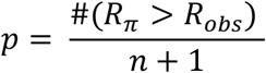 which estimates the probability of obtaining Robs under the null hypothesis that Y is not correlated to Xstatus.The null hypothesis is rejected if p < .05 FDR-corrected for multiple comparisons [49], for the 12 tests performed (3 tissue classes with 4 qMRI parameters).
Two-tailed t-tests were applied post-hoc on the significant results of permutation tests to compare the ARoC distribution between disease status, i.e., Xstatus = 0 against Xstatus = 1. Inferences were conducted at a significance level of .05.
The same pipeline was applied to the brain volumetric changes (BPF, GMF and LF) to test their correlation to the disease activity status.
2.6 Analysis of lesions and peripheral tissues
For white matter lesions analysis, we did not use ARoC but exploited directly the qMRI parameters voxel values. Importantly, with USwL, the prior lesion mask is only used in a probabilistic way and the estimated posterior lesion map, obtained using MTsat, PD, R1 and FLAIR images, typically showed more extended lesion than clinically visible on the FLAIR image alone, as obtained with LST.
Therefore, we separated focal lesions clinically detected on FLAIR images from their peripheral regions detected on qMRI maps. Two different peripheral regions were considered: one for each time point (T0 and T1). Therefore, at T0, three distinct lesion-related regions were isolated:
The lesions, as clinically defined, corresponding to hyperintensity on conventional FLAIR MR image acquired at T0 (referred to as ‘focal FLAIR lesion’).
The peripheral region detected on qMRI maps at T0, not including the FLAIR lesion (referred to as ‘initial peripheral lesion’).
The peripheral region detected with the qMRI maps at T1, computed by masking out the T1 lesion mask with the T0 lesion mask (referred to as ‘later peripheral lesion’). This region allows us to determine whether its microstructure at T0 forebodes a full-blown plaque, detectable during follow up.
The three areas were compared between each other and with NAWM, in order to characterize them on a microstructural basis (Figure 2). Only enlarging lesions were considered for these comparisons.

Schematic illustration of the NAWM and 3 lesions-related areas: Focal FLAIR lesion (dark gray area), Initial peripheral lesion detected at T0 (medium gray area), Later peripheral lesion detected at T1 (dashed, left, and light gray, right, area)
NAWM region consisted of all white matter voxels which did not belong to any of the three lesion-related regions. The four areas were independent as no voxel could belong to more than one class at the same time.
For all participants, MTsat, PD, R1 and R2* median values were extracted from each lesion area, considering lesions individually (between 2 and 66 measurements per subject). Similarly, the median qMRI values within NAWM were also extracted (one measurement per subject). These values were extracted from T0 and T1 scans separately. Statistical analyses were performed in SAS 9.4 (SAS Institute, Cary, NC). None of the qMRI parameter was normally distributed, therefore we applied a log transformation on each of them prior to statistical analysis. For each qMRI parameter, a separate Generalized Linear Mixed Model (GLMM) tested the effect of areas (NAWM and the three lesion-related areas), and time points (T0 and T1), as well as their interaction (i.e., area*time), on the median qMRI parameter value, with a first-order autoregressive variance/covariance model and participants as a random factor (intercept). The degrees of freedom were estimated using Kenward-Roger’s method. Statistical significance was estimated at p < .05 after adjustment for multiple comparison using Tukey’s procedure.
3. Results
3.1 Volume changes
BPF annually decreased between T0 and T1 by -0.67 ± 1.12% (significantly different from zero; paired-sample t-tests; t(16) = 2.57; p = .0204) whereas LF increased by 22.88 ± 26.13% (t(16) = −3.70; p = .0019). GMF non-significantly decreased by -0.30 ± 1.44%.
3.2 Analysis of normal-appearing tissues
As expected, changes in MTsat and R2* within NABT between T0 and T1 varied across subjects (Figure 3). PD and R1 exhibited similar behaviors, see Supplementary data.

Line plots illustrating individual ARoC’s for MTsat (left) and R2* (right) in NAWM. Each line corresponds to one subject. Dotted lines represent increasing rates.
At the group level, with the GLM analysis and permutation inference, we observed that the ARoC of MTsat and R2* positively regressed with disease status as follows (Table 2): MTsat in NAWM and NACGM and R2* in NAWM significantly increase in patients who fare well (Xstatus = 1).
Regression coefficients and their associated p-values (in parentheses) for the effects of Xstatus on the individual ARoC for each qMRI parameter (MTsat, PD, R1 and R2*) and for volumetric measurements (BPF and LF). * Results significant at p < .05, FDR corrected.
Post-hoc t-tests applied on these significant results for a clearer illustration of the difference in disease status (Figure 4) were all significant at a level of .05.

Violin plots of significant change rates in microstructure with respect to Xstatus. From left to right: MT in NAWM, MT in NACGM, R2* in NAWM. * P < .05.
Regarding BPF and LF, their correlation to the disease activity status was not significant (Table 2), suggesting that qMRI parameters are more sensitive to subtle microstructural changes in NABT over time than global morphological measurements
3.3 Analysis of lesion microstructure
The number of enlarging WM lesions between T0 and T1 varied from 2 to 66 across patients, corresponding on average to 63% (±31%) of the number of initial focal lesions. The number of enlarging lesions did not significantly differ between patients’ disease status groups (t(15) = .244, p = .811). GLMMs found a significant effect of areas (3 lesion regions and NAWM) for MT, R1, R2* and PD median (MT: F3 = 35.34, p < .0001, PD: F3 = 68.03, p < .0001, R1: F3 = 40.26, p < .0001, R2*: F3 = 32.32, p < .0001). By contrast, neither time effect (T0 vs T1; MT: F3 = 0.36, p = .5481, PD: F3 = 1.20, p = .2735, R1: F3 = 2.05, p = .1520, R2*: F3 = 2.86, p = .0911), nor the area*time interaction (MT: F3 = 0.09, p = .9671, PD: F3 = 0.14, p = .9346, R1: F3 = 0.14, p = .9331, R2*: F3 = 0.40, p = .7565) were significant, suggesting the microstructural stability of the initial lesion core. Post-hoc tests confirmed significant differences between the four tissue areas.
MTsat, R1 and R2* were significantly larger from FLAIR lesion to initial peripheral lesion, from initial to later peripheral lesion and from later peripheral lesion to NAWM. The reverse was observed for PD. The significant difference in parameters between initial and later peripheral lesion at T0 suggests that subtle microstructural changes appear in the periphery of the initial lesion, months before their detection as focal FLAIR lesions at T1. Adjusted p-values appear in Figure 5. Detailed statistical results of the GLMM’s appear in Supplementary data.

Microstructural parameters in NAWM and the 3 lesion-related areas, for each scanning time T0 and T1. P-values were obtained with post-hoc tests on the tissue area effect. * P < .05.
4. Discussion
This longitudinal study followed up volumetric data and qMRI brain metrics (MTsat, PD, R1, R2*) in 17 MS patients for a median time interval of 30 months. The main results are threefold. First, the microstructure of normal appearing brain tissues changes over time and these modifications concur with, and potentially drive, clinical evolution. This critical finding suggests that repair mechanism and edema resorption can be monitored in vivo. Second, the microstructure within WM plaques is remarkably heterogeneous. Importantly, at their periphery, microstructural alterations foreshadow their expansion, as detected by conventional MRI. Third, as expected, we observed a small but significant brain atrophy and lesion load increase with time.
Quantitative MRI parameter time course within NABT
In this study, we used a multiparameter mapping protocol that was gradually optimized and validated for multi-centric studies [12]. It provides high-resolution maps of multiple qMRI parameters from data acquired during a single scanning session of acceptable duration. A number of cross-sectional studies using a combination of MT, R1, R2* or PD parameters reported significant changes in the microstructure of NABT in MS [26, 50–56]. By contrast, longitudinal analyses of multiparameter qMRI data are scarce. A progressive shortening of T2/T2* [57] or increase in R2* [58–60] was reported within the basal ganglia, suggesting increased of myelin and/or iron contents as well as edema resorption. Likewise, PD and T1 increased within a year, suggesting a demyelination and/or axonal loss [61]. MTR progressively decreases in NAWM of MS patients over one [62] or two years [63]. These abnormalities tend to be more pronounced in progressive phenotypes [64] and were associated to a slow, diffuse and global myelin pathology.
Here, we showed that MTsat within NAWM and NACGM and R2* values within NAWM increase in clinically stable or improving patients. Because MTsat and R2* both correlate with myelin content [14, 30, 31, 65–67], our results suggest repair mechanisms within NABT of patients who are responding to disease modifying treatments, despite the initial myelin/axonal loss and independently from WM focal lesion evolution. These results echo cross-sectional analyses showing that healthy controls (HC) have higher MTsat and R2* values within the same tissue classes compared to MS patients [26]. Annual rates of change of R1 and PD within NABT were not significantly associated with the individual clinical status in this study, although R1 reduction within NABT has already been reported in cross sectional [26, 53, 54] and longitudinal [61] studies comparing MS subjects to HC.
Lesion microstructure
Focal inflammatory demyelinating lesions have been extensively characterized and are traditionally classified as active, chronic active (smoldering) or inactive plaques according to the presence and distribution of plaque-infiltrating macrophages/microglia [68–70]. Focal WM pathology is a constantly evolving process including episodes of demyelination and remyelination but also accumulation of irreversible axonal damage. Age, disease duration, clinical phenotype as well as disease modifying treatment all contribute to the dynamic nature of focal WM pathology [69, 71]. This accounts for the large inter- and intra-individual heterogeneity of MS, which conventional MRI is largely unable to capture. By contrast, quantitative MRI parameters are sensitive to myelin, axonal as well as iron contents and appear as promising markers of plaque dynamics. For instance, MTR was shown to sharply decrease within gadolinium enhancing lesions before recovering during the subsequent months [72–74]. Likewise, reduction of MTR within NAWM, days to weeks before the formation of a new active lesion was also demonstrated [75, 76], and long-term MTR changes in WM plaques were observed in relation with disease progression [64, 77]. The present study broadens the quantitative characterization of plaque dynamics, in keeping with previous longitudinal studies [57, 78]. Two important findings emerge from the results. First, qMRI refines lesion segmentation, as compared to the processing based on the sole FLAIR image. In consequence, the initial lesion revealed by qMRI is typically wider that the plaque detected in FLAIR. Its periphery is characterized by a decrease in MTsat and R2* as compared to NAWM, suggesting an incipient demyelination, reminiscent of the so-called ‘periplaques’ [79]. Moreover, MTsat, R2* and R1 values progressively decrease from NAWM to plaque core, suggesting a centripetal loss of myelin content. Second, plaque microstructure is altered in plaque periphery before any observable change in conventional MRI signals. This finding suggests, in keeping with neuropathological observations [69, 71, 80, 81] that subclinical ongoing inflammation and/or demyelination takes place in the periphery of an active plaque, well before it is detectable on FLAIR or T1 post-gadolinium sequences. If confirmed on larger population samples, this finding might significantly modify treatment management in MS patients.
Oddly enough, plaque qMRI parameters did not significantly change across time. Because iron concentration increases within chronic active or smoldering lesions [82, 83], we were expecting a progressive increase in R2* value. This negative result might be due to the small sample size, the short period of follow up or the limited sensitivity of R2* to local iron concentration as compared to the combined use of R2* and quantitative susceptibility mapping (QSM) [21].
Volumetric Data
CNS atrophy occurs in all stages of MS, since the preclinical phase of the disease and progresses throughout its course, at a much higher rate than one reported in normal aging [84–87]. In this study, the annual brain percentage volume loss at the group level was > 0.4%, which is in line with previous publications [88]. We also showed a significant increase in lesion fraction. Volumetric data (ARoC’s) were highly variable across subjects: changes in BPF range from -2.52 to 1.17% and LF from -0.78 to 103.06%. This variability arises from a large number of factors which do or do not relate to MS: age, disease duration, disease phenotype, disease modifying treatment, circadian rhythm, hydration… [86, 87]. Moreover, annual changes in brain parenchymal fraction as well as lesion fraction only partially correlated to patients’ disease status, in accordance with a large amount of publications [61, 89]. This highlights the lack of specificity and sensitivity of volumetric measurements, at least at the individual level.
Study limitation
As mentioned earlier, the main limitation of the study was the small size and heterogeneous aspect of the present dataset. Indeed, it is composed of only 17 patients, with a rather broad range of characteristics such as age, disease duration, disease phenotype, disease modifying treatment, etc., which are known to influence the disability state of the patient and thus their ability to put together repair mechanisms within cerebral tissues [1, 69–71, 90, 91]. In addition, the time interval between two scanning sessions varied quite a lot across patients (between 14 and 61 months), although it was brought back to an annual rate where possible. All of these features were imposed by standard clinical follow up. Therefore, these results should not be over-interpreted but are nevertheless promising and call for a replication with a larger and more homogeneous or controlled set of MS patients.
5. Conclusion
These preliminary results highlight the relevance of multiple qMRI data in the monitoring of subtle changes within NABT and plaque dynamics in relation with repair or disease progression. Of course, large scale longitudinal study would be needed to reproduce these findings and better exploit the full potential of qMRI parameters.
Data Availability
All data produced in the present study are available upon reasonable request to the authors
6. Declarations of interest
None.
7. Funding
NV, EL and CP are supported by the Fonds de la Recherche Scientifique (F.R.S-FNRS Belgium)
Footnotes
Abbreviations: ARoC: Annual Rate of Change, BPF: Brain Parenchymal Fraction, CDP: Confirmed Disability Progression, cMRI: Conventional Magnetic Resonance Imaging, CNS: Central Nervous System, GMF: Grey Matter Fraction, GLMM: General Linear Mixed Model, LF: Lesion Fraction, MPM: Multiparameter mapping, MRI: Magnetic Resonance Imaging, MS: Multiple Sclerosis, MT: Magnetization Transfer, MTR: Magnetization Transfer Ratio, MTsat: Saturated Magnetization Transfer, NABT: Normal Appearing Brain Tissue, NACGM: Normal Appearing Cortical Grey Matter, NADGM: Normal Appearing Deep Grey matter, NAWM: Normal Appearing White Matter, NEDA-3: No Evidence of Disease Activity, RRMS: Relapsing-Remitting Multiple Sclerosis, PD: Proton Density, PMS: Progressive Multiple Sclerosis, qMRI: Quantitative Magnetic Resonance Imaging, R1: Longitudinal Relaxation Rate (1/T1), R2*: Transverse Relaxation Rate (1/T2*), TIV: Total Intracranial Volume, TPM: Tissue Probability Map, US: Unified Segmentation, USwL: Unified Segmentation with Lesion
8. References
- [1].↵
- [2].↵
- [3].↵
- [4].↵
- [5].↵
- [6].↵
- [7].↵
- [8].↵
- [9].↵
- [10].↵
- [11].↵
- [12].↵
- [13].↵
- [14].↵
- [15].↵
- [16].↵
- [17].↵
- [18].↵
- [19].↵
- [20].↵
- [21].↵
- [22].↵
- [23].
- [24].↵
- [25].↵
- [26].↵
- [27].↵
- [28].↵
- [29].↵
- [30].↵
- [31].↵
- [32].↵
- [33].↵
- [34].
- [35].↵
- [36].↵
- [37].↵
- [38].↵
- [39].↵
- [40].↵
- [41].↵
- [42].↵
- [43].↵
- [44].↵
- [45].↵
- [46].↵
- [47].↵
- [48].↵
- [49].↵
- [50].↵
- [51].
- [52].
- [53].↵
- [54].↵
- [55].
- [56].↵
- [57].↵
- [58].↵
- [59].
- [60].↵
- [61].↵
- [62].↵
- [63].↵
- [64].↵
- [65].↵
- [66].
- [67].↵
- [68].↵
- [69].↵
- [70].↵
- [71].↵
- [72].↵
- [73].
- [74].↵
- [75].↵
- [76].↵
- [77].↵
- [78].↵
- [79].↵
- [80].↵
- [81].↵
- [82].↵
- [83].↵
- [84].↵
- [85].
- [86].↵
- [87].↵
- [88].↵
- [89].↵
- [90].↵
- [91].↵


