medRxiv
  • Home
  • About
  • Submit
  • ALERTS / RSS
Advanced Search

Causal and Candidate Gene Variants in a Large Cohort of Women with Primary Ovarian Insufficiency

B Gorsi, EJ Hernandez, B Moore, M Moriwaki, CY Chow, E Coelho, E Taylor, C Lu, A Walker, P Touraine, LM Nelson, AR Cooper, ER Mardis, A Rajkovic, M Yandell, CK Welt
doi: https://doi.org/10.1101/2021.06.02.21258048
B Gorsi
1Utah Center for Genetic Discovery, Department of Human Genetics, University of Utah, Salt Lake City, UT, USA
  • Find this author on Google Scholar
  • Find this author on PubMed
  • Search for this author on this site
EJ Hernandez
1Utah Center for Genetic Discovery, Department of Human Genetics, University of Utah, Salt Lake City, UT, USA
  • Find this author on Google Scholar
  • Find this author on PubMed
  • Search for this author on this site
B Moore
1Utah Center for Genetic Discovery, Department of Human Genetics, University of Utah, Salt Lake City, UT, USA
  • Find this author on Google Scholar
  • Find this author on PubMed
  • Search for this author on this site
M Moriwaki
2Division of Endocrinology, Metabolism and Diabetes, University of Utah, Salt Lake City, UT, USA
  • Find this author on Google Scholar
  • Find this author on PubMed
  • Search for this author on this site
CY Chow
3Department of Human Genetics, University of Utah School of Medicine, Salt Lake City, UT, USA
  • Find this author on Google Scholar
  • Find this author on PubMed
  • Search for this author on this site
E Coelho
3Department of Human Genetics, University of Utah School of Medicine, Salt Lake City, UT, USA
  • Find this author on Google Scholar
  • Find this author on PubMed
  • Search for this author on this site
E Taylor
4University of Utah School of Medicine, Salt Lake City, UT, USA
  • Find this author on Google Scholar
  • Find this author on PubMed
  • Search for this author on this site
C Lu
4University of Utah School of Medicine, Salt Lake City, UT, USA
  • Find this author on Google Scholar
  • Find this author on PubMed
  • Search for this author on this site
A Walker
4University of Utah School of Medicine, Salt Lake City, UT, USA
  • Find this author on Google Scholar
  • Find this author on PubMed
  • Search for this author on this site
P Touraine
5Sorbonne Universite, Hôpital Universitaire Pitié Salpêtrière-Charles Foix, Service d’Endocrinologie et Médecine de la Reproduction, Centre de Maladies Endocriniennes Rares de la Croissance et du Développement, Centre de Pathologies Gynécologiques Rares, Paris, France
  • Find this author on Google Scholar
  • Find this author on PubMed
  • Search for this author on this site
LM Nelson
6Conover Foundation, McLean, VA, USA
  • Find this author on Google Scholar
  • Find this author on PubMed
  • Search for this author on this site
AR Cooper
7Vios Fertility Institute, St. Louis, MO, USA
  • Find this author on Google Scholar
  • Find this author on PubMed
  • Search for this author on this site
ER Mardis
8Institute for Genomic Medicine, Nationwide Children’s Hospital, Ohio State University College of Medicine, Columbus, OH 43215
  • Find this author on Google Scholar
  • Find this author on PubMed
  • Search for this author on this site
A Rajkovic
9Department of Pathology, University of California San Francisco School of Medicine, San Francisco, CA, 94143 USA
  • Find this author on Google Scholar
  • Find this author on PubMed
  • Search for this author on this site
M Yandell
1Utah Center for Genetic Discovery, Department of Human Genetics, University of Utah, Salt Lake City, UT, USA
  • Find this author on Google Scholar
  • Find this author on PubMed
  • Search for this author on this site
CK Welt
2Division of Endocrinology, Metabolism and Diabetes, University of Utah, Salt Lake City, UT, USA
  • Find this author on Google Scholar
  • Find this author on PubMed
  • Search for this author on this site
  • For correspondence: cwelt{at}genetics.utah.edu
  • Abstract
  • Full Text
  • Info/History
  • Metrics
  • Supplementary material
  • Data/Code
  • Preview PDF
Loading

Abstract

A genetic etiology accounts for unexplained primary ovarian insufficiency (POI; amenorrhea with an elevated FSH level). Subjects with POI (n=291) and controls recruited for health in old age or 1000 Genomes (n=233) underwent whole exome or whole genome sequencing. Data were analyzed using a rare variant scoring method and a Bayes factor-based framework for identifying genes harboring pathogenic variants. Candidate heterozygous variants were identified in known genes and genes with functional evidence. Gene sets with increased burden of deleterious alleles included the categories transcription and translation, DNA damage and repair, meiosis and cell division. Variants were found in novel genes from the enhanced categories. Functional evidence supported 7 new risk genes for POI (USP36, VCP, WDR33, PIWIL3, NPM2, LLGL1 and BOD1L1). Aggregating clinical data and genetic risk with a categorical approach may expand the genetic architecture of heterozygous rare gene variants causing risk for POI.

Introduction

Primary ovarian insufficiency (POI) encompasses a continuum from infertility in women with ovarian dysfunction to early menopause1. The cause of POI remains unknown in the majority of women, making intervention impossible to initiate until it is too late1.

Data overwhelmingly support a genetic cause in women with POI2–4. Twin studies estimate heritability from 53-71%2–4. There is a strong relationship between age at menopause in mothers and daughters, with an odds ratio of 6 (95% confidence intervals 3.4, 10.7) for early menopause in daughters whose mothers had early menopause5. In small studies it has been estimated that up to 30% of POI cases are familial6. The most common known genetic causes include X chromosome defects, FMR1 premutations and autoimmune causes1. Nevertheless, the additive effect of these and known iatrogenic causes explain less than 30% of POI. A remarkable number of new POI-associated genes have been discovered, facilitated by whole exome sequencing (WES) in consanguineous and large families7–15. The women in these families typically develop POI before puberty, also termed ovarian dysgenesis. Mutations in the DNA of these women have been identified in genes important for mitochondrial function, meiosis, homologous recombination and DNA damage repair7,8,10,12,16.

The inheritance pattern for POI is not recessive in all cases. Heterozygous mutations in genes such as eIF4ENIF1 cause POI in women in the mid reproductive years9. Recessive gene mutations found to cause POI and primary amenorrhea also cause POI or earlier menopause in heterozygous mothers, demonstrating that dominant and semi-dominant mutations may be causal17,18, with heterozygous damaging variants in known genes or in two or more candidate genes causing POI19,20. One study of POI suggested an additive effect from common variants contributing to age at menopause, with a recent study suggesting that common variation may explain a portion of earlier age at menopause, as low as age 34 years21,22.

Most previous WES analyses in large numbers of women with sporadic POI used a candidate gene approach to identify gene mutations most likely to cause POI. However, a variant-centric approach has identified novel POI candidate genes19,23. We used an unbiased approach and a new prioritization algorithm (GEM)24–26 to identify damaging gene variants in known POI genes. We then used a category-wide association approach to test the hypothesis that additional candidate mutations could be found in clustered gene sets created using known genes and gene candidates from model organisms27,28. We demonstrated a significant enhancement in identified gene sets in women with POI compared to controls. These gene sets revealed additional candidate genes in POI, with seven genes confirmed by functional studies to play a role in oocyte or ovary development. These findings improve our understanding of the genetic architecture of POI, an extremely heterogeneous disorder.

Subjects

All subjects were diagnosed with POI as defined by at least 4 months of amenorrhea and an FSH level in the menopausal range. All women were 18 years or older, and had a 46XX karyotype, and normal FMR1 repeat number. Subjects (n=35) were recruited in Boston. Additional subjects were recruited from the Partners Biobank (n=63). A second cohort (n=98) was recruited at the National Institute of Health (NIH) for a study of non-syndromic POI29. These subjects were re-consented to have their DNA undergo WES at Washington University (LMN, ARC and ERM). A third cohort was recruited from Pittsburgh (n=20), Italy (n=43) and France (n=32)(AR and PT). All Boston subjects underwent a medical history and physical exam and family history. Subjects from the NIH, Pittsburgh, Italy and France had limited phenotypic data.

Control subjects for category-wide association using GEM24 included 96 unrelated, unaffected subjects recruited for health in old age and 137 CEU, FIN and GBR samples from the 1000 Genomes Project (total n=233 controls)30,31. The majority of CEU samples are from Utah families recruited for large family size (n=47 of 61)32. The control subjects underwent whole genome sequencing, as previously described33. All subjects provided written, informed consent from the University of Utah, Washington University, University of Pittsburgh or the Sorbonne Universite IRB.

Methods

DNA samples were extracted (Qiagen) and subjected to WES. The Boston cohort was sequenced using the Illumina HiSeq 2000 (Illumina). All candidate susceptibility variants in the Boston cohort were Sanger sequenced for verification. Sequencing of the NIH/Washington University cohort was performed using the Roche NimbleGen VCRome 2.1 (HGSC design) exome capture and the Illumina HiSeq 2500 for sequencing at the McDonnell Genome Institute at Washington University. The dataset was accessed through dbGAP (NIH approved request #47895-1, Project #11971). The Pittsburgh, French and Italian cohorts were sequenced at the Pittsburgh Clinical Genomics Laboratory using the Haloplex Exome Target Enrichment System or the Agilent SureSelect V5 Capture Kit (Agilent Technologies, Santa Clara, CA), and 2× 100 bp paired-end WES was performed on an Illumina HiSeq 2500 (San Diego, CA, USA).

The control subjects’ DNA underwent whole genome sequencing (WGS) using the Illumina X Ten sequencing platform (Nantomics, Culver City, CA). The comparison of variants in cases using WES versus controls using WGS would result in a conservative estimate of variants in cases based on the higher coverage expected from WGS.

Alignment and Variant Calling

Alignment and variant calling were performed by the Utah Center for Genetic Discovery (UCGD) core services. Fastq files were downloaded from the Pittsburgh Clinical Genomics Laboratory and dbGAP. Variants were called through the UCGD pipeline using the Sentieon software package (https://www.sentieon.com) 34. Reads were aligned to the human reference build GRCh37 using BWA-MEM (Burrows-Wheeler Aligner). SAMBLASTER was used to mark duplicate reads and de-duplicate aligned BAM files. Aligned BAM files underwent INDEL realignment and base recalibration using Realigner and QualCal algorithms from the Sentieon software package3 to produce polished BAM files. Each polished BAM file was processed using the Sentieon’s Haplotyper algorithm to produce gVCF files35. Sample gVCF files were combined and jointly genotyped with 728 samples comprised of the 1000 genomes project (CEU) samples and samples unrelated to reproduction or cancer phenotypes to produce a multi-sample VCF file. To produce the final VCF variant quality scores, VCF files were recalibrated using Sentieon’s VarCal algorithm to estimate the accuracy of variant calls and reduce potential false positive calls.

Quality Control

Quality control algorithms were applied to sequence reads (Fastq files), aligned reads (BAM files) and variants (VCF files)36. Fastp was used to evaluate read quality, read duplication rate, presence of adapter and overrepresented sequences in Fastq files37. Indexcov was used to estimate depth and coverage of aligned sequence data using BAM indexes. Further alignment quality metrics were calculated on BAM files with samtools stats. The 291 cases were sequenced using different exome capture kits, we therefore standardized QC analysis regions with a bed file made up of exonic regions from coding gene models from RefSeq and Ensemble gene sets. These regions were used to obtain the total number of reads, percentage aligned reads and mean and median coverage for all samples.

Variant quality metrics were calculated by running bcftools stats38–40. The overall quality of VCF callsets were evaluated using Peddy to confirm sex, relatedness, heterozygosity and ancestry of each individual and identifying potential sample-level data quality issues41.

Identification of Damaging Gene Variants

For each case, the uploaded VCF file was scored with VAAST Variant Prioritizer (VVP) and Variant Annotation Analysis and Search Tool (VAAST) to prioritize potentially deleterious variants and damaged genes25,42. VVP and VAAST use a likelihood ratio test (LRT) to score each variant and the aggregate burden of variants for each gene in affected individuals relative to a set of 2,492 control genomes of healthy individuals from the 1000 Genomes Project43. The LRT incorporates three components of each variant; the severity of amino acid substitution, phylogenetic conservation of the variant, and the frequency of the variant relative to the control population. The sum of the top scoring variant(s) based (one variant for dominant inheritance and two variants for recessive inheritance) represents the cumulative likelihood ratio (CLR) for a given gene. The significance of each gene’s VAAST CLRT score is evaluated by a permutation test that randomizes the case/control status of individuals in each of 1e6 permutations and generates a permutation p value for the gene. The output from VAAST is an ordered gene list ranked for the probability of being damaged relative to the control genomes.

Variants identified in the VAAST analysis above were further refined by selecting only variants found at a minor allele frequency (MAF) <0.001 and with no homozygotes found in gnomAD44,45. The choice of a MAF <0.001 cutoff was based on the frequency of the fragile X premutation. The prevalence of the premutation in the population is 0.004 and it is the most common single gene cause of POI identified to date. A fragile X premutation accounts for only 6% of sporadic POI cases46, making 0.001 a conservative upper bound for the risk attributable to any one gene. We also removed variants in genes known to tolerate a large burden of genetic variation such as olfactory receptors, snoRNAs, mucins and T cell receptors47. Finally, we required an Omicia score of >0.7; a meta-classifier that combines scores from SIFT, PolyPhen, MutationTaster and PhyloP to predict pathogenicity48–53. A range of 4-25 damaging gene variants were found per person.

GEM Analysis

We also used GEM to identify gene variants in each subject that were most likely to be pathogenic24. GEM is an Electronic Clinical Decision Support System (eCDSS) framework that aggregates and adjudicates data from multiple algorithms and clinical datasets to provide rapid and accurate diagnosis of individual genomes24. GEM generates a Bayes Factor-based score that calculates the degree of support for and against a given model (a gene allele is pathogenic vs. benign) considering multiple lines of evidence from the following variant analysis tools and data sources: VVP, VAAST, Phevor, mode of inheritance for disease genes from Online Mendelian Inheritance in Man (OMIM), pathogenicity of variants (ClinVar), population specific allele frequencies (gnomAD), quality of variants and the overall genome (data) and quality of the genomic location (gnomAD)25,26,42,44, 54–56. Using this data, GEM identifies potentially pathogenic genotypes and evaluates support for their association with disease. Gene variants for each subject were considered candidates if they had a GEM score ≥ 1 (strong support for the model of pathogenicity)57, together with genes having a GEM score ≥ 0.69 (substantial support for the model of pathogenicity), and a Phevor Bayes factor ≥ 0.9 (genes with a strong association with POI)54. One to twenty-four gene variants were identified for each subject.

Creating Categories for Enrichment Analysis

We used the Database for Annotation, Visualization and Integrated Discovery (DAVID) to functionally annotate known POI genes and candidate genes identified in model organisms 14,58. The analysis yielded 47 clusters with one removed for too few genes (<20; Cluster 37)(Supplementary Table 1). Cluster 37 contained mismatch repair genes, but the genes were also found in other clusters and was therefore redundant.

Calculating Gene Burden on Resampled Gene Lists and Housekeeping Genes

We randomly selected 146 genes from a list of housekeeping genes that are constitutively expressed over many developmental time points in 16 tissue types (Supplementary Table 2). We ensured that none of the housekeeping genes were found in our gene sets identified in the GEM analysis. In addition, we created a burden-matched set of genes. For this, we created a burden ratio for every gene by summing the number of rare variants (MAF ≤ 0.005) in the longest coding transcript of each gene and dividing by transcript length. For each decile in the distribution of this burden ratio, we determined the mean and standard deviation of the burden ratio. We then used these mean and standard deviations to generate randomly sampled gene sets for each decile that had matched mean and standard deviation for burden ratio. These burden matched gene sets were used to test the significance of gene set enrichment in the subjects.

Permutation Tests and Case/Control Comparison

For the permutation tests and analysis, we used the GEM results with a GEM score ≥ 1 generated from the POI cases and control individuals to test for enrichment in individuals with POI compared to controls. Both sets of data generated GEM results using two different HPO terms: POI (HP:0008209) and phenotypic abnormality (HP:0000118) to create 4 sets of data: Cases POI, Cases Phenotypic Abnormality (root), Controls POI, Controls Phenotypic Abnormality (root). The reason we ran GEM using the root of the HPO ontology (Phenotypic Abnormality) was to control for overly connected genes that might have inflated Phevor scores, thus reducing biases due to the nature of the ontology. We then determined the number of damaged genes found in GEM results (number of successes) from genes listed in the individual pathways, a burden-matched gene list and a housekeeping gene list.

Permutation analyses were performed using a random sampling strategy to evaluate enrichment of the POI dataset against gene lists related to functional aspects of the disease. A gene list containing 18,876 RefSeq genes was first created, excluding mucins and olfactory receptor genes. For each functional gene list of size N, random samples of equal size were drawn from the 18,876 genes. This process was repeated 100,000 times, each time with an independently generated random gene list, to create an empirical distribution of the number of damaged genes (GEM score ≥ 1) for each functional gene list. To test whether the probands show enrichment in the functional gene lists, the actual number of damaged genes found for each list was compared to the distribution of damaged genes found using burden-matched gene lists and the housekeeping genes list. To test for statistical significance, we used Fisher’s exact test to calculate a p value using the 2×2 contingency table testing the hypothesis that the number of damaging genes that matched the functional list was significantly larger in the POI GEM runs than in the root Phenotypic Abnormality GEM runs. All p values were adjusted for multiple testing (False Discovery Rate, FDR). To generate a final score depicting the most significantly enriched pathways, adjusted POI p values were divided by Phenotypic Abnormality p values to generate a normalized score that represents enrichment. The higher the ratio, the more enriched the pathway. Pathways with a corrected p value <0.05 and a log2 ratio of greater than 2 for the POI p value/Phenotypic Abnormality p value were considered significant pathways.

Oocyte Expression

To determine whether candidate genes are expressed in mammalian oocytes, 35 day-old female mice were treated with an intraperitoneal injection of 5 IU PMSG to initiate follicular development and 5 IU hCG 48 hours later to induce ovulation60. Eighteen hours later, mice were sacrificed, oviducts dissected to remove oocytes and cumulus cells manually removed. RNA was isolated from oocytes using RNeasy (Qiagen, Valencia, CA)9. Reverse transcription was performed with SuperScript Master Mix (Life Technologies, Carlsbad, CA) using SuperScript III RT and random primers. Quantitative real-time polymerase chain reaction was performed for the expression of candidate genes and glyceraldehyde-3-phosphate dehydrogenase (Gapdh) as an endogenous control using PowerUp SYBR Green Master Mix (Applied Biosystems, Foster City, CA). Primers were designed to span two exons to avoid amplifying genomic DNA. Primer sequences are provided (Supplementary Table 3). Samples were examined in triplicate and at three dilutions. mRNA levels were determined using the 2-ΔΔCT method to calculate relative quantification and to correct for expression of endogenous controls.

Functional Analysis

Flies were raised at 25° C on standard diet based on the Bloomington Drosophila Stock Center standard medium with malt. We obtained 20 RNAi lines from the Bloomington Drosophila Stock Center. Ovary/germline specific RNAi knockdowns were performed using Gal4 DNA-binding protein and Upstream Activator Sequence (GAL4/UAS) technology, as previously described61. We crossed flies carrying the Maternal Triple Driver-GAL4 (MTD-GAL4; BDSC 31777) transgene to flies carrying each respective UAS-RNAi transgene to generate female flies with ovary specific knockdown of each gene. Control flies were generated by crossing flies carrying the MTD-GAL4 transgene to the appropriate AttP RNAi background strain (does not carry UAS-RNAi transgene). Virgin female RNAi knockdown (and control) flies were collected on CO2 anesthesia and aged 3-5 days on standard media supplemented with dry yeast. Female knockdown flies were singly mated with a 3-5 day old Canton S male. Individual mating pairs were observed to ensure successful mating. Males were removed after mating. We measured four female reproductive phenotypes: 1) egg number: number of eggs laid in first 8 hrs post mating; 2) hatchability: number of adults that hatched from those eggs; 3) total fertility over 10 days post mating; 4) overall ovary appearance and morphology62. For egg number, newly mated females were place in vials for 8 hrs and egg number was counted. For hatchability, all the progeny that eclosed from the egg number vial were counted (progeny #/egg #). To measure total fertility, mated females were transferred to new vials every two days for ten days and all the progeny were summed over the entire period. For ovary images, adult females were collected under CO2 anesthesia, dissected and immediately imaged. Ovaries were imaged at 3X magnification using a Leica EC3 camera. We assayed at 8-10 females per RNAi knockdown. Statistical analysis was performed using R software. P-values were determined using ANOVA. A p value < 0.05 was used for significance.

Results

Whole exome sequencing produced a mean of 96 million reads per individual (range 31-186 million) and an average of 99.6% mapped/aligned reads to the GRCh37 human reference genome with an average duplication rate of 8.4% (Supplementary Figure 1A-C) for the 280 samples that passed QC metric cutoffs. Fastp identified read quality and insert sizes within the normal ranges. Variant calling produced an average of 21,923 SNVs and 576 indels per sample, with an average depth of 55x per sample (Supplementary Figure 1G-I). Peddy was used to infer the sex, heterozygosity, ancestry, and relatedness of subjects, and compared to known metadata about samples (Supplementary Figure 2A-D)41. From these quality control metrics we identified and removed four samples (Supplementary Table 4) that had very low heterozygosity and low coverage. Three samples were removed due to high duplication rates. One sample was removed due to excess heterozygosity. We also discovered a previously unidentified deletion of the long arm of the X chromosome (93.7% homozygous X:130678467-X:155171537) in sample IPOF32. In total, we removed 9 samples from analysis for the quality issues described above leaving 282 samples for the analysis. Peddy confirmed the sex of the POI subjects (Supplementary Figure 2A), and the known relatedness of a few individuals (Supplementary Table 5, Supplementary Figure 2D-E). An additional four sib pairs were identified in the cohort and one family with dominant inheritance was included (Supplementary Figure 2E). For these related individuals, only one subject was included in the joint analyses. PCA projection of the samples together with data from the 1000 genomes identified individuals of European descent (n=235), admixed American (n=18), African (n=10), South Asian (n=4), East Asian (n=3), and unknown (n=12) ancestry (Supplementary Table 6, Supplementary Figure 2C).

In 19 subjects, we identified variants in genes previously determined to cause POI, including confirmation of previously identified variants in 12 subjects with primary amenorrhea (6.7%; Table 1)19,63–66. Five of these variants were found as heterozygous genotypes in the genes NR5A1, PTPN22 and eIF4ENIF19,17,67.

View this table:
  • View inline
  • View popup
Table 1.

Candidate variants in previously identified genes causing POI.

Sixty-four subjects (23%) carried at least one variant in a previously identified POI gene that was determined to cause ovarian dysgenesis or primary amenorrhea with autosomal recessive inheritance (Table 2). Twenty-seven subjects (10%) carried a heterozygous variant in a gene for which there was a previously identified functional model (Table 3)68,69. Variants at genomic loci that were not conserved across species were not considered for analysis, although variants impacting conserved amino acids that were found only in mammalian species were included. One subject carried two variants in FANCM and one subject carried two variants in RECQL4, however, it was not possible to confirm whether these variants were in cis or trans. Fourteen subjects carried 2, one subject carried 3 and one subject carried 4 candidate POI risk variants in different genes.

View this table:
  • View inline
  • View popup
Table 2.

Candidate variants found as heterozygotes in previously identified genes causing POI or associated with age at natural menopause.

View this table:
  • View inline
  • View popup
Table 3.

Candidate variants in genes from pathways with functional models

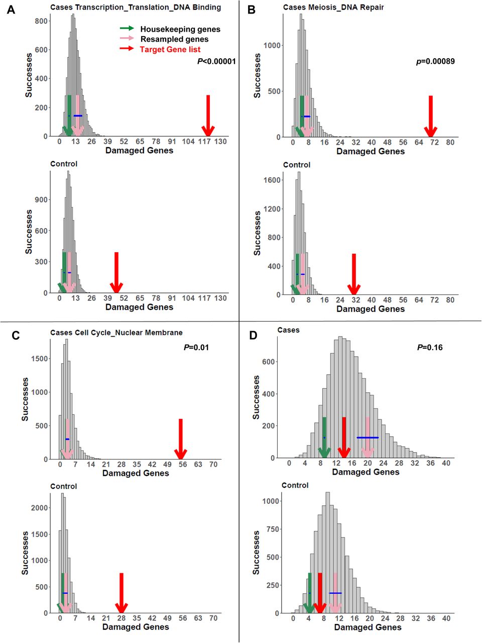
We next determined gene clusters for known genes for POI in women and candidates from animal models. DAVID analysis identified 47 gene list clusters with enrichment >2 (Supplementary Table 1). We then examined enrichment of these 47 gene sets in women with POI compared to controls and found 13 significant gene sets. These gene sets encompassed GO term biological processes including transcription/translation, DNA damage and repair, oogenesis, cell proliferation, hormone regulation, growth factors, regulation of gene expression, embryogenesis, cytoplasmic signaling, male gonad development, chromatin binding, cell division and protein phosphorylation (Table 4, Figure 1). Further, there was significant enhancement compared to housekeeping genes and burden-matched gene lists in POI cases compared to controls (Figure 1A-C and Supplementary Table 2). The majority of the causal or candidate genes were found in the enriched gene sets (Tables 1-3). The two genes that were not found in the gene lists are important for meiosis (MARF1 and ANKRD31).

View this table:
  • View inline
  • View popup
Table 4.

Enhanced biological pathways or clusters in women with POI compared to controls.

Figure 1.
  • Download figure
  • Open in new tab
Figure 1.

Three examples of enriched pathways in the POI data set as determined by the permutation tests in cases (upper panels) and controls (lower panels). Enriched pathways that encompassed novel POI genes (Table 5) included: A) Transcription/Translation/DNA binding, B) Meiosis/DNA Repair/Homologous Recombination and C) Cell Division/Meiosis, compared to D) Housekeeping Genes. The number of damaged genes from the target gene list in the pathways of interest (red arrow) is compared to the distribution of damaged genes in random gene lists of equal number to the lists of interest (gray bars), burden-matched control genes (pink arrows) and housekeeping genes (green arrows). The burden-matched genes and housekeeping genes are not significantly enriched for any gene set. p values are controlled for the false discovery rate.

We examined the remaining candidate genes that had not yet been implicated in a woman with POI or in an animal model. We identified several deleterious variants in genes found in the implicated gene sets (Table 5)70–77. None of these gene variants was identified in the control groups that we assessed. Additional candidates were identified in these and other gene sets, although their pathogenicity was not as strong based on conservation, allele frequency or gene constraint (Supplementary Table 7).

View this table:
  • View inline
  • View popup
Table 5.

Variants in candidate genes and candidate pathways with no previous model for primary ovarian insufficiency.

Functional Studies

The potential pathogenicity of the variants not previously identified is outlined in the Supplementary Data (Supplementary Information). For genes with variants not previously identified in POI or in animal models of POI or not previously examined in oocytes (Table 5), RTPCR in mouse oocytes that have resumed and/or completed meiosis I was performed to ensure that the candidate gene was expressed. Of the 24 genes tested, four were present but not highly expressed in the oocyte (Table 5 and Supplementary Tables 3 and 8).

D. melanogaster orthologues were identified for 20 of 35 candidate genes queried (Supplemental Table 9). Thirteen candidates could not be obtained based on availability or could not be tested based on lack of orthology or absence of ovarian expression. Two of the candidates had multiple weak orthologues and were not pursued (POLK and ANKRD31).

Five knockdowns (USP36, VCP, WDR33, PIWIL3 and NPM2) were completely infertile with atrophic ovaries (Table 6 and Supplementary Figures 18 and 19). Two gene knockdowns demonstrated decreased hatchability and fertility, with abnormal (LLGL1) or normal ovaries (BOD1L1). Two gene knockdowns had variable or mild ovarian defects that were not statistically significant (CDK7 and BRIP1). One gene knockdown was lethal (RUVBL2).

View this table:
  • View inline
  • View popup
  • Download powerpoint
Table 6.

Ovary and fertility phenotypes in D. melanogaster RNAi knockdown in ovaries/germline.

Discussion

We performed WES in 291 subjects with POI from three cohorts. Using two methods, a broad and unbiased discovery method and a more robust prioritization algorithm (GEM), we identified the most likely pathogenic variants in these women with POI. Our data suggest that the candidate genes for POI in individual women are highly heterogeneous. However, when the most likely candidate genes were categorized into functionally related groups, the genes aligned into 13 clusters that were enriched in cases compared to controls after correcting for multiple testing, gene size and pathogenic specificity for POI. New candidate genes were found in enhanced gene sets that included genes important for transcription/translation, DNA damage and repair, meiosis and cell division. Functional analysis in D. melanogaster supported a role in oocyte or ovary development for seven genes not previously associated with POI. Taken together, the data support a categorical approach to understanding the genetic architecture for POI.

After an initial broad search for damaging variants, we used an AI-based eCDSS tool, GEM, that employs variant impact (VAAST and VVP), patient phenotypes (Phevor), known Mendelian and pathogenic variants (OMIM, ClinVar) and ancestry to identify disease-causing genotypes15,24,25,42,54. Using GEM, we supplemented our data with previously analyzed WESs and have replicated genetic findings in 11 out of 12 subjects with primary amenorrhea, demonstrating the utility of the new software. The only gene variant that was not identified was in MARF1, which has not yet been associated with POI in OMIM. GEM identified additional homozygous or compound heterozygous mutations (HFM1, DCAF17) or heterozygous mutations (NR5A1) in previously identified genes78–80.

GEM also identified heterozygous deleterious variants in known genes, particularly in women with POI and secondary amenorrhea. GEM uncovered variants in 28 genes previously demonstrated to cause POI with recessive inheritance, out of 42 gene variants total (67%). GEM also identified 13 of 20 candidate variants in genes with evidence for ovarian insufficiency in an animal or other experimental species model (65%). The results are not surprising based on the use of HPO POI terms in the algorithm, which emphasizes phenotype in GEM54. Further tool development will encompass gene pathways for discovery.

With the exception of subjects with POI and primary amenorrhea, the majority of candidate variants identified in the current study are heterozygous, arguing for a dominant, semi-dominant, or complex inheritance pattern for POI that occurs later in the reproductive years. Genome-wide association studies (GWASs) of age at natural menopause support the concept that menopause has a complex inheritance pattern68. Further, the largest GWAS of early menopause, defined as menopause before the age of 45 years, replicated 4 common variants associated with age at natural menopause and demonstrated that menopause risk alleles have an additive contribution to age at menopause21. Recent data also suggest that common variants contribute to menopause occurring as early as 34 years22. However, the contribution of common variation explains only a small portion of the genetic risk for menopause under age 40 years22. The current study using WES was not able to assess common variants, but did demonstrate overlap between common GWA variants and rare, deleterious variants in the same candidate genes. For example, nonsynonymous variants in BRCA1 are associated with earlier menopause by approximately 6 months68. In the current study, we identified a frameshift mutation, expected to result in early protein termination, possibly causative for POI. Additional genes with deleterious variants (MSH6, CHD7) and some with rare missense variants (RAD54L, HELQ, POLG) also overlap with candidate genes associated with age at natural menopause (Table 2). The apparent overlap of common variants associated with menopause age and deleterious variants in the same candidate genes is consistent with the hypothesis that mutations in these genes play a causative role in POI.

Further support for the causative role of heterozygous gene variants in POI comes from the reproductive history of the mothers of girls with primary amenorrhea. A heterozygous MND1 gene mutation in a mother resulted in POI at age 35 years45. Similarly, a heterozygous mutation in MCM8 caused POI in a mother at age 29 years18. In both families, the daughters with homozygous mutations presented with primary amenorrhea. With the exception of a few reports, age at menopause is rarely mentioned or may not yet have occurred for mothers of girls with POI. However, age at menopause is heritable supporting the segregation of ovarian damaging genes with an effect on age at menopause through the mother3. It is also not surprising that heterozygous variants that relatively decrease fertility would be removed from the population through decreased progeny81, and might therefore be inherited from the father since reproductive lifespan is not limited in men. Taken together, these cases also support the hypothesis that heterozygous mutations can result in earlier age at menopause.

Although the number of subjects in the current study is not sufficient to replicate the genes individually, we were able to demonstrate significantly enriched gene clusters controlled for multiple testing. Previous work in autism and congenital heart disease has used a similar category-wide association study approach27,28. Our approach was unbiased; first examining the most deleterious variants in women with POI to identify known genes and candidates with previous functional models, and subsequently determining whether additional genes were found in the clustered gene sets. Interestingly, a MAGENTA analysis of age at natural menopause variants identified similar enhanced categories for candidate genes inferred from genome-wide associated variants68. In addition, known genes causing male azoospermia were enriched in comparable pathways82. Taken together, a category enhanced approach identifies consistent gene sets across reproductive studies. Genes falling into gene sets including oogenesis, spermatogenesis, meiosis, DNA damage and repair, transcription and translation, chromatin binding, regulation of gene expression, growth factors, embryo development, cell division, extracellular to cytoplasmic signaling, protein kinase phophorylation, and vasoactivity and hormone regulation were enriched compared to controls in our unbiased candidate gene search for damaging mutations across the genome (Figure 1)83. New candidate genes were identified within these gene sets demonstrating that the category approach provides a mechanism for new candidate gene discovery.

Our D. melanogaster knockdown model affords a mechanism to determine an oocyte and ovarian phenotype at scale for genes in enhanced pathways. The genes and developmental processes involved in oogenesis in D. melanogaster overlap with those in the mouse84. We chose quantifiable fertility assays including egg laying rates, hatchability and ovarian morphology62. The use of RNAi technology also presumes that the gene is not fully deleted and serves as an excellent model for heterozygous gene variants. Using our D. melanogaster model, we identified five genes that are critical for ovarian or oocyte development and that fall into the enriched pathways we defined: transcription/translation, meiosis, DNA repair and DNA damage. RNAi knockdown resulted in atrophic ovaries with no eggs or progeny (Table 6 and Supplementary Figures 18 and 19).

USP36 is a deubiquitinase demonstrated to promote RNA polymerase I stability for the ribosomal RNA processing and translation85. Previous studies found that the scny D. melanogaster homologue also acts as a histone H2B ubiquitin protease86. The atrophic ovary in the knockdown shows that the ribosomal RNA translation and/or the chromatin modification function may affect oocyte or ovarian development in addition to its role in embryogenesis85.

WDR33 plays a role as one of 4 proteins that recognize the polyadenylation signal in the 3’-end processing of mRNA precursors87. The gene is highly expressed in testes and we have now demonstrated that it is also highly expressed in mouse oocytes (Supplementary Table 8). RNAi knockdown results in an atrophic ovary. Thus, WDR33 may also play critical role in ovarian or oocyte development.

PIWIL3 is a P-element induced wimpy testis protein short RNA found in human, nonhuman primate and bovine oocytes. It is specifically expressed in maturing human oocytes during oogenesis88 and in bovine oocytes from the GV stage onward89. It is critical for germline integrity from DNA transposable element activity90. The affected subject carries two PIWIL3 variants; a frameshift mutation and a stop gain mutation that both remove the PIWI domain from the protein90. We also identified a stop gain mutation in PIWIL2, a family member that is expressed in fetal human germ cells89. Although knockouts of the mouse PIWIL2 homologue Mili were described as fertile, there were no details provided across the reproductive lifespan91. These data demonstrate the importance of the PIWIL genes in the ovary in addition to the testes.

NPM2 is found in oocytes before germinal vesicle breakdown92. The Npm2 knockout females are infertile, with normal sized pronuclei that lack nucleoli92,93. Although previous studies suggest that infertility is caused by failure of zygote development, our data suggest that NPM2 is critical for oocyte and ovary development.

VCP, or valosin-containing protein, is an ATPase associated with a variety of activities94. It is expressed in GV oocytes and preimplantation embryos in the mouse and controls germinal vesicle breakdown. Vcp knockout mice demonstrate no homozygotes because they have a defect in early embryonic development. Our model demonstrates atrophic ovaries. A missense variant in a highly conserved threonine in the N terminal was found in two sisters and a mother with POI (Supplementary Figure 2E). The N terminal is the portion that interacts with other proteins. Therefore, VCP should be considered a new candidate for POI.

In contrast to genes described above, there were only subtle phenotypes identified using our model in genes associated with DNA damage and repair pathways. Two genes in the pathway, LLGL1 and BOD1L1, were highly expressed in the oocyte and demonstrated decreased hatchability and decreased fertility. The LLGL1 cytoskeletal network is involved in maintaining cell polarity and epithelial integrity95. Mutations including the gene region on chr 17 cause Smith Magenis syndrome, a disorder of developmental delay, behavioral abnormalities, sleep disturbance and abdominal obesity. An indel upstream of LLGL1 in Shaanbei White Cashmere goats is associated with change in litter size96. BOD1L1 stabilizes RAD51 at the site of DNA replication forks97. The frameshift variant in our subject would remove all ATM phosphorylation sites, along with the majority of the protein. Other genes in the DNA damage and repair pathway with no previous functional models or human mutations had no phenotype in our D. melanogaster model and more sensitive functional models may be needed. Nevertheless, a number of previously well validated variants involved in the homologous recombination steps in meiosis were discovered in our cohort (Figure 2)83. Many of these gene mutations may result in meiotic failure and oocyte loss. Given the large number of gene mutations falling into the DNA damage and repair pathway, intervening to rescue meiosis for development of normal gametes may be a treatment opportunity in POI.

Figure 2.
  • Download figure
  • Open in new tab
Figure 2.

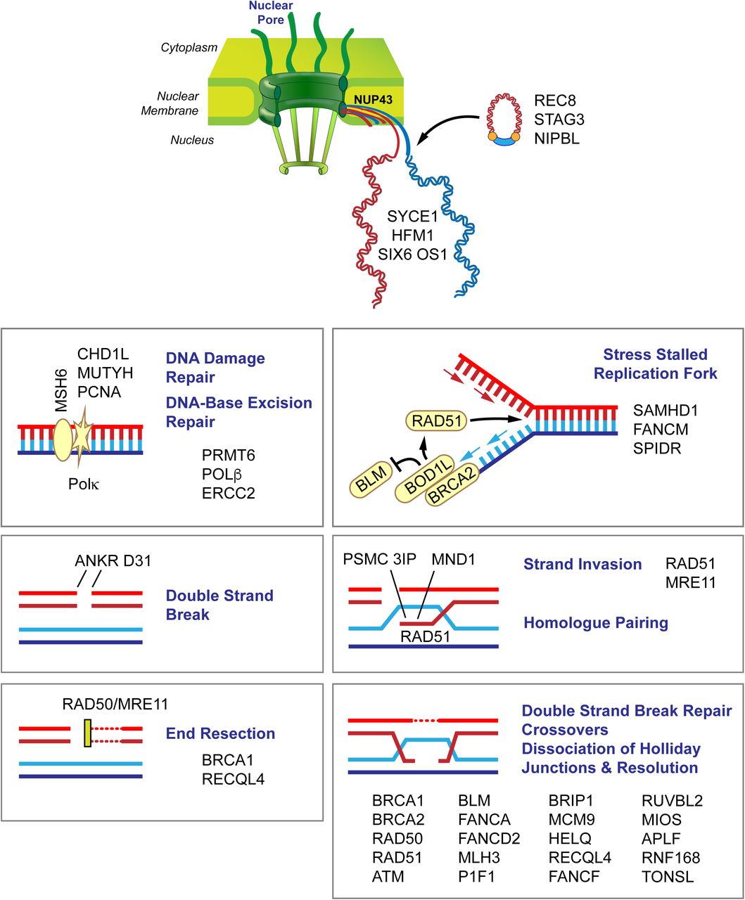
Candidate genes in women with POI. Variants in a number of genes involved in chromosome pairing and DNA damage and repair are involved in meiosis. The figure depicts candidate genes that are involved in chromosome movement, double strand breaks, end resection, double strand break repair, crossovers and dissociation and resolution of Holliday junctions. Members of the nuclear pore complex (NUP43) play a role in chromosome movement and organization. After DNA replication (ORC6), the synaptonemal complex pairs homologous chromosomes (PSMC3IP) loaded with condensin and cohesion complex proteins (STAG3, REC8, NIPBL) and connects the synaptonemal complex to DNA repair proteins (SYCE1). During recombination, double strand breaks form (ATM, ANKRD3, PIF1), ends are resected (BRCA1, SAMHD1, BOD1L1), and crossovers occur (HFM1) through strand invasion (PSMC3IP, MND1, RAD51). Subsequently, DNA double strand break repair (CHD1L, POLG, POLK, MSH6, PCNA, NUPR1, APLF, NBN, RAD50, RUVBL2, MRE11), DNA repair (CDK7, MLH3, PRMT6, HELQ, TONSL), strand annealing (RECQL4) and repair via homologous recombination (BRCA2, BRIP1, FANCD2, HELQ, FANCM, FANCF, BLM, MCM9, USP36) take place. Kinetochore/chromosome assembly, orientation and segregation (HAUS6, CENPF, NUP43, NCAPG2, LLGL1, NINL, ATRX) follow recombination.

An association between autoimmune oophoritis, with POI as the end-stage, has been demonstrated only with adrenal autoimmunity98. We identified a novel PTPN22 variant, a gene associated with adrenal insufficiency, and inTARBP1, a gene associated with autoimmune syndromes99,100. A final subject carried a variant in IL1B, which has been associated with ovarian inflammation. Further delineation of the associated autoimmune risk genes and diseases will clarify the relationship between autoimmunity, genetics and POI.

Our study is limited by whole exome sequencing. We were not able to evaluate common variation, some promoter regions and could not evaluate copy number variants. We did not have trios for the majority of subjects and did not recruit family members to clarify segregation or de novo mutations. Future studies will also be needed to more carefully analyze the mitochondrial genome.

The current cohort forms one of the largest WES datasets analyzed for POI. We used an unbiased approach and a new AI-based algorithm to identify the most likely pathogenic variants. We also demonstrated new genes important for oogenesis and ovarian development using a model D. melanogaster system. Our more global approach contrasts to previous studies that examined individual consanguineous families and/or were restricted to candidate gene lists. Collectively, our results identify not only disease-causing variants, but also gene categories involved in POI. These results should prove useful for precision medicine efforts aimed at early identification of gene variants increasing a woman’s likelihood to experience infertility or a shortened reproductive lifespan. The early identification of women at risk for POI may enable fertility preserving measures. More broadly, better understanding of the genetic architecture of POI might also aid in identifying additional comorbid risks in a subset of the subjects.

Data Availability

The data from NIH is available in dbGAP (Project #11971).

https://www.ncbi.nlm.nih.gov/gap/

Supplementary Table 1. Annotation clusters identified using the Database for Annotation, Visualization and Integrated Discovery (DAVID). Data were organized into 47 clusters with enrichment scores of >2.

Supplementary Table 2. Housekeeping genes. Genes that are constantly and uniformly expressed over many developmental and adult time points in 16 tissue types were chosen as housekeeping genes to examine enrichment.

Supplementary Table 3. PCR primers used to analyze gene expression in super ovulated mouse oocytes using RTPCR.

Supplementary Table 4. Samples removed by Peddy for very low heterozygosity and low coverage.

Supplementary Table 5. Related subjects identified by Peddy.

Supplementary Table 6. Ancestry identified by PCA plot and projection onto 1000 genomes data.

Supplementary Table 7. GEM results for all subjects. The GEM results for all genes with a GEM score greater than 0 are presented.

Supplementary Table 8. Oocyte expression. RTPCR was performed in superovulated mouse oocytes for gene targets with no previous functional studies.

Supplementary Table 9. Genes chosen for RNAi knockdown in a D. melanogaster model with Bloomington Drosophila Stock Center Number (BDSC#).

Supplementary Figure 1. Quality control metrics for 283 POI cases.

Box whisker plots of the alignment statistics and vcf statistics for 283 cram and vcf files that passed QC metrics: A) Total number of reads per sample, B) Percentage of Aligned reads, C) Percentage of duplicate reads, D) Mean coverage per sample, E) Median coverage per sample, F) Percentage of Coverage over 20 bases, G) Number of SNPs per sample extracted from the Bcftools statistics, H) Number of Indels found per sample, and I) Average Depth per sample.

Supplementary Figure 2. Peddy analysis of 283 samples from the final VCF files of women with POI. A) The predicted sex was female for all cases. B) The proportion of heterozygous calls ranged from 0.12 to 0.18 at a median depth of 30 to 65. C) PCA projection of the 283 cases onto ancestry of 1000 Genomes data. The majority of subjects were of European ancestry as expected. D) Coefficient of relatedness between two samples plotted by sampling 25K sites in the genome and comparing the relatedness reported in the ped file to the relatedness inferred from the genotypes. Thus, five sib pairs were confirmed, along with grandparent-parent and parent-child relationships. E) Five pedigrees of relationships confirmed by Peddy and investigators.

Supplementary Figure 3-16. Enriched pathways in the POI data set as determined by the permutation tests in cases (upper left panels) and controls (lower left panels) and compared to data from the same pathways for the root phenotypic abnormality (upper right and lower right panels). The number of damaged genes from the pathways of interest (red arrow) is compared to the distribution of damaged genes in random gene lists of equal number to the lists of interest (gray bars), burden-matched control genes (pink arrows), and housekeeping genes (green arrows). The burden-matched genes and 16A) housekeeping genes are not significantly enriched for any gene set.

Supplementary Figure 17. PA-1 cells were transfected using PolyJet transfection reagent (SignaGen Laboratories, Rockville, MD) with WT eIF4ENIF1 or eIF4ENIF1 containing the c.603T>G variant created using the QuikChange II Site-Directed Mutagenesis kit (Agilent Technologies, Santa Clara, CA) into a pcDNA3.1(-) expression vector (Invitrogen, Carlsbad, CA) using the NEBuilder HiFi DNA Assembly Cloning Kit (New England Biolabs, Ipswich, MA). Stable cell lines were generated by selection of colonies resistant to 750 μg/ml G418 (Life Technologies, Carlsbad, CA). Cells were seeded at 3 × 10⁴ cells/well in an 8-well chamber slide. After 48 hours, cells were fixed with ice-cold 100% methanol for 5 minutes at room temperature (RT), followed by washing with PBST comprising 0.1% Tween-20 in 1× PBS (Fisher Scientific, Waltham, MA). Cells were blocked with 1% BSA and 22.52 mg/ml glycine (Fisher Scientific, Waltham, MA) in PBST for 30 minutes at RT and then incubated with an N-terminal antibody (Novus Biologicals, Centennial, CO) diluted in 1% BSA in PBST at 4℃ overnight. After another wash with PBST, cells were labeled with an anti-rabbit Alexa Fluor 594 (Invitrogen, Carlsbad, CA) for 1 hour at RT, washed with PBST as before, counterstained with DAPI (Southern Biotech, Birmingham, AL), and mounted with glycerol mounting medium with DABCO (Electron Microscopy Sciences, Hatfield, PA). The Nikon fluorescent microscope was used for image acquisition. The c.603T>G variant, p.S201R, is located in the nuclear import signal of eIF4ENIF1. The top panels show the N terminal eIF4ENIF1 images with DAPI staining of the nucleus, while the bottom panels show the N terminal eIF4ENIF1 images. Compared to the A) wild type eIF4ENIF1, the B) S201R variant transfected cells demonstrated disorganized localization of eIF4ENIF1 with increased intranuclear protein.

Supplementary Figure 18. Drosophila melanogaster phenotypes.

Hatchability and total fertility values are plotted for all genes tested by RNAi in Drosophila. P values for genes with significantly different phenotypic values are highlighted in red. N= 8-10 for all measurements. C= control; KD= RNAi knockdown

Supplementary Figure 19. Drosophila melanogaster ovarian phenotype.

Representative images of ovaries from RNAi knockdowns that produced atrophic ovaries and a control. All other RNAi knockdowns that produced normal ovaries appear identical to the control and are not shown.

Acknowledgements

The work in this publication was supported by the Center for Genomic Medicine and its Functional Analysis Service. The work was also supported by R56HD090159 and R01HD099487 (CKW) and R35GM124780 (CYC and EC). The content is solely the responsibility of the authors and does not necessarily represent the official views of the National Institutes of Health.

Footnotes

  • Disclosure: MY is a stock holder or has received stock option awards from Fabric Genomics Inc. BM and MY have received consulting fees from Fabric Genomics Inc. ARC is a consultant for Ferring and a scientific advisory board member for INVObioscience and Celmatix.

  • All authors approved the final version.

References

  1. ↵
    Welt, C. K. Primary ovarian insufficiency: a more accurate term for premature ovarian failure. Clinical endocrinology 68, 499–509, doi:10.1111/j.1365-2265.2007.03073.x (2008).
    OpenUrlCrossRefPubMed
  2. ↵
    de Bruin, J. P. et al. The role of genetic factors in age at natural menopause. Human reproduction 16, 2014–2018 (2001).
    OpenUrlCrossRefPubMedWeb of Science
  3. ↵
    Treloar, S. A., Do, K. A. & Martin, N. G. Genetic influences on the age at menopause. Lancet 352, 1084–1085, doi:10.1016/S0140-6736(05)79753-1 (1998).
    OpenUrlCrossRefPubMedWeb of Science
  4. ↵
    Snieder, H., MacGregor, A. J. & Spector, T. D. Genes control the cessation of a woman’s reproductive life: a twin study of hysterectomy and age at menopause. The Journal of clinical endocrinology and metabolism 83, 1875–1880, doi:10.1210/jcem.83.6.4890 (1998).
    OpenUrlCrossRefPubMedWeb of Science
  5. ↵
    Torgerson, D. J., Thomas, R. E. & Reid, D. M. Mothers and daughters menopausal ages: is there a link? European journal of obstetrics, gynecology, and reproductive biology 74, 63–66 (1997).
    OpenUrlCrossRefPubMedWeb of Science
  6. ↵
    Vegetti, W. et al. Inheritance in idiopathic premature ovarian failure: analysis of 71 cases. Human reproduction 13, 1796–1800 (1998).
    OpenUrlCrossRefPubMedWeb of Science
  7. ↵
    Vries, L. D. et al. Exome sequencing reveals SYCE1 mutation associated with autosomal recessive primary ovarian insufficiency. The Journal of clinical endocrinology and metabolism, jc20141268, doi:10.1210/jc.2014-1268 (2014).
    OpenUrlCrossRefPubMed
  8. ↵
    Caburet, S. et al. Mutant cohesin in premature ovarian failure. The New England journal of medicine 370, 943–949, doi:10.1056/NEJMoa1309635 (2014).
    OpenUrlCrossRefPubMed
  9. ↵
    Kasippillai, T. et al. Mutations in eIF4ENIF1 are associated with primary ovarian insufficiency. The Journal of clinical endocrinology and metabolism 98, E1534–1539, doi:10.1210/jc.2013-1102 (2013).
    OpenUrlCrossRefPubMedWeb of Science
  10. ↵
    Wood-Trageser, M. A. et al. MCM9 mutations are associated with ovarian failure, short stature, and chromosomal instability. American journal of human genetics 95, 754–762, doi:10.1016/j.ajhg.2014.11.002 (2014).
    OpenUrlCrossRefPubMed
  11. Katari, S. et al. Novel Inactivating Mutation of the FSH Receptor in Two Siblings of Indian Origin With Premature Ovarian Failure. The Journal of clinical endocrinology and metabolism 100, 2154–2157, doi:10.1210/jc.2015-1401 (2015).
    OpenUrlCrossRefPubMed
  12. ↵
    AlAsiri, S. et al. Exome sequencing reveals MCM8 mutation underlies ovarian failure and chromosomal instability. The Journal of clinical investigation 125, 258–262, doi:10.1172/JCI78473 (2015).
    OpenUrlCrossRefPubMed
  13. Qin, Y., Jiao, X., Simpson, J. L. & Chen, Z. J. Genetics of primary ovarian insufficiency: new developments and opportunities. Human reproduction update 21, 787–808, doi:10.1093/humupd/dmv036 (2015).
    OpenUrlCrossRefPubMed
  14. ↵
    Tucker, E. J., Grover, S. R., Bachelot, A., Touraine, P. & Sinclair, A. H. Premature Ovarian Insufficiency: New perspectives on genetic cause and phenotypic spectrum. Endocrine reviews, er20161047, doi:10.1210/er.2016-1047 (2016).
    OpenUrlCrossRefPubMed
  15. ↵
    Al-Agha, A. E. et al. Primary Ovarian Insufficiency and Azoospermia in Carriers of a Homozygous PSMC3IP Stop Gain Mutation. The Journal of clinical endocrinology and metabolism 103, 555–563, doi:10.1210/jc.2017-01966 (2018).
    OpenUrlCrossRefPubMed
  16. ↵
    Ahmed, S. et al. Exome analysis identified a novel missense mutation in the CLPP gene in a consanguineous Saudi family expanding the clinical spectrum of Perrault Syndrome type-3. J Neurol Sci 353, 149–154, doi:10.1016/j.jns.2015.04.038 (2015).
    OpenUrlCrossRef
  17. ↵
    Lourenco, D. et al. Mutations in NR5A1 associated with ovarian insufficiency. The New England journal of medicine 360, 1200–1210, doi:10.1056/NEJMoa0806228 (2009).
    OpenUrlCrossRefPubMedWeb of Science
  18. ↵
    Heddar, A., Beckers, D., Fouquet, B., Roland, D. & Misrahi, M. A Novel Phenotype Combining Primary Ovarian Insufficiency Growth Retardation and Pilomatricomas With MCM8 Mutation. The Journal of clinical endocrinology and metabolism 105, doi:10.1210/clinem/dgaa155 (2020).
    OpenUrlCrossRefPubMed
  19. ↵
    Tucker, E. J. et al. TP63-truncating variants cause isolated premature ovarian insufficiency. Hum Mutat 40, 886–892, doi:10.1002/humu.23744 (2019).
    OpenUrlCrossRef
  20. ↵
    Patino, L. C. et al. New mutations in non-syndromic primary ovarian insufficiency patients identified via whole-exome sequencing. Human reproduction 32, 1512–1520, doi:10.1093/humrep/dex089 (2017).
    OpenUrlCrossRef
  21. ↵
    Murray, A. et al. Common genetic variants are significant risk factors for early menopause: results from the Breakthrough Generations Study. Human molecular genetics 20, 186–192, doi:10.1093/hmg/ddq417 (2011).
    OpenUrlCrossRefPubMedWeb of Science
  22. ↵
    https://doi.org/10.1101/2021.01.11.20248322.
  23. ↵
    Jaillard, S. et al. New insights into the genetic basis of premature ovarian insufficiency: Novel causative variants and candidate genes revealed by genomic sequencing. Maturitas 141, 9–19, doi:10.1016/j.maturitas.2020.06.004 (2020).
    OpenUrlCrossRef
  24. ↵
    De La Vega, F. M. et al. Artificial intelligence enables comprehensive genome interpretation and nomination of candidate diagnoses for rare genetic diseases. medRxiv, 2021.2002.2009.21251456, doi:10.1101/2021.02.09.21251456 (2021).
    OpenUrlAbstract/FREE Full Text
  25. ↵
    Hu, H. et al. VAAST 2.0: improved variant classification and disease-gene identification using a conservation-controlled amino acid substitution matrix. Genetic epidemiology 37, 622–634, doi:10.1002/gepi.21743 (2013).
    OpenUrlCrossRefPubMed
  26. ↵
    1. Jonathan L. Haines
    Kennedy, B. et al. Using VAAST to Identify Disease-Associated Variants in Next-Generation Sequencing Data. Current protocols in human genetics / editorial board, Jonathan L. Haines … [et al.] 81, 6 14 11–16 14 25, doi:10.1002/0471142905.hg0614s81 (2014).
    OpenUrlCrossRef
  27. ↵
    Watkins, W. S. et al. De novo and recessive forms of congenital heart disease have distinct genetic and phenotypic landscapes. Nat Commun 10, 4722, doi:10.1038/s41467-019-12582-y (2019).
    OpenUrlCrossRef
  28. ↵
    Werling, D. M. et al. An analytical framework for whole-genome sequence association studies and its implications for autism spectrum disorder. Nature genetics 50, 727–736, doi:10.1038/s41588-018-0107-y (2018).
    OpenUrlCrossRefPubMed
  29. ↵
    Popat, V. B., Vanderhoof, V. H., Calis, K. A., Troendle, J. F. & Nelson, L. M. Normalization of serum luteinizing hormone levels in women with 46,XX spontaneous primary ovarian insufficiency. Fertility and sterility 89, 429–433, doi:10.1016/j.fertnstert.2007.02.032 (2008).
    OpenUrlCrossRefPubMed
  30. ↵
    Kerber, R. A., O’Brien, E., Boucher, K. M., Smith, K. R. & Cawthon, R. M. A genome-wide study replicates linkage of 3p22-24 to extreme longevity in humans and identifies possible additional loci. PLoS One 7, e34746, doi:10.1371/journal.pone.0034746 (2012).
    OpenUrlCrossRefPubMed
  31. ↵
    Smith, K. R. et al. Familial aggregation of survival and late female reproduction. J Gerontol A Biol Sci Med Sci 64, 740–744, doi:10.1093/gerona/glp055 (2009).
    OpenUrlCrossRefPubMed
  32. ↵
    Malhotra, A., Cromer, K., Leppert, M. F. & Hasstedt, S. J. The power to detect genetic linkage for quantitative traits in the Utah CEPH pedigrees. J. Hum. Genet. 50, 69–75, doi:10.1007/s10038-004-0222-8 (2005).
    OpenUrlCrossRefPubMedWeb of Science
  33. ↵
    Moriwaki, M. M., B.; Mosbruger, T.; Neklason, D.W.; Yandell, M.; Jorde, L.B.; Welt, C.K. POLR2C Mutations are Associated with Primary Ovarian Insufficiency in Women. J Endocr Soc 1, 162–173 (2017).
    OpenUrl
  34. ↵
    Freed, D., Aldana, R. Weber, J.A., Edwards, J.S. The Sentieon Genomics Tools - A fast and accurate solution to variant calling from next-generation sequence data. bioRxiv (2017).
  35. ↵
    Faust, G. G. & Hall, I. M. SAMBLASTER: fast duplicate marking and structural variant read extraction. Bioinformatics 30, 2503–2505, doi:10.1093/bioinformatics/btu314 (2014).
    OpenUrlCrossRefPubMedWeb of Science
  36. ↵
    Ewels, P., Magnusson, M., Lundin, S. & Kaller, M. MultiQC: summarize analysis results for multiple tools and samples in a single report. Bioinformatics 32, 3047–3048, doi:10.1093/bioinformatics/btw354 (2016).
    OpenUrlCrossRefPubMed
  37. ↵
    Chen, S., Zhou, Y., Chen, Y. & Gu, J. fastp: an ultra-fast all-in-one FASTQ preprocessor. Bioinformatics 34, i884–i890, doi:10.1093/bioinformatics/bty560 (2018).
    OpenUrlCrossRefPubMed
  38. ↵
    Pedersen, B. S., Collins, R. L., Talkowski, M. E. & Quinlan, A. R. Indexcov: fast coverage quality control for whole-genome sequencing. Gigascience 6, 1–6, doi:10.1093/gigascience/gix090 (2017).
    OpenUrlCrossRefPubMed
  39. Li, H. et al. The Sequence Alignment/Map format and SAMtools. Bioinformatics 25, 2078–2079, doi:10.1093/bioinformatics/btp352 (2009).
    OpenUrlCrossRefPubMedWeb of Science
  40. ↵
    Li, H. A statistical framework for SNP calling, mutation discovery, association mapping and population genetical parameter estimation from sequencing data. Bioinformatics 27, 2987–2993, doi:10.1093/bioinformatics/btr509 (2011).
    OpenUrlCrossRefPubMedWeb of Science
  41. ↵
    Pedersen, B. S. & Quinlan, A. R. Who’s Who? Detecting and Resolving Sample Anomalies in Human DNA Sequencing Studies with Peddy. American journal of human genetics 100, 406–413, doi:10.1016/j.ajhg.2017.01.017 (2017).
    OpenUrlCrossRefPubMed
  42. ↵
    Flygare, S. et al. The VAAST Variant Prioritizer (VVP): ultrafast, easy to use whole genome variant prioritization tool. BMC Bioinformatics 19, 57, doi:10.1186/s12859-018-2056-y (2018).
    OpenUrlCrossRefPubMed
  43. ↵
    Genomes Project, C. et al. An integrated map of genetic variation from 1,092 human genomes. Nature 491, 56–65, doi:10.1038/nature11632 (2012).
    OpenUrlCrossRefPubMedWeb of Science
  44. ↵
    Lek, M. et al. Analysis of protein-coding genetic variation in 60,706 humans. Nature 536, 285–291, doi:10.1038/nature19057 (2016).
    OpenUrlCrossRefPubMedWeb of Science
  45. ↵
    Jolly, A. et al. Exome Sequencing of a Primary Ovarian Insufficiency Cohort Reveals Common Molecular Etiologies for a Spectrum of Disease. The Journal of clinical endocrinology and metabolism 104, 3049–3067, doi:10.1210/jc.2019-00248 (2019).
    OpenUrlCrossRef
  46. ↵
    Hantash, F. M. et al. FMR1 premutation carrier frequency in patients undergoing routine population-based carrier screening: insights into the prevalence of fragile X syndrome, fragile X-associated tremor/ataxia syndrome, and fragile X-associated primary ovarian insufficiency in the United States. Genet. Med. 13, 39–45, doi:10.1097/GIM.0b013e3181fa9fad (2011).
    OpenUrlCrossRefPubMed
  47. ↵
    Fuentes Fajardo, K. V. et al. Detecting false-positive signals in exome sequencing. Hum Mutat 33, 609–613, doi:10.1002/humu.22033 (2012).
    OpenUrlCrossRefPubMed
  48. ↵
    Coonrod, E. M., Margraf, R. L., Russell, A., Voelkerding, K. V. & Reese, M. G. Clinical analysis of genome next-generation sequencing data using the Omicia platform. Expert Rev Mol Diagn 13, 529–540, doi:10.1586/14737159.2013.811907 (2013).
    OpenUrlCrossRef
  49. Ng, P. C. & Henikoff, S. Predicting deleterious amino acid substitutions. Genome research 11, 863–874, doi:10.1101/gr.176601 (2001).
    OpenUrlAbstract/FREE Full Text
  50. Adzhubei, I. A. et al. A method and server for predicting damaging missense mutations. Nature methods 7, 248–249, doi:10.1038/nmeth0410-248 (2010).
    OpenUrlCrossRefPubMedWeb of Science
  51. Schwarz, J. M., Rodelsperger, C., Schuelke, M. & Seelow, D. MutationTaster evaluates disease-causing potential of sequence alterations. Nature methods 7, 575–576, doi:10.1038/nmeth0810-575 (2010).
    OpenUrlCrossRefPubMedWeb of Science
  52. Pollard, K. S., Hubisz, M. J., Rosenbloom, K. R. & Siepel, A. Detection of nonneutral substitution rates on mammalian phylogenies. Genome research 20, 110–121, doi:10.1101/gr.097857.109 (2010).
    OpenUrlAbstract/FREE Full Text
  53. ↵
    Breiman, l. Random forests. Machine Learning. 45, 5–32 (2001).
    OpenUrlCrossRef
  54. ↵
    Singleton, M. V. et al. Phevor combines multiple biomedical ontologies for accurate identification of disease-causing alleles in single individuals and small nuclear families. American journal of human genetics 94, 599–610, doi:10.1016/j.ajhg.2014.03.010 (2014).
    OpenUrlCrossRefPubMed
  55. Landrum, M. J. et al. ClinVar: improvements to accessing data. Nucleic acids research 48, D835–D844, doi:10.1093/nar/gkz972 (2020).
    OpenUrlCrossRef
  56. ↵
    Online Mendelian Inheritance in Man, OMIM®. (McKusick-Nathans Institute of Genetic Medicine, Johns Hopkins University).
  57. ↵
    https://en.wikipedia.org/wiki/Bayes_factor.
  58. ↵
    Huang da, W., Sherman, B. T. & Lempicki, R. A. Systematic and integrative analysis of large gene lists using DAVID bioinformatics resources. Nat Protoc 4, 44–57, doi:10.1038/nprot.2008.211 (2009).
    OpenUrlCrossRefPubMedWeb of Science
  59. Karczewski, K. J. et al. The mutational constraint spectrum quantified from variation in 141,456 humans. Nature 581, 434–443, doi:10.1038/s41586-020-2308-7 (2020).
    OpenUrlCrossRefPubMed
  60. ↵
    Luo, C. et al. Superovulation strategies for 6 commonly used mouse strains. J Am Assoc Lab Anim Sci 50, 471–478 (2011).
    OpenUrlPubMed
  61. ↵
    Palu, R. A. S. & Chow, C. Y. Baldspot/ELOVL6 is a conserved modifier of disease and the ER stress response. PLoS genetics 14, e1007557, doi:10.1371/journal.pgen.1007557 (2018).
    OpenUrlCrossRef
  62. ↵
    Wolfner, M. F. Battle and ballet: molecular interactions between the sexes in Drosophila. J. Hered. 100, 399–410, doi:10.1093/jhered/esp013 (2009).
    OpenUrlCrossRefPubMedWeb of Science
  63. ↵
    Yang, X. et al. Gene variants identified by whole-exome sequencing in 33 French women with premature ovarian insufficiency. Journal of assisted reproduction and genetics 36, 39–45, doi:10.1007/s10815-018-1349-4 (2019).
    OpenUrlCrossRef
  64. Meduri, G. et al. Delayed puberty and primary amenorrhea associated with a novel mutation of the human follicle-stimulating hormone receptor: clinical, histological, and molecular studies. The Journal of clinical endocrinology and metabolism 88, 3491–3498, doi:10.1210/jc.2003-030217 (2003).
    OpenUrlCrossRefPubMedWeb of Science
  65. Touraine, P. et al. New natural inactivating mutations of the follicle-stimulating hormone receptor: correlations between receptor function and phenotype. Molecular endocrinology 13, 1844–1854, doi:10.1210/mend.13.11.0370 (1999).
    OpenUrlCrossRefPubMedWeb of Science
  66. ↵
    Gurbuz, F. et al. Novel inactivating mutations of the DCAF17 gene in American and Turkish families cause male infertility and female subfertility in the mouse model. Clin. Genet. 93, 853–859, doi:10.1111/cge.13183 (2018).
    OpenUrlCrossRef
  67. ↵
    Zhernakova, A., Withoff, S. & Wijmenga, C. Clinical implications of shared genetics and pathogenesis in autoimmune diseases. Nat Rev Endocrinol 9, 646–659, doi:10.1038/nrendo.2013.161 (2013).
    OpenUrlCrossRefPubMed
  68. ↵
    Day, F. R. et al. Large-scale genomic analyses link reproductive aging to hypothalamic signaling, breast cancer susceptibility and BRCA1-mediated DNA repair. Nature genetics 47, 1294–1303, doi:10.1038/ng.3412 (2015).
    OpenUrlCrossRefPubMed
  69. ↵
    Stolk, L. et al. Meta-analyses identify 13 loci associated with age at menopause and highlight DNA repair and immune pathways. Nature genetics 44, 260–268, doi:10.1038/ng.1051 (2012).
    OpenUrlCrossRefPubMed
  70. ↵
    Wang, H. & Hoog, C. Structural damage to meiotic chromosomes impairs DNA recombination and checkpoint control in mammalian oocytes. J Cell Biol 173, 485–495, doi:10.1083/jcb.200512077 (2006).
    OpenUrlAbstract/FREE Full Text
  71. Qin, Y. et al. CSB-PGBD3 Mutations Cause Premature Ovarian Failure. PLoS genetics 11, e1005419, doi:10.1371/journal.pgen.1005419 (2015).
    OpenUrlCrossRefPubMed
  72. Guzeloglu-Kayisli, O. et al. Embryonic poly(A)-binding protein (EPAB) is required for oocyte maturation and female fertility in mice. Biochem J 446, 47–58, doi:10.1042/BJ20120467 (2012).
    OpenUrlAbstract/FREE Full Text
  73. Song, Z. H. et al. Germ cell-specific Atg7 knockout results in primary ovarian insufficiency in female mice. Cell Death Dis. 6, e1589, doi:10.1038/cddis.2014.559 (2015).
    OpenUrlCrossRefPubMed
  74. Pangas, S. A. et al. Intraovarian activins are required for female fertility. Molecular endocrinology 21, 2458–2471, doi:10.1210/me.2007-0146 (2007).
    OpenUrlCrossRefPubMedWeb of Science
  75. Tomic, D. et al. Ovarian follicle development requires Smad3. Molecular endocrinology 18, 2224–2240, doi:10.1210/me.2003-0414 (2004).
    OpenUrlCrossRefPubMedWeb of Science
  76. Ying, Y., Liu, X. M., Marble, A., Lawson, K. A. & Zhao, G. Q. Requirement of Bmp8b for the generation of primordial germ cells in the mouse. Molecular endocrinology 14, 1053–1063, doi:10.1210/mend.14.7.0479 (2000).
    OpenUrlCrossRefPubMedWeb of Science
  77. ↵
    Myllymaa, S. et al. Inhibition of oocyte growth factors in vivo modulates ovarian folliculogenesis in neonatal and immature mice. Reproduction 139, 587–598, doi:10.1530/REP-09-0391 (2010).
    OpenUrlAbstract/FREE Full Text
  78. ↵
    Wang, J., Zhang, W., Jiang, H., Wu, B. L. & Primary Ovarian Insufficiency, C. Mutations in HFM1 in recessive primary ovarian insufficiency. The New England journal of medicine 370, 972–974, doi:10.1056/NEJMc1310150 (2014).
    OpenUrlCrossRefPubMed
  79. Robevska, G. et al. Functional characterization of novel NR5A1 variants reveals multiple complex roles in disorders of sex development. Hum Mutat 39, 124–139, doi:10.1002/humu.23354 (2018).
    OpenUrlCrossRef
  80. ↵
    Camats, N. et al. Ten novel mutations in the NR5A1 gene cause disordered sex development in 46,XY and ovarian insufficiency in 46,XX individuals. The Journal of clinical endocrinology and metabolism 97, E1294–1306, doi:10.1210/jc.2011-3169 (2012).
    OpenUrlCrossRefPubMedWeb of Science
  81. ↵
    Zeng, J. et al. Signatures of negative selection in the genetic architecture of human complex traits. Nature genetics 50, 746–753, doi:10.1038/s41588-018-0101-4 (2018).
    OpenUrlCrossRef
  82. ↵
    Kasak, L. & Laan, M. Monogenic causes of non-obstructive azoospermia: challenges, established knowledge, limitations and perspectives. Human genetics, doi:10.1007/s00439-020-02112-y (2020).
    OpenUrlCrossRef
  83. ↵
    Lascarez-Lagunas, L., Martinez-Garcia, M. & Colaiacovo, M. SnapShot: Meiosis - Prophase I. Cell 181, 1442–1442 e1441, doi:10.1016/j.cell.2020.04.038 (2020).
    OpenUrlCrossRef
  84. ↵
    Lei, L. & Spradling, A. C. Mouse oocytes differentiate through organelle enrichment from sister cyst germ cells. Science 352, 95–99, doi:10.1126/science.aad2156 (2016).
    OpenUrlAbstract/FREE Full Text
  85. ↵
    Fraile, J. M. et al. Loss of the deubiquitinase USP36 destabilizes the RNA helicase DHX33 and causes preimplantation lethality in mice. The Journal of biological chemistry 293, 2183–2194, doi:10.1074/jbc.M117.788430 (2018).
    OpenUrlAbstract/FREE Full Text
  86. ↵
    Lee, M. C., Skora, A. D. & Spradling, A. C. Identification of Genes Mediating Drosophila Follicle Cell Progenitor Differentiation by Screening for Modifiers of GAL4::UAS Variegation. G3 (Bethesda) 7, 309–318, doi:10.1534/g3.116.036038 (2017).
    OpenUrlAbstract/FREE Full Text
  87. ↵
    Sun, Y. et al. Molecular basis for the recognition of the human AAUAAA polyadenylation signal. Proceedings of the National Academy of Sciences of the United States of America 115, E1419–E1428, doi:10.1073/pnas.1718723115 (2018).
    OpenUrlAbstract/FREE Full Text
  88. ↵
    Gomes Fernandes, M. et al. Human-specific subcellular compartmentalization of P-element induced wimpy testis-like (PIWIL) granules during germ cell development and spermatogenesis. Human reproduction 33, 258–269, doi:10.1093/humrep/dex365 (2018).
    OpenUrlCrossRef
  89. ↵
    Roovers, E. F. et al. Piwi proteins and piRNAs in mammalian oocytes and early embryos. Cell Rep 10, 2069–2082, doi:10.1016/j.celrep.2015.02.062 (2015).
    OpenUrlCrossRefPubMed
  90. ↵
    Tan, M. et al. PIWIL3 Forms a Complex with TDRKH in Mammalian Oocytes. Cells 9, doi:10.3390/cells9061356 (2020).
    OpenUrlCrossRef
  91. ↵
    Kuramochi-Miyagawa, S. et al. Mili, a mammalian member of piwi family gene, is essential for spermatogenesis. Development 131, 839–849, doi:10.1242/dev.00973 (2004).
    OpenUrlAbstract/FREE Full Text
  92. ↵
    Burns, K. H. et al. Roles of NPM2 in chromatin and nucleolar organization in oocytes and embryos. Science 300, 633–636, doi:10.1126/science.1081813 (2003).
    OpenUrlAbstract/FREE Full Text
  93. ↵
    Inoue, A., Ogushi, S., Saitou, M., Suzuki, M. G. & Aoki, F. Involvement of mouse nucleoplasmin 2 in the decondensation of sperm chromatin after fertilization. Biol Reprod 85, 70–77, doi:10.1095/biolreprod.110.089342 (2011).
    OpenUrlCrossRefPubMedWeb of Science
  94. ↵
    Peng, H. et al. Valosin-containing protein is associated with maintenance of meiotic arrest in mouse oocytesdagger. Biol Reprod 100, 963–970, doi:10.1093/biolre/ioy244 (2019).
    OpenUrlCrossRef
  95. ↵
    Beattie, R. et al. Mosaic Analysis with Double Markers Reveals Distinct Sequential Functions of Lgl1 in Neural Stem Cells. Neuron 94, 517–533 e513, doi:10.1016/j.neuron.2017.04.012 (2017).
    OpenUrlCrossRefPubMed
  96. ↵
    Liu, N. et al. A 21-bp indel within the LLGL1 gene is significantly associated with litter size in goat. Anim. Biotechnol., 1–6, doi:10.1080/10495398.2019.1677682 (2019).
    OpenUrlCrossRef
  97. ↵
    Higgs, M. R. et al. BOD1L Is Required to Suppress Deleterious Resection of Stressed Replication Forks. Mol Cell 59, 462–477, doi:10.1016/j.molcel.2015.06.007 (2015).
    OpenUrlCrossRefPubMed
  98. ↵
    Hoek, A., Schoemaker, J. & Drexhage, H. A. Premature ovarian failure and ovarian autoimmunity. Endocrine reviews 18, 107–134, doi:10.1210/edrv.18.1.0291 (1997).
    OpenUrlCrossRefPubMedWeb of Science
  99. ↵
    Frommer, L. & Kahaly, G. J. Autoimmune Polyendocrinopathy. The Journal of clinical endocrinology and metabolism 104, 4769–4782, doi:10.1210/jc.2019-00602 (2019).
    OpenUrlCrossRef
  100. ↵
    Mandel, M., Gurevich, M., Pauzner, R., Kaminski, N. & Achiron, A. Autoimmunity gene expression portrait: specific signature that intersects or differentiates between multiple sclerosis and systemic lupus erythematosus. Clin. Exp. Immunol. 138, 164–170, doi:10.1111/j.1365-2249.2004.02587.x (2004).
    OpenUrlCrossRefPubMedWeb of Science
  101. Desai, S. et al. MCM8 and MCM9 Nucleotide Variants in Women with Primary Ovarian Insufficiency. The Journal of clinical endocrinology and metabolism, jc20162565, doi:10.1210/jc.2016-2565 (2016).
    OpenUrlCrossRef
  102. Eskenazi, S. B., A.; Hugon-Rodin, J.; Plu-Bureau, G.; Gompel, A.; Catteau-Jonard, S.; Molina-Gomes, D.; Dewailly, D.; Dode, C.; Christin-Maitre, S.; Touraine, P. Next generation sequencing should be proposed to every woman with “idiopathic” premature ovarian insufficiency. J Endocr Soc (2021).
  103. Vuillaumier-Barrot, S. et al. Expanding the Spectrum of PMM2-CDG Phenotype. JIMD Rep 5, 123–125, doi:10.1007/8904_2011_114 (2012).
    OpenUrlCrossRef
  104. Zhao, M., Feng, F., Chu, C., Yue, W. & Li, L. A novel EIF4ENIF1 mutation associated with a diminished ovarian reserve and premature ovarian insufficiency identified by whole-exome sequencing. Journal of ovarian research 12, 119, doi:10.1186/s13048-019-0595-0 (2019).
    OpenUrlCrossRef
  105. Perry, J. R. et al. DNA mismatch repair gene MSH6 implicated in determining age at natural menopause. Human molecular genetics 23, 2490–2497, doi:10.1093/hmg/ddt620 (2014).
    OpenUrlCrossRefPubMedWeb of Science
  106. Chen, B. et al. Consanguineous familial study revealed biallelic FIGLA mutation associated with premature ovarian insufficiency. Journal of ovarian research 11, 48, doi:10.1186/s13048-018-0413-0 (2018).
    OpenUrlCrossRef
  107. Morita, Y. et al. Caspase-2 deficiency prevents programmed germ cell death resulting from cytokine insufficiency but not meiotic defects caused by loss of ataxia telangiectasia-mutated (Atm) gene function. Cell Death Differ. 8, 614–620, doi:10.1038/sj.cdd.4400845 (2001).
    OpenUrlCrossRefPubMedWeb of Science
  108. Pierce, S. B. et al. Mutations in LARS2, encoding mitochondrial leucyl-tRNA synthetase, lead to premature ovarian failure and hearing loss in Perrault syndrome. American journal of human genetics 92, 614–620, doi:10.1016/j.ajhg.2013.03.007 (2013).
    OpenUrlCrossRefPubMed
  109. Chen, A. et al. Mutations in the mitochondrial ribosomal protein MRPS22 lead to primary ovarian insufficiency. Human molecular genetics 27, 1913–1926, doi:10.1093/hmg/ddy098 (2018).
    OpenUrlCrossRef
  110. Adelman, C. A. et al. HELQ promotes RAD51 paralogue-dependent repair to avert germ cell loss and tumorigenesis. Nature 502, 381–384, doi:10.1038/nature12565 (2013).
    OpenUrlCrossRefPubMedWeb of Science
  111. Renault, L. et al. BMPR1A and BMPR1B Missense Mutations Cause Primary Ovarian Insufficiency. The Journal of clinical endocrinology and metabolism 105, doi:10.1210/clinem/dgz226 (2020).
    OpenUrlCrossRef
  112. Norling, A. et al. Identification of a duplication within the GDF9 gene and novel candidate genes for primary ovarian insufficiency (POI) by a customized high-resolution array comparative genomic hybridization platform. Human reproduction 29, 1818–1827, doi:10.1093/humrep/deu149 (2014).
    OpenUrlCrossRefPubMed
  113. Qin, Y. et al. NOBOX homeobox mutation causes premature ovarian failure. American journal of human genetics 81, 576–581, doi:10.1086/519496 (2007).
    OpenUrlCrossRefPubMedWeb of Science
  114. Smirin-Yosef, P. et al. A Biallelic Mutation in the Homologous Recombination Repair Gene SPIDR Is Associated With Human Gonadal Dysgenesis. The Journal of clinical endocrinology and metabolism 102, 681–688, doi:10.1210/jc.2016-2714 (2017).
    OpenUrlCrossRef
  115. Chrzanowska, K. H. et al. High prevalence of primary ovarian insufficiency in girls and young women with Nijmegen breakage syndrome: evidence from a longitudinal study. The Journal of clinical endocrinology and metabolism 95, 3133–3140, doi:10.1210/jc.2009-2628 (2010).
    OpenUrlCrossRefPubMed
  116. Wang, L. L. et al. Association between osteosarcoma and deleterious mutations in the RECQL4 gene in Rothmund-Thomson syndrome. Journal of the National Cancer Institute 95, 669–674, doi:10.1093/jnci/95.9.669 (2003).
    OpenUrlCrossRefPubMedWeb of Science
  117. Siitonen, H. A. et al. The mutation spectrum in RECQL4 diseases. Eur J Hum Genet 17, 151–158, doi:10.1038/ejhg.2008.154 (2009).
    OpenUrlCrossRefPubMed
  118. Subramanian, G. N., Lavin, M. & Homer, H. A. Premature ovarian ageing following heterozygous loss of Senataxin. Molecular human reproduction 27, doi:10.1093/molehr/gaaa080 (2021).
    OpenUrlCrossRef
  119. Bakker, S. T. et al. Fancf-deficient mice are prone to develop ovarian tumours. J. Pathol. 226, 28–39, doi:10.1002/path.2992 (2012).
    OpenUrlCrossRefPubMed
  120. Huang, H. L. et al. Mutant ZP1 in familial infertility. The New England journal of medicine 370, 1220–1226, doi:10.1056/NEJMoa1308851 (2014).
    OpenUrlCrossRefPubMed
  121. Savitsky, K. et al. A single ataxia telangiectasia gene with a product similar to PI-3 kinase. Science 268, 1749–1753, doi:10.1126/science.7792600 (1995).
    OpenUrlAbstract/FREE Full Text
  122. Weinberg-Shukron, A. et al. Essential Role of BRCA2 in Ovarian Development and Function. The New England journal of medicine 379, 1042–1049, doi:10.1056/NEJMoa1800024 (2018).
    OpenUrlCrossRefPubMed
  123. Xu, H., Beasley, M. D., Warren, W. D., van der Horst, G. T. & McKay, M. J. Absence of mouse REC8 cohesin promotes synapsis of sister chromatids in meiosis. Dev. Cell 8, 949–961, doi:10.1016/j.devcel.2005.03.018 (2005).
    OpenUrlCrossRefPubMedWeb of Science
  124. Bouilly, J. et al. Identification of Multiple Gene Mutations Accounts for a new Genetic Architecture of Primary Ovarian Insufficiency. The Journal of clinical endocrinology and metabolism 101, 4541–4550, doi:10.1210/jc.2016-2152 (2016).
    OpenUrlCrossRef
  125. Fouquet, B. et al. A homozygous FANCM mutation underlies a familial case of non-syndromic primary ovarian insufficiency. Elife 6, doi:10.7554/eLife.30490 (2017).
    OpenUrlCrossRef
  126. Fan, S. et al. Homozygous mutations in C14orf39/SIX6OS1 cause non-obstructive azoospermia and premature ovarian insufficiency in humans. American journal of human genetics 108, 324–336, doi:10.1016/j.ajhg.2021.01.010 (2021).
    OpenUrlCrossRef
  127. Gomez, H. L. et al. C14ORF39/SIX6OS1 is a constituent of the synaptonemal complex and is essential for mouse fertility. Nat Commun 7, 13298, doi:10.1038/ncomms13298 (2016).
    OpenUrlCrossRefPubMed
  128. Fogli, A. et al. Ovarian failure related to eukaryotic initiation factor 2B mutations. American journal of human genetics 72, 1544–1550, doi:10.1086/375404 (2003).
    OpenUrlCrossRefPubMedWeb of Science
  129. Maddirevula, S. et al. A genomics approach to females with infertility and recurrent pregnancy loss. Human genetics 139, 605–613, doi:10.1007/s00439-020-02143-5 (2020).
    OpenUrlCrossRef
  130. Bekheirnia, M. R. et al. POLG mutation in a patient with cataracts, early-onset distal muscle weakness and atrophy, ovarian dysgenesis and 3-methylglutaconic aciduria. Gene 499, 209–212, doi:10.1016/j.gene.2012.02.034 (2012).
    OpenUrlCrossRef
  131. Arora, H., et al. Bloom syndrome. Int. J. Dermatol. 53, 798–802, doi:10.1111/ijd.12408 (2014).
    OpenUrlCrossRefPubMed
  132. Bashamboo, A. et al. Loss of Function of the Nuclear Receptor NR2F2, Encoding COUP-TF2, Causes Testis Development and Cardiac Defects in 46,XX Children. American journal of human genetics 102, 487–493, doi:10.1016/j.ajhg.2018.01.021 (2018).
    OpenUrlCrossRef
  133. Zangen, D. et al. XX ovarian dysgenesis is caused by a PSMC3IP/HOP2 mutation that abolishes coactivation of estrogen-driven transcription. American journal of human genetics 89, 572–579, doi:10.1016/j.ajhg.2011.09.006 (2011).
    OpenUrlCrossRefPubMed
  134. Alvaro Mercadal, B. et al. AMH mutations with reduced in vitro bioactivity are related to premature ovarian insufficiency. Human reproduction 30, 1196–1202, doi:10.1093/humrep/dev042 (2015).
    OpenUrlCrossRefPubMed
  135. Baronchelli, S. et al. Investigating the role of X chromosome breakpoints in premature ovarian failure. Mol. Cytogenet. 5, 32, doi:10.1186/1755-8166-5-32 (2012).
    OpenUrlCrossRefPubMed
  136. Ylikallio, E. et al. MCM3AP in recessive Charcot-Marie-Tooth neuropathy and mild intellectual disability. Brain 140, 2093–2103, doi:10.1093/brain/awx138 (2017).
    OpenUrlCrossRef
  137. Murray, A., Webb, J., Dennis, N., Conway, G. & Morton, N. Microdeletions in FMR2 may be a significant cause of premature ovarian failure. Journal of medical genetics 36, 767–770, doi:10.1136/jmg.36.10.767 (1999).
    OpenUrlAbstract/FREE Full Text
  138. Pitman, J. L. et al. The fate of granulosa cells following premature oocyte loss and the development of ovarian cancers. Int. J. Dev. Biol. 56, 949–958, doi:10.1387/ijdb.120144jp (2012).
    OpenUrlCrossRefPubMed
  139. Moniruzzaman, M. & Miyano, T. KIT-KIT ligand in the growth of porcine oocytes in primordial follicles. J Reprod Dev 53, 1273–1281, doi:10.1262/jrd.19107 (2007).
    OpenUrlCrossRefPubMedWeb of Science
  140. Matikainen, T. et al. Caspase-3 gene knockout defines cell lineage specificity for programmed cell death signaling in the ovary. Endocrinology 142, 2468–2480, doi:10.1210/endo.142.6.8078 (2001).
    OpenUrlCrossRefPubMedWeb of Science
  141. Fotovati, A., Abu-Ali, S., Nakayama, K. & Nakayama, K. I. Impaired ovarian development and reduced fertility in female mice deficient in Skp2. J. Anat. 218, 668–677, doi:10.1111/j.1469-7580.2011.01370.x (2011).
    OpenUrlCrossRefPubMed
  142. Weng, K. A., Jeffreys, C. A. & Bickel, S. E. Rejuvenation of meiotic cohesion in oocytes during prophase I is required for chiasma maintenance and accurate chromosome segregation. PLoS genetics 10, e1004607, doi:10.1371/journal.pgen.1004607 (2014).
    OpenUrlCrossRefPubMed
  143. Boekhout, M. et al. REC114 Partner ANKRD31 Controls Number, Timing, and Location of Meiotic DNA Breaks. Mol Cell 74, 1053–1068 e1058, doi:10.1016/j.molcel.2019.03.023 (2019).
    OpenUrlCrossRef
  144. Papanikos, F. et al. Mouse ANKRD31 Regulates Spatiotemporal Patterning of Meiotic Recombination Initiation and Ensures Recombination between X and Y Sex Chromosomes. Mol Cell 74, 1069–1085 e1011, doi:10.1016/j.molcel.2019.03.022 (2019).
    OpenUrlCrossRef
  145. Juneja, S. C., Barr, K. J., Enders, G. C. & Kidder, G. M. Defects in the germ line and gonads of mice lacking connexin43. Biol Reprod 60, 1263–1270, doi:10.1095/biolreprod60.5.1263 (1999).
    OpenUrlCrossRefPubMedWeb of Science
  146. Iida, T. & Lilly, M. A. missing oocyte encodes a highly conserved nuclear protein required for the maintenance of the meiotic cycle and oocyte identity in Drosophila. Development 131, 1029–1039, doi:10.1242/dev.01001 (2004).
    OpenUrlAbstract/FREE Full Text
  147. Hsieh, M. et al. Mice null for Frizzled4 (Fzd4-/-) are infertile and exhibit impaired corpora lutea formation and function. Biol Reprod 73, 1135–1146, doi:10.1095/biolreprod.105.042739 (2005).
    OpenUrlCrossRefPubMedWeb of Science
  148. Inagaki, A., Roset, R. & Petrini, J. H. Functions of the MRE11 complex in the development and maintenance of oocytes. Chromosoma 125, 151–162, doi:10.1007/s00412-015-0535-8 (2016).
    OpenUrlCrossRef
  149. Lipkin, S. M. et al. Meiotic arrest and aneuploidy in MLH3-deficient mice. Nature genetics 31, 385–390, doi:10.1038/ng931 (2002).
    OpenUrlCrossRefPubMedWeb of Science
  150. Lim, A. K. et al. The nuage mediates retrotransposon silencing in mouse primordial ovarian follicles. Development 140, 3819–3825, doi:10.1242/dev.099184 (2013).
    OpenUrlAbstract/FREE Full Text
  151. Arafat, M. et al. Mutation in TDRD9 causes non-obstructive azoospermia in infertile men. Journal of medical genetics 54, 633–639, doi:10.1136/jmedgenet-2017-104514 (2017).
    OpenUrlAbstract/FREE Full Text
  152. Aboura, A. et al. Array comparative genomic hybridization profiling analysis reveals deoxyribonucleic acid copy number variations associated with premature ovarian failure. The Journal of clinical endocrinology and metabolism 94, 4540–4546, doi:10.1210/jc.2009-0186 (2009).
    OpenUrlCrossRefPubMedWeb of Science
  153. Million Passe, C. M. et al. Loss of the protein NUPR1 (p8) leads to delayed LHB expression, delayed ovarian maturation, and testicular development of a sertoli-cell-only syndrome-like phenotype in mice. Biol Reprod 79, 598–607, doi:10.1095/biolreprod.108.068304 (2008).
    OpenUrlCrossRefPubMed
  154. Ratts, V. S., Flaws, J. A., Kolp, R., Sorenson, C. M. & Tilly, J. L. Ablation of bcl-2 gene expression decreases the numbers of oocytes and primordial follicles established in the post-natal female mouse gonad. Endocrinology 136, 3665–3668, doi:10.1210/endo.136.8.7628407 (1995).
    OpenUrlCrossRefPubMedWeb of Science
  155. de Boer, J. et al. Premature aging in mice deficient in DNA repair and transcription. Science 296, 1276–1279, doi:10.1126/science.1070174 (2002).
    OpenUrlAbstract/FREE Full Text
  156. Xu, B. et al. Proliferating cell nuclear antigen (PCNA) regulates primordial follicle assembly by promoting apoptosis of oocytes in fetal and neonatal mouse ovaries. PLoS One 6, e16046, doi:10.1371/journal.pone.0016046 (2011).
    OpenUrlCrossRefPubMed
  157. Shindo, T. et al. ADAMTS-1: a metalloproteinase-disintegrin essential for normal growth, fertility, and organ morphology and function. The Journal of clinical investigation 105, 1345–1352, doi:10.1172/JCI8635 (2000).
    OpenUrlCrossRefPubMedWeb of Science
  158. Fischer-Vize, J. A., Rubin, G. M. & Lehmann, R. The fat facets gene is required for Drosophila eye and embryo development. Development 116, 985–1000 (1992).
    OpenUrlAbstract/FREE Full Text
  159. Baumann, C., Viveiros, M. M. & De La Fuente, R. Loss of maternal ATRX results in centromere instability and aneuploidy in the mammalian oocyte and pre-implantation embryo. PLoS genetics 6, e1001137, doi:10.1371/journal.pgen.1001137 (2010).
    OpenUrlCrossRefPubMed
  160. Messiaen, S. et al. Rad54 is required for the normal development of male and female germ cells and contributes to the maintainance of their genome integrity after genotoxic stress. Cell Death Dis. 4, e774, doi:10.1038/cddis.2013.281 (2013).
    OpenUrlCrossRefPubMed
  161. Luo, M. et al. Protein Arginine Methyltransferase 6 Involved in Germ Cell Viability during Spermatogenesis and Down-Regulated by the Androgen Receptor. Int. J. Mol. Sci. 16, 29467–29481, doi:10.3390/ijms161226186 (2015).
    OpenUrlCrossRef
  162. Liu, S. S., et al. A Novel Regulatory Axis, CHD1L-MicroRNA 486-Matrix Metalloproteinase 2, Controls Spermatogonial Stem Cell Properties. Molecular and cellular biology 39, doi:10.1128/MCB.00357-18 (2019).
    OpenUrlAbstract/FREE Full Text
  163. Iles, N., Rulten, S., El-Khamisy, S. F. & Caldecott, K. W. APLF (C2orf13) is a novel human protein involved in the cellular response to chromosomal DNA strand breaks. Molecular and cellular biology 27, 3793–3803, doi:10.1128/MCB.02269-06 (2007).
    OpenUrlAbstract/FREE Full Text
  164. Bohgaki, T. et al. Genomic instability, defective spermatogenesis, immunodeficiency, and cancer in a mouse model of the RIDDLE syndrome. PLoS genetics 7, e1001381, doi:10.1371/journal.pgen.1001381 (2011).
    OpenUrlCrossRefPubMed
  165. Wang, H. et al. Inhibition of CDK7 bypasses spindle assembly checkpoint via premature cyclin B degradation during oocyte meiosis. Biochim. Biophys. Acta 1863, 2993–3000, doi:10.1016/j.bbamcr.2016.09.020 (2016).
    OpenUrlCrossRef
  166. Stern, H. R., Sefcikova, J., Chaparro, V. E. & Beuning, P. J. Mammalian DNA Polymerase Kappa Activity and Specificity. Molecules 24, doi:10.3390/molecules24152805 (2019).
    OpenUrlCrossRef
  167. Shapouri, F. et al. Tob1 is expressed in developing and adult gonads and is associated with the P-body marker, Dcp2. Cell Tissue Res. 364, 443–451, doi:10.1007/s00441-015-2328-z (2016).
    OpenUrlCrossRef
  168. Consortium, G. T. The Genotype-Tissue Expression (GTEx) project. Nature genetics 45, 580–585, doi:10.1038/ng.2653 (2013).
    OpenUrlCrossRefPubMed
  169. Xu, C. et al. Crystal structure of human nuclear pore complex component NUP43. FEBS Lett. 589, 3247–3253, doi:10.1016/j.febslet.2015.09.008 (2015).
    OpenUrlCrossRefPubMed
  170. Bui, K. H. et al. Integrated structural analysis of the human nuclear pore complex scaffold. Cell 155, 1233–1243, doi:10.1016/j.cell.2013.10.055 (2013).
    OpenUrlCrossRefPubMedWeb of Science
  171. Srivastava, S. et al. BRAT1 mutations present with a spectrum of clinical severity. Am J Med Genet A 170, 2265–2273, doi:10.1002/ajmg.a.37783 (2016).
    OpenUrlCrossRef
  172. Khan, T. N. et al. Mutations in NCAPG2 Cause a Severe Neurodevelopmental Syndrome that Expands the Phenotypic Spectrum of Condensinopathies. American journal of human genetics 104, 94–111, doi:10.1016/j.ajhg.2018.11.017 (2019).
    OpenUrlCrossRef
  173. Chang, H. R. et al. Hypomorphic Mutations in TONSL Cause SPONASTRIME Dysplasia. American journal of human genetics 104, 439–453, doi:10.1016/j.ajhg.2019.01.009 (2019).
    OpenUrlCrossRef
  174. Burrage, L. C. et al. Bi-allelic Variants in TONSL Cause SPONASTRIME Dysplasia and a Spectrum of Skeletal Dysplasia Phenotypes. American journal of human genetics 104, 422–438, doi:10.1016/j.ajhg.2019.01.007 (2019).
    OpenUrlCrossRef
  175. Larsson, V. J., Jafferali, M. H., Vijayaraghavan, B., Figueroa, R. A. & Hallberg, E. Mitotic spindle assembly and gamma-tubulin localisation depend on the integral nuclear membrane protein Samp1. J Cell Sci 131, doi:10.1242/jcs.211664 (2018).
    OpenUrlAbstract/FREE Full Text
  176. Wang, N., Zhang, P., Guo, X., Zhou, Z. & Sha, J. Hnrnpk, a protein differentially expressed in immature rat ovarian development, is required for normal primordial follicle assembly and development. Endocrinology 152, 1024–1035, doi:10.1210/en.2010-0797 (2011).
    OpenUrlCrossRefPubMedWeb of Science
  177. Mendez, R. & Richter, J. D. Translational control by CPEB: a means to the end. Nat. Rev. Mol. Cell Biol. 2, 521–529, doi:10.1038/35080081 (2001).
    OpenUrlCrossRefPubMedWeb of Science
  178. Catchpoole, D. et al. Mutation analysis of H19 and NAP1L4 (hNAP2) candidate genes and IGF2 DMR2 in Beckwith-Wiedemann syndrome. Journal of medical genetics 37, 212–215, doi:10.1136/jmg.37.3.212 (2000).
    OpenUrlFREE Full Text
  179. Dunwell, T. L. & Holland, P. W. H. A sister of NANOG regulates genes expressed in pre-implantation human development. Open Biol 7, doi:10.1098/rsob.170027 (2017).
    OpenUrlCrossRefPubMed
  180. Zheng, Y. et al. The Seckel syndrome and centrosomal protein Ninein localizes asymmetrically to stem cell centrosomes but is not required for normal development, behavior, or DNA damage response in Drosophila. Mol Biol Cell 27, 1740–1752, doi:10.1091/mbc.E15-09-0655 (2016).
    OpenUrlAbstract/FREE Full Text
  181. de Munnik, S. A. et al. Meier-Gorlin syndrome: growth and secondary sexual development of a microcephalic primordial dwarfism disorder. Am J Med Genet A 158A, 2733–2742, doi:10.1002/ajmg.a.35681 (2012).
    OpenUrlCrossRefPubMed
  182. Sun, X. et al. FancJ (Brip1) loss-of-function allele results in spermatogonial cell depletion during embryogenesis and altered processing of crossover sites during meiotic prophase I in mice. Chromosoma 125, 237–252, doi:10.1007/s00412-015-0549-2 (2016).
    OpenUrlCrossRef
  183. Li, J. et al. Differential display identifies overexpression of the USP36 gene, encoding a deubiquitinating enzyme, in ovarian cancer. Int. J. Med. Sci. 5, 133–142, doi:10.7150/ijms.5.133 (2008).
    OpenUrlCrossRefPubMed
  184. Chapman, K. M. et al. Linking spermatid ribonucleic acid (RNA) binding protein and retrogene diversity to reproductive success. Mol. Cell. Proteomics 12, 3221–3236, doi:10.1074/mcp.M113.030585 (2013).
    OpenUrlAbstract/FREE Full Text
  185. Virant-Klun, I., Leicht, S., Hughes, C. & Krijgsveld, J. Identification of Maturation-Specific Proteins by Single-Cell Proteomics of Human Oocytes. Mol. Cell. Proteomics 15, 2616–2627, doi:10.1074/mcp.M115.056887 (2016).
    OpenUrlAbstract/FREE Full Text
  186. Celichowski, P. et al. “Positive Regulation of RNA Metabolic Process” Ontology Group Highly Regulated in Porcine Oocytes Matured In Vitro: A Microarray Approach. Biomed Res Int 2018, 2863068, doi:10.1155/2018/2863068 (2018).
    OpenUrlCrossRef
Back to top
PreviousNext
Posted June 04, 2021.
Download PDF

Supplementary Material

Data/Code
Email

Thank you for your interest in spreading the word about medRxiv.

NOTE: Your email address is requested solely to identify you as the sender of this article.

Enter multiple addresses on separate lines or separate them with commas.
Causal and Candidate Gene Variants in a Large Cohort of Women with Primary Ovarian Insufficiency
(Your Name) has forwarded a page to you from medRxiv
(Your Name) thought you would like to see this page from the medRxiv website.
CAPTCHA
This question is for testing whether or not you are a human visitor and to prevent automated spam submissions.
Share
Causal and Candidate Gene Variants in a Large Cohort of Women with Primary Ovarian Insufficiency
B Gorsi, EJ Hernandez, B Moore, M Moriwaki, CY Chow, E Coelho, E Taylor, C Lu, A Walker, P Touraine, LM Nelson, AR Cooper, ER Mardis, A Rajkovic, M Yandell, CK Welt
medRxiv 2021.06.02.21258048; doi: https://doi.org/10.1101/2021.06.02.21258048
Twitter logo Facebook logo LinkedIn logo Mendeley logo
Citation Tools
Causal and Candidate Gene Variants in a Large Cohort of Women with Primary Ovarian Insufficiency
B Gorsi, EJ Hernandez, B Moore, M Moriwaki, CY Chow, E Coelho, E Taylor, C Lu, A Walker, P Touraine, LM Nelson, AR Cooper, ER Mardis, A Rajkovic, M Yandell, CK Welt
medRxiv 2021.06.02.21258048; doi: https://doi.org/10.1101/2021.06.02.21258048

Citation Manager Formats

  • BibTeX
  • Bookends
  • EasyBib
  • EndNote (tagged)
  • EndNote 8 (xml)
  • Medlars
  • Mendeley
  • Papers
  • RefWorks Tagged
  • Ref Manager
  • RIS
  • Zotero
  • Tweet Widget
  • Facebook Like
  • Google Plus One

Subject Area

  • Obstetrics and Gynecology
Subject Areas
All Articles
  • Addiction Medicine (349)
  • Allergy and Immunology (668)
  • Allergy and Immunology (668)
  • Anesthesia (181)
  • Cardiovascular Medicine (2648)
  • Dentistry and Oral Medicine (316)
  • Dermatology (223)
  • Emergency Medicine (399)
  • Endocrinology (including Diabetes Mellitus and Metabolic Disease) (942)
  • Epidemiology (12228)
  • Forensic Medicine (10)
  • Gastroenterology (759)
  • Genetic and Genomic Medicine (4103)
  • Geriatric Medicine (387)
  • Health Economics (680)
  • Health Informatics (2657)
  • Health Policy (1005)
  • Health Systems and Quality Improvement (985)
  • Hematology (363)
  • HIV/AIDS (851)
  • Infectious Diseases (except HIV/AIDS) (13695)
  • Intensive Care and Critical Care Medicine (797)
  • Medical Education (399)
  • Medical Ethics (109)
  • Nephrology (436)
  • Neurology (3882)
  • Nursing (209)
  • Nutrition (577)
  • Obstetrics and Gynecology (739)
  • Occupational and Environmental Health (695)
  • Oncology (2030)
  • Ophthalmology (585)
  • Orthopedics (240)
  • Otolaryngology (306)
  • Pain Medicine (250)
  • Palliative Medicine (75)
  • Pathology (473)
  • Pediatrics (1115)
  • Pharmacology and Therapeutics (466)
  • Primary Care Research (452)
  • Psychiatry and Clinical Psychology (3432)
  • Public and Global Health (6527)
  • Radiology and Imaging (1403)
  • Rehabilitation Medicine and Physical Therapy (814)
  • Respiratory Medicine (871)
  • Rheumatology (409)
  • Sexual and Reproductive Health (410)
  • Sports Medicine (342)
  • Surgery (448)
  • Toxicology (53)
  • Transplantation (185)
  • Urology (165)